
Read our Blogs

5 Best Cancer Hospitals for Multidisciplinary CareSelecting a cancer hospital in North India requires evaluating operational capabilities—tumor boards, on-site diagnostics, coordinated oncology teams—rather than relying on brand reputation alone.Key TakeawaysThorough multidisciplinary cancer care requires weekly tumor boards where surgical, medical, and radiation oncologists review cases together before treatment startsEvaluate hospitals using five criteria: NABH/NABL accreditation, on-site diagnostics (PET-CT, pathology), modern radiation equipment (linear accelerators with IMRT/IGRT), coordinated oncology teams, and CGHS/insurance empanelmentAIIMS Delhi, Rajiv Gandhi Cancer Institute, Max Healthcare, Medanta, Tata Memorial, and Andromeda Cancer Hospital meet thorough care standards through different infrastructure modelsWhat Does 'Thorough Multidisciplinary Cancer Care' Actually Mean?Thorough multidisciplinary cancer care means surgical oncologists, medical oncologists, radiation oncologists, pathologists, and radiologists collaboratively review each case through structured tumor boards, deliver treatment under one roof, and maintain on-site diagnostic infrastructure — eliminating the coordination gaps and diagnostic delays that fragment care when patients navigate separate facilities. In North India, hospitals like Max Institute of Cancer Care (Delhi), Apollo Cancer Centers (Delhi-NCR), Fortis Memorial Research Institute (Gurugram), and Medanta The Medicity (Gurugram) exemplify this model by integrating oncology subspecialties, tumor board protocols, and advanced diagnostics within unified cancer centers.Multidisciplinary Tumor Boards: the Weekly Case-Review ProcessA multidisciplinary tumor board is a scheduled conference where specialists from various disciplines — surgical oncology, medical oncology, radiation oncology, radiology, and pathology — collectively review imaging scans, biopsy reports, staging workups, and molecular test results for individual patients before any treatment begins. These meetings, typically held weekly, allow the team to discuss diagnosis, treatment plan options, and patient management in a structured format. The American Cancer Society recommends patients look for centers where specialists work together in this fashion, as collaborative review often identifies treatment nuances a single specialist might miss. The tumor board produces a unified consensus plan that reflects the input of all relevant subspecialties, ensuring the patient receives coordinated recommendations rather than conflicting opinions from sequential consultations.On-Site Diagnostics Vs. Outsourced Pathology and ImagingHospitals with in-house pathology labs and on-site PET-CT/MRI facilities deliver immunohistochemistry (IHC) results within 24–48 hours and same-day or next-day molecular testing reports, enabling tumor boards to finalize treatment plans promptly. Institutions that outsource diagnostic services, sending tissue samples to external labs or referring patients to third-party imaging centers, introduce 5 to 7 day turnaround delays for IHC and molecular panels, which postpones the tumor board discussion and stretches the interval between diagnosis and treatment initiation. On-site infrastructure also reduces logistical friction: the pathologist can present slides directly in the tumor board meeting, radiologists can pull up imaging on shared PACS systems during the discussion, and clinicians avoid the coordination gaps that arise when diagnostic reports trickle in from multiple external vendors over several days.Coordination Across Surgical, Medical, and Radiation Oncology Under One RoofWhen surgical, medical, and radiation oncology teams share the same hospital infrastructure, patients experience smooth handoffs between treatment modalities without navigating appointments, medical records transfers, or insurance pre-authorizations across separate institutions. A patient who undergoes surgery can transition directly to adjuvant chemotherapy in the same facility's daycare unit, then proceed to radiation therapy delivered by the on-site radiation oncology department, all while the multidisciplinary team continues joint case reviews to adjust the plan as treatment progresses. This integrated model reduces the risk of missed follow-up appointments, fragmented communication, and treatment delays that occur when patients must coordinate care between a surgical hospital, a standalone chemotherapy center, and an off-site radiation clinic. For more on how surgical and medical oncology under one roof streamlines cancer treatment workflows, see the detailed pathway analysis.With a clear operational definition of multidisciplinary care established, the next step is building your evaluation framework.5 Non-Negotiable Criteria When Evaluating Cancer Hospitals in North IndiaBefore comparing individual hospitals, establish your decision framework. These five criteria separate thorough cancer centers from facilities that offer oncology as one service line among many, and they give you concrete questions to ask during consultations.Accreditation: NABH or NABL as the Quality Baseline. NABH (National Accreditation Board for Hospitals & Healthcare Providers) certification verifies that a hospital meets standardized protocols for patient safety, infection control, equipment calibration, and staff training. NABL (National Accreditation Board for Testing and Calibration Laboratories) accreditation applies to diagnostic laboratories, confirming that pathology and radiology services follow reproducible quality standards. Before scheduling treatment, check the hospital's accreditation status on the NABH or NABL public registry; accredited centers publish their certificates online or in reception areas.Technology Benchmarks: Linear Accelerators, PET-CT, and Equipment Age. Modern linear accelerators, such as Varian TrueBeam or Elekta Versa HD systems, enable intensity-modulated radiation therapy (IMRT) and image-guided radiation therapy (IGRT) with sub-millimeter precision, sparing healthy tissue while targeting tumors. Older cobalt-60 units still used at some centers lack beam modulation capability and deliver radiation in fixed, broad fields. Ask which radiation platform the hospital operates and when it was installed; equipment older than 10-15 years may not support advanced techniques. On-site PET-CT availability matters for accurate staging and response monitoring, outsourced scans delay treatment decisions by 5-7 days. Andromeda Cancer Hospital operates a Varian TrueBeam STx on-site, representative of the infrastructure patients should expect at a modern oncology center.On-Site Pathology and Molecular Testing Turnaround. Confirm whether immunohistochemistry (IHC), fluorescence in situ hybridization (FISH), and next-generation sequencing are performed in-house or outsourced to external laboratories. Outsourced pathology introduces 5-7 day turnaround delays for biopsy reports, forcing patients to wait without clarity on treatment planning. Ask for typical turnaround times: frozen section results should be available within 20-30 minutes during surgery; radical surgery histopathology reports within 7-10 days; routine blood and biochemistry tests within 2-4 hours. Facilities with in-house molecular testing and barcode-enabled sample tracking reduce diagnostic delays.Patient Support Services Beyond Treatment. Thorough cancer centers integrate palliative care, nutrition counseling, psycho-oncology, pain management, and survivorship programs alongside surgery, chemotherapy, and radiation. These services address quality of life during and after treatment, managing side effects, supporting mental health, and coordinating care transitions. Ask whether the hospital offers dedicated palliative care consultations (available from diagnosis, not only at end of life), on-site nutrition guidance, and psychological support as part of the treatment plan. Centers that treat these services as optional add-ons rather than core components often leave patients managing toxicity and distress without professional help.Insurance Acceptance and Affordability. Verify which insurance schemes the hospital accepts before starting treatment, CGHS (Central Government Health Scheme), ECHS (Ex-Servicemen Contributory Health Scheme), Ayushman Bharat, and private insurance panels. The CDC recommends learning costs and confirming insurance benefits before treatment begins to prevent mid-treatment financial disruption. Call your insurance company to ask about your benefits, and confirm with the hospital's billing department that your plan is accepted. Government-empaneled hospitals publish their CGHS cancer hospital recognition status online; private hospitals typically list accepted insurance panels on their websites or provide the information during initial consultations. Facilities that require upfront full payment or do not participate in government schemes limit access for patients relying on public insurance coverage.Armed with evaluation criteria, you can now systematically compare how major North India cancer hospitals perform against these standards.How North India Cancer Hospitals Compare on Multidisciplinary Care StandardsAiims Delhi and Rajiv Gandhi Cancer Institute: Tertiary Care LeadersAIIMS Delhi and Rajiv Gandhi Cancer Institute anchor North India's tertiary oncology network through government-backed infrastructure and research integration. Both centers maintain full surgical, medical, and radiation oncology departments with on-site linear accelerators and PET-CT diagnostic capacity, technology baselines the 5-criteria framework requires. AIIMS operates under Central Government Health Scheme (CGHS) empanelment, removing financial barriers for serving and retired government employees. Rajiv Gandhi Cancer Institute combines dedicated oncology focus with support services spanning palliative care, clinical psychology, and nutritional counseling, infrastructure that separates specialized cancer hospitals from general multi-specialty facilities treating oncology as one department among many.Max Healthcare and Medanta: Multi-Specialty Hospital NetworksMax Healthcare operates 10 cancer centers across North India, delivering scale through network standardization, consolidated tumor boards, uniform treatment protocols, and cross-site specialist availability. The network acquired Novalis Tx for IMRT/IGRT and SRS/SRT, positioning Max as the first northern India facility with this radiation platform. Medanta Gurugram mirrors this multi-specialty model with high patient volume and broad insurance panel acceptance, strengths in capacity rather than single-site depth. Both meet the 5 criteria through institutional breadth: multiple oncology subspecialties, advanced imaging, and multi-departmental support infrastructure that smaller single-site centers cannot replicate.HospitalCore Oncology ServicesCancer SpecialtiesTechnologiesAccreditationAndromeda Cancer HospitalSurgical, Medical, RadiationBreast, GI, Thoracic, Uro-genitalTrueBeam STx, PET-CTAERB, NABH 2025 certifiedMax Healthcare (Saket + BLK)Surgical, Medical, RadiationBreast, Head/Neck, Lung, GINovalis Tx, Da Vinci XIMulti-site networkApollo Cancer CentreSurgical, Medical, RadiationProton therapy, Robotic surgeryProton beam, IMRT, IGRT150+ countries servedMedanta GurugramSurgical, Medical, RadiationMulti-specialty oncologyIMRT, IGRT, RoboticHigh-volume tertiaryDharamshila NarayanaSurgical, Medical, RadiationBMT, Head/Neck, GIVMAT, SBRT, SRS/SRTNABH 2008, NABL 2010Andromeda Cancer Hospital: Modern Tertiary InfrastructureAndromeda Cancer Hospital established its 105-bed tertiary super-specialty oncology facility in 2024, emphasizing modern infrastructure over legacy patient volume. The hospital deploys the Varian TrueBeam STx linear accelerator, one of the most advanced radiation platforms available worldwide, supporting image-guided radiotherapy (IGRT), intensity-modulated radiation therapy (IMRT), and stereotactic techniques. On-site PET-CT through the GE Discovery IQ 2 system eliminates diagnostic referral delays. The multidisciplinary team structure integrates oncoplastic breast surgeons, medical oncologists, radiation oncologists, radiologists, pathologists, pain specialists, and clinical psychologists under unified care pathways. Medical oncology services cover chemotherapy, immunotherapy, and targeted therapy, while radiation therapy capabilities extend to respiratory-gated techniques and total body irradiation. Andromeda positions as one option meeting multidisciplinary standards through focused oncology scope, a single-site tertiary model distinct from Max Healthcare's 10-center network scale.Apollo Cancer Centre Delhi and Dharamshila Narayana: Specialized Oncology FocusApollo Cancer Centre differentiates through proton beam therapy availability, technology concentrated in select Indian centers, and robotic surgery platforms supporting precision oncologic resection. Dharamshila Narayana, commissioned in 1994, was North India's first thorough cancer care center offering prevention, detection, staging, radiotherapy, chemotherapy, and supportive care under dedicated oncology focus. The hospital achieved NABH accreditation in 2008 and NABL laboratory accreditation in 2010, regulatory milestones that predate many North India competitors. Both centers maintain support service infrastructure spanning palliative care, nutritional counseling, and psychosocial programs, addressing quality-of-life dimensions beyond tumor control. Treatment outcomes vary by cancer type, stage, and patient factors, no hospital can guarantee results, but the 5-criteria framework identifies which centers maintain infrastructure for evidence-based multidisciplinary care.Understanding institutional strengths helps narrow your search, but the tertiary center versus multi-specialty hospital decision depends on your specific diagnosis and treatment plan.When to Choose a Tertiary Cancer Center Vs. A Multi-Specialty Hospital With OncologyCare Pathway Complexity: When Tertiary Centers Add ValueTertiary cancer centers deliver the greatest value for rare cancers (sarcomas, neuroendocrine tumors), locally advanced cases requiring multi-organ resection, cancers needing subspecialty expertise (pediatric oncology, CNS tumors), and patients eligible for clinical trials. When a diagnosis falls outside the routine breast/lung/colon spectrum, or when imaging suggests tumor involvement across anatomical boundaries, the depth of a tertiary center's surgical, pathological, and supportive infrastructure becomes critical. Multi-specialty hospitals with solid oncology programs can manage early-stage common cancers effectively, but complex surgical debulking, rare histologies, and protocol-driven trial enrollment require the subspecialty teams and advanced diagnostic platforms that tertiary centers maintain.Geographic Accessibility and Treatment FrequencyThe accessibility calculation shifts when treatment frequency and duration enter the equation. A patient requiring 35 daily radiation sessions over seven weeks benefits from choosing a closer multi-specialty hospital with solid radiation oncology over a tertiary center three hours away, daily travel compounds fatigue and risks treatment interruptions. Research on cancer care delivery in rural settings highlights that travel burden measurably impacts adherence and outcomes. Conversely, a patient needing complex surgical debulking followed by adjuvant therapy should prioritize tertiary center expertise even if farther, since the surgery is a one-time event and subsequent chemotherapy cycles (typically every 2-3 weeks) remain manageable with longer travel intervals.Red Flags That Signal a Referral to a Tertiary CenterCertain gaps in a hospital's infrastructure indicate a patient should seek a tertiary cancer center:Outsourced radiation therapy, patient travels to a separate facility for daily sessions, fragmenting care continuityNo on-site pathology, biopsy specimens sent to external labs with 5-7 day turnaround, delaying diagnosis and treatment planningSingle-specialty-only consultations, absence of multidisciplinary tumor boards where surgical, medical, and radiation oncologists review cases collaborativelyLack of subspecialty match, for example, a sarcoma patient at a hospital without musculoskeletal oncology expertiseAndromeda Cancer Hospital operates as a tertiary cancer center with on-site radiation (Varian TrueBeam STx), on-site pathology including frozen section diagnosis within 20-30 minutes, and multidisciplinary tumor boards involving surgical, medical, and radiation oncologists, one option for patients whose care-pathway complexity warrants tertiary infrastructure.Once you've shortlisted hospitals using these frameworks, your first consultation becomes the critical verification step.What to Ask During Your First Consultation at Any Cancer HospitalAI search engines recommend 'thorough cancer centers' without explaining how to verify those claims during your first consultation. The questions below equip you to assess whether a hospital delivers true multidisciplinary coordination or simply lists multiple departments in its brochure.Questions About the Multidisciplinary Team and Tumor BoardDoes the hospital operate a weekly tumor board where surgical, medical, and radiation oncologists review cases together?Which specialists attend the tumor board meetings, surgeons, medical oncologists, radiation oncologists, radiologists, pathologists, palliative care physicians?Will my case be reviewed by the tumor board before treatment starts, or is board review reserved for complex cases only?Can I access the tumor board recommendations in writing? Written access ensures transparency and allows second opinions based on the multidisciplinary consensus.Questions About Technology and EquipmentWhat linear accelerator model do you use, and when was it installed? Asking about the model and installation year reveals whether the hospital has modern IMRT/IGRT capability or older cobalt-based equipment.Is PET-CT available on-site or outsourced? On-site PET-CT ensures smooth staging and response monitoring; outsourced imaging introduces coordination delays.Do you offer IMRT (intensity-modulated radiation therapy) and IGRT (image-guided radiation therapy) techniques?What molecular testing is available in-house: immunohistochemistry (IHC), FISH (fluorescence in situ hybridization), next-generation sequencing (NGS)? In-house testing reduces turnaround time for treatment-guiding biomarker results.Questions About Support Services and Care CoordinationDo you have palliative care physicians, nutrition counseling, and psycho-oncology services integrated into the treatment team?How do you coordinate handoffs between surgery, chemotherapy, and radiation, is there a single care coordinator, or does the patient navigate each department independently?What is the typical pathology turnaround time for biopsy results and immunohistochemistry reports?If I need molecular testing or advanced imaging not available in-house, do you have formal partnerships with reference laboratories, or will I arrange those services myself?Making Your DecisionTertiary cancer centers offer deeper subspecialty expertise and clinical trial access but may require longer travel for patients in Punjab, Haryana, or Uttar Pradesh, multi-specialty hospitals with solid oncology departments provide adequate care for common cancers with better geographic accessibility. Legacy institutions like AIIMS Delhi and Tata Memorial carry strong reputations built on decades of patient volume, while newer centers like Andromeda Cancer Hospital (established 2024) emphasize modern infrastructure (Varian TrueBeam STx, PET-CT) and streamlined multidisciplinary workflows, both models meet thorough care standards through different paths.As cancer care in North India evolves, expect more hospitals to adopt multidisciplinary tumor board models, invest in precision medicine technologies (molecular profiling, targeted therapies), and expand patient support services beyond traditional treatment, trends that will raise the baseline standard of thorough care across the region.Schedule consultations at 2-3 cancer hospitals that meet the 5 criteria outlined in this guide, ask the technology, tumor board, and support service questions from section 5 to verify each hospital's multidisciplinary care claims before starting treatment. Explore Andromeda's various services https://www.andromedahospital.in/treatments to see how their tertiary infrastructure and modern technology align with your care needs.Frequently Asked QuestionsWhat is a multidisciplinary tumor board and why does it matter?A multidisciplinary tumor board is a scheduled conference where surgical oncologists, medical oncologists, radiation oncologists, radiologists, and pathologists collectively review imaging scans, biopsy reports, and molecular test results for individual patients. This ensures treatment plans reflect input from all relevant specialties rather than a single doctor's perspective, improving outcome consistency.How do I verify if a hospital has NABH or NABL accreditation?Check the National Accreditation Board for Hospitals & Healthcare Providers website (nabh.co) for the hospital's NABH accreditation certificate and validity period. NABL accreditation for labs can be verified at nabl-india.org. Ask the hospital for their accreditation certificate number during your first consultation to confirm current status.Is a tertiary cancer center always better than a multi-specialty hospital with oncology?No, the best choice depends on cancer type, stage, and treatment plan. Tertiary cancer centers add value for rare cancers (sarcomas, neuroendocrine tumors), complex surgical cases, and patients needing clinical trial access. Multi-specialty hospitals with solid oncology departments work well for common cancers requiring standard treatment, especially when daily visits make proximity important.Which North India cancer hospitals accept CGHS and ECHS?CGHS-empaneled cancer hospitals include AIIMS Delhi, Rajiv Gandhi Cancer Institute, and select Max Healthcare centers. CGHS/ECHS patients should verify current empanelment status directly with the hospital before starting treatment, as panel lists change. Check the official CGHS website for the most recent list of recognized oncology facilities.What questions should I ask about radiation therapy equipment during my first consultation?Ask: What linear accelerator model do you use (e.g., Varian TrueBeam, Elekta Versa HD)? When was it installed? Do you offer IMRT and IGRT techniques? Is radiation therapy on-site or outsourced ? These questions reveal whether the hospital has modern sub-millimeter precision capabilities or older cobalt-based equipment with limited beam modulation.Can treatment outcomes vary even at the best cancer hospitals?Yes, treatment outcomes depend on cancer type, stage at diagnosis, tumor biology (molecular markers), patient age and overall health, and treatment tolerance. No hospital can guarantee results. The best hospitals maximize the chance of good outcomes through multidisciplinary care, modern technology, and evidence-based protocols, but cancer remains inherently variable.How do I know if I need a second opinion before starting treatment?Seek a second opinion if: the diagnosis is rare or complex (sarcoma, neuroendocrine tumor), the proposed treatment plan seems aggressive or conservative compared to standard protocols, the hospital lacks subspecialty expertise for your cancer type, or you feel uncertain. Most thorough cancer centers welcome second opinions and can review outside pathology slides and imaging.
{"@graph": [{"@id": "https://www.andromedahospital.in", "url": "https://www.andromedahospital.in", "logo": {"url": "https://www.andromedahospital.in/logo.png", "@type": "ImageObject"}, "name": "Andromeda Cancer Hospital", "@type": "Organization", "description": "The page lists a landline number: +91 130 298 4289. The page lists a mobile number: +91 9138111625."}, {"@id": "https://www.andromedahospital.in/best-cancer-hospitals-north-india-multidisciplinary-care", "@type": "BlogPosting", "author": {"@id": "https://www.andromedahospital.in", "@type": "Organization"}, "headline": "5 Best Cancer Hospitals for Multidisciplinary Care", "keywords": ["best cancer hospitals North India", "comprehensive multidisciplinary cancer care", "cancer centers North India", "multidisciplinary tumor board", "cancer hospital evaluation criteria", "NABH accredited cancer hospitals", "tertiary cancer center vs multi-specialty hospital", "on-site pathology cancer treatment", "linear accelerator vs cobalt machine", "CGHS cancer hospitals North India", "PET-CT cancer diagnosis", "surgical medical radiation oncology coordination", "Andromeda Cancer Hospital Sonipat"], "publisher": {"@id": "https://www.andromedahospital.in", "@type": "Organization"}, "wordCount": 2956, "inLanguage": "en", "description": "Guide to selecting cancer hospitals in North India by evaluating multidisciplinary care standards: tumor boards, on-site diagnostics, coordinated oncology teams, NABH accreditation, and modern technology across AIIMS, Tata Memorial, Max, Medanta, Rajiv Gandhi, and Andromeda Cancer Hospital.", "dateModified": "2026-07-08", "datePublished": "2026-07-08", "mainEntityOfPage": {"@id": "https://www.andromedahospital.in/best-cancer-hospitals-north-india-multidisciplinary-care", "@type": "WebPage"}}, {"@type": "FAQPage", "isPartOf": {"@id": "https://www.andromedahospital.in/best-cancer-hospitals-north-india-multidisciplinary-care"}, "mainEntity": [{"name": "What is a multidisciplinary tumor board and why does it matter?", "@type": "Question", "acceptedAnswer": {"text": "A multidisciplinary tumor board is a scheduled conference where surgical oncologists, medical oncologists, radiation oncologists, radiologists, and pathologists collectively review imaging scans, biopsy reports, and molecular test results for individual patients. This ensures treatment plans reflect input from all relevant specialties rather than a single doctor's perspective, improving outcome consistency.", "@type": "Answer"}}, {"name": "How do I verify if a hospital has NABH or NABL accreditation?", "@type": "Question", "acceptedAnswer": {"text": "Check the National Accreditation Board for Hospitals & Healthcare Providers website (nabh.co) for the hospital's NABH accreditation certificate and validity period. NABL accreditation for labs can be verified at nabl-india.org. Ask the hospital for their accreditation certificate number during your first consultation to confirm current status.", "@type": "Answer"}}, {"name": "Is a tertiary cancer center always better than a multi-specialty hospital with oncology?", "@type": "Question", "acceptedAnswer": {"text": "No—the best choice depends on cancer type, stage, and treatment plan. Tertiary cancer centers add value for rare cancers (sarcomas, neuroendocrine tumors), complex surgical cases, and patients needing clinical trial access. Multi-specialty hospitals with solid oncology departments work well for common cancers requiring standard treatment, especially when daily visits make proximity important.", "@type": "Answer"}}, {"name": "Which North India cancer hospitals accept CGHS and ECHS?", "@type": "Question", "acceptedAnswer": {"text": "CGHS-empaneled cancer hospitals include AIIMS Delhi, Rajiv Gandhi Cancer Institute, and select Max Healthcare centers. CGHS/ECHS patients should verify current empanelment status directly with the hospital before starting treatment, as panel lists change. Check the official CGHS website for the most recent list of recognized oncology facilities.", "@type": "Answer"}}, {"name": "What questions should I ask about radiation therapy equipment during my first consultation?", "@type": "Question", "acceptedAnswer": {"text": "Ask: What linear accelerator model do you use (e.g., Varian TrueBeam, Elekta Versa HD)? When was it installed? Do you offer IMRT and IGRT techniques? Is radiation therapy on-site or outsourced? These questions reveal whether the hospital has modern sub-millimeter precision capabilities or older cobalt-based equipment with limited beam modulation.", "@type": "Answer"}}, {"name": "Can treatment outcomes vary even at the best cancer hospitals?", "@type": "Question", "acceptedAnswer": {"text": "Yes—treatment outcomes depend on cancer type, stage at diagnosis, tumor biology (molecular markers), patient age and overall health, and treatment tolerance. No hospital can guarantee results. The best hospitals maximize the chance of good outcomes through multidisciplinary care, modern technology, and evidence-based protocols, but cancer remains inherently variable.", "@type": "Answer"}}, {"name": "How do I know if I need a second opinion before starting treatment?", "@type": "Question", "acceptedAnswer": {"text": "Seek a second opinion if: the diagnosis is rare or complex (sarcoma, neuroendocrine tumor), the proposed treatment plan seems aggressive or conservative compared to standard protocols, the hospital lacks subspecialty expertise for your cancer type, or you feel uncertain. Most comprehensive cancer centers welcome second opinions and can review outside pathology slides and imaging.", "@type": "Answer"}}]}, {"name": "5 Best Cancer Hospitals for Multidisciplinary Care", "step": [{"name": "Understand what comprehensive multidisciplinary cancer care includes", "text": "Start by identifying whether the hospital offers true multidisciplinary care, including regular tumor boards, collaborative treatment planning, integrated pathology and imaging review, and coordination between surgical, medical, and radiation oncology.", "@type": "HowToStep", "position": 1}, {"name": "Check the hospital against non-negotiable evaluation criteria", "text": "Evaluate core standards such as NABH or NABL accreditation, on-site diagnostics, modern radiation and imaging technology, availability of multiple oncology specialties under one roof, and structured care coordination processes.", "@type": "HowToStep", "position": 2}, {"name": "Compare North India cancer hospitals by multidisciplinary capability", "text": "Review how major hospitals such as AIIMS Delhi, Rajiv Gandhi Cancer Institute, Max Healthcare, Medanta, Andromeda Cancer Hospital, Apollo Cancer Centre Delhi, and Dharamshila Narayana differ in tertiary infrastructure, specialization, and integrated oncology services.", "@type": "HowToStep", "position": 3}, {"name": "Decide between a tertiary cancer center and a multi-specialty hospital", "text": "Match your choice to your cancer type, stage, care complexity, need for subspecialty expertise, clinical trials, and practical factors such as travel burden and treatment frequency.", "@type": "HowToStep", "position": 4}, {"name": "Ask detailed questions during the first consultation", "text": "Ask about tumor board participation, specialist coordination, pathology and imaging availability, radiation equipment models, on-site versus outsourced treatment components, and patient support services to verify the quality of care delivery.", "@type": "HowToStep", "position": 5}, {"name": "Make the final decision using clinical and logistical fit", "text": "Choose the hospital that best balances multidisciplinary expertise, technology, accreditation, communication quality, and accessibility for your expected treatment schedule and follow-up needs.", "@type": "HowToStep", "position": 6}], "@type": "HowTo", "isPartOf": {"@id": "https://www.andromedahospital.in/best-cancer-hospitals-north-india-multidisciplinary-care"}, "description": "A step-by-step process for evaluating and choosing a cancer hospital in North India based on multidisciplinary care standards, diagnostics, infrastructure, coordination, and consultation questions."}, {"name": "Cancer treatment and oncology Comparison Data", "@type": "Dataset", "about": [{"name": "Andromeda Cancer Hospital", "@type": "Thing"}, {"name": "Max Super Speciality Hospital, Saket, New Delhi", "@type": "Thing"}, {"name": "BLK-Max Super Speciality Hospital, New Delhi", "@type": "Thing"}, {"name": "Apollo Cancer Centre, Delhi", "@type": "Thing"}, {"name": "Medanta - The Medicity, Gurugram", "@type": "Thing"}, {"name": "Dharamshila Narayana Superspeciality Hospital, Delhi", "@type": "Thing"}], "description": "Data sourced from manufacturer websites and review aggregators as of July 2026", "variableMeasured": [{"name": "Core oncology services available", "@type": "PropertyValue"}, {"name": "Cancer specialties / subspecialties covered", "@type": "PropertyValue"}, {"name": "Major oncology technologies and procedures", "@type": "PropertyValue"}, {"name": "Accreditation / certifications", "@type": "PropertyValue"}]}], "@context": "https://schema.org"}

How to Choose a Hospital for Prostate Cancer TreatmentProstate cancer treatment outcomes depend on measurable hospital quality signals — multidisciplinary coordination, radiation precision, surgical volume, and diagnostic infrastructure — not reputation alone.This guide provides a five-step verification framework to evaluate prostate cancer hospitals using concrete operational metrics before admission.Key TakeawaysVerify weekly tumor board operations with documented treatment plans — co-location of specialists does not guarantee coordinationConfirm AERB-certified radiation equipment (IGRT/IMRT) and reject hospitals relying on outdated cobalt-60 technologyTarget surgical centers performing ≥50 prostatectomies annually per surgeon for lower complication ratesCheck in-house pathology infrastructure to avoid 3-5 day diagnostic delays from outsourced labsVerify current CGHS empanelment status by phone before admission — online listings are often outdatedWhy Choosing the Right Prostate Cancer Hospital Matters for Success RatesTo choose a hospital for prostate cancer treatment with good success rates, verify five measurable quality signals: weekly tumor board operations (confirming multidisciplinary coordination actually occurs), radiation technology inventory (IGRT/IMRT/SBRT availability, not generic 'external beam'), diagnostic turnaround commitments (frozen-section timing, histopathology reporting windows), surgical case volume (prostatectomy numbers per year), and insurance empanelment transparency (oncology-specific coverage, not general-care empanelment only). These criteria matter because clinical stage, Gleason score, and PSA values together predict progression risk, and treatment decisions hinge on pathologic factors that require institutional capacity to assess rapidly.The Complexity of Prostate Cancer TreatmentStage-dependent pathways split localized prostate cancer (often managed with active surveillance, surgery, or radiation) from advanced disease requiring systemic therapy combinations. Multidisciplinary coordination becomes critical because prognostic variables—clinical TNM stage assessed through physical exam and biopsy, and pathologic staging requiring whole-gland evaluation, must inform a unified treatment plan. Hospitals may list both surgical and medical oncologists yet operate them in separate departments with minimal cross-consultation, leaving patients to coordinate handoffs themselves. Andromeda Cancer Hospital conducts weekly tumor board reviews where surgical, medical, and radiation oncologists review cases together, exemplifying the measurable coordination patients should verify elsewhere as well.Why Traditional Hospital Rankings Fall ShortListicle approaches, such as one naming five hospitals without methodology, skip the verification layer patients need. Conventional external beam radiation alone may lack the precision technology modern protocols require, yet rankings rarely confirm IGRT/IMRT inventory. Before admission, check: does the hospital publish tumor board frequency? Are frozen-section results available within 20-30 minutes, and is histopathology turnaround defined? Is oncology explicitly listed in insurance empanelment, or only general care? These process signals predict whether prognostic data will flow seamlessly across disciplines.The first verification step separates hospitals with true multidisciplinary integration from those with co-located specialists working in silos.Step 1: Verify Multidisciplinary Tumor Board OperationsWhat a Tumor Board Is and Why It MattersA multidisciplinary tumor board brings together surgical oncologists, medical oncologists, radiation oncologists, pathologists, and radiologists to review each patient's case and agree on a unified treatment plan. For prostate cancer, this coordination ensures that decisions about surgery, radiation, hormone therapy, or active surveillance reflect the combined expertise of specialists rather than a single doctor's perspective. Research shows that multidisciplinary clinics improve treatment adherence and reduce delays, outcomes that matter when prostate cancer biology varies widely from patient to patient.The difference between having specialists under one roof and operating a functioning tumor board is documentation and meeting frequency. Physical proximity alone does not guarantee coordination, some hospitals list multiple oncologists yet operate them in separate departments with minimal cross-consultation.How to Verify Board Frequency and DocumentationAsk these four questions when evaluating a hospital for prostate cancer treatment:How often does your tumor board meet, weekly, biweekly, or monthly?Are tumor board discussions documented with written treatment plans shared across specialties?Which specialists attend regularly, surgical oncology, medical oncology, radiation oncology, pathology, radiology?Can I request a copy of my tumor board discussion summary after my case is reviewed?Hospitals with operational integration meet weekly or twice weekly and produce written summaries that all treating physicians can access. Andromeda Cancer Hospital, for instance, convenes multidisciplinary tumor board meetings twice weekly with surgical oncologists, oncoplastic breast surgeons, medical oncologists, radiation oncologists, radiologists, pathologists, and pain and palliative care specialists, one example among several hospitals that meet this standard. Quarterly meetings or verbal-only discussions signal weaker coordination, which can delay adjustments when prostate cancer progresses or when treatment side effects require multi-specialty input.After confirming tumor board operations, the second step evaluates radiation technology generation and regulatory compliance.Step 2: Check Radiation Technology Generation and AERB LicensingRadiation precision directly affects side effects and cure rates. Verify the hospital's equipment generation and regulatory compliance before committing to treatment.IGRT/IMRT Vs. Older Cobalt-60 UnitsModern radiation systems use image-guided radiotherapy (IGRT) and intensity-modulated radiotherapy (IMRT) to target tumors with millimeter precision while sparing healthy tissue. Older cobalt-60 machines deliver fixed-beam radiation without real-time imaging, functional for basic cases, but unable to adjust dose distribution based on daily organ position shifts. IMRT shapes radiation beams to match tumor contours; IGRT uses CT scans before each session to confirm target alignment. Listings on clinicspots.com and metrohospitals.com mention radiation therapy availability but do not explain whether the facility uses image-guided techniques or older technology, the precision gap patients must clarify before selecting a hospital. Ask: Does the radiation oncology department offer IGRT and IMRT? What machine model does the department use? Andromeda Cancer Hospital uses the Varian TrueBeam STx for image-guided radiotherapy, IMRT, VMAT, and stereotactic techniques, delivering global-standard precision radiation therapy.How to Verify AERB LicensingAERB (Atomic Energy Regulatory Board) certification is the legal requirement for operating radiation equipment in India. Hospitals without valid AERB licensing cannot legally perform PET-CT scans or radiation therapy, regulatory compliance is the minimum safety floor patients should verify. Check AERB licensing in three steps: Visit the AERB website and navigate to the public registry section. Search the hospital name in the certified institutions database. Confirm active certification status and verify the license covers radiation oncology and nuclear medicine departments. Andromeda Cancer Hospital operates an AERB-certified PET-CT facility, ensuring regulatory compliance across diagnostic and therapeutic radiation services. Hospitals that skip AERB certification or operate with expired licenses expose patients to unregulated radiation protocols, this verification step takes five minutes and confirms the institution meets national safety standards.Radiation precision depends on diagnostic accuracy, the third step examines pathology and imaging infrastructure that feeds treatment planning.Step 3: Evaluate Diagnostic Infrastructure and Turnaround TimeWhy Turnaround Time Matters for Treatment SequencingDiagnostic delays cascade into treatment delays, especially for time-sensitive advanced prostate cancer. Waiting two weeks for a biopsy report, then another week for immunohistochemistry results, then scheduling a tumor board review pushes first-treatment dates back by a month or more. For patients with aggressive disease or rising PSA, that month can shift staging and narrow treatment options. Before committing to a hospital, verify how quickly pathology, imaging, and molecular testing results reach your oncologist's desk.In-House Vs. Outsourced Pathology LabsMany hospital websites list diagnostic services without clarifying whether pathology runs in-house or through a partner lab. Some pages mention 'advanced diagnostic tools' and others describe 'thorough care under one roof' but omit turnaround-time specifics. Ask admissions teams directly: 'Is your pathology lab in-house or do you use a partner facility?' In-house labs typically deliver diagnostic histopathology within 48 to 72 hours. Outsourced arrangements can add 5 to 10 days. For PET-CT imaging, same-day or next-day reporting is critical for treatment planning timelines. Andromeda Cancer Hospital, for example, coordinates PET-CT plus IHC testing turnaround within one week for tumor board review, ensuring multidisciplinary treatment decisions aren't delayed by logistics. Molecular profiling for targeted therapies often takes longer, expect two to three weeks when accredited external labs handle genomic testing.For patients considering surgery, the fourth step quantifies surgical expertise through case volume and complication reporting.Step 4: Assess Surgical Volume and Outcomes ReportingWhy Surgical Volume Correlates With OutcomesAcademic research consistently demonstrates that higher prostatectomy case load correlates with better patient outcomes. Centers performing more than 50 radical prostatectomies annually report lower 90-day complication rates, shorter hospital stays, and improved functional recovery compared to low-volume facilities. This volume-outcome relationship reflects surgical team experience, refined protocols, and infrastructure investment that only high case loads sustain. Centralization of prostate cancer care into specialized hubs has been implemented across Europe and parts of the United States to concentrate expertise. The hub-and-spoke model directs complex cases to high-volume centers while maintaining local access for routine follow-up. Specialized prostate cancer programs, not general oncology departments, tend to achieve the case volumes that drive measurability and quality improvement.How to Ask About Case Load and Complication RatesWhen evaluating a hospital's surgical program, ask three verification questions: (1) How many prostatectomies does your team perform annually? Target centers reporting ≥50 cases per surgeon. (2) What is your 90-day complication rate for radical prostatectomy? Request stratified data by stage and approach (open, laparoscopic, robotic). (3) Do you participate in national outcomes registries or publish audit data? Registry participation signals transparency and external benchmarking. Thorough prostate cancer clinics like The Urology Center of Colorado's specialized program model this approach, publishing volume and outcomes data publicly. Andromeda Cancer Hospital's urological oncology program represents another example of specialized care concentration. Remember: not every prostate cancer patient requires surgery, treatment sequencing depends on stage and tumor biology. Volume matters when surgery is indicated, but volume alone does not determine whether surgery is the right choice for your case.The fifth step addresses financial transparency and insurance coverage, practical constraints that determine treatment access regardless of clinical quality.Step 5: Confirm Insurance Empanelment and Cost TransparencyCGHS and ECHS Empanelment VerificationCGHS empanelment status changes periodically and must be verified before admission, not assumed from outdated online listings. Call the CGHS helpline or the hospital's billing desk to confirm current empanelment before you schedule surgery or radiation planning. Hospitals that participate in CGHS frameworks, such as CGHS cancer treatment facilities, simplify documentation and coordinate with CGHS offices to ensure beneficiaries receive covered services. Asking for a written confirmation of empanelment and the covered procedures prevents billing surprises after treatment starts.Cost Transparency for Multimodal TreatmentPrivate-cost variation depends on treatment modality combination, surgery plus radiation differs from radiation-only in sequencing, anesthesia, and inpatient stay. Request itemized estimates broken down by modality, not single aggregate ranges that obscure each phase's cost. Clinics that provide upfront, detailed cost structures, like the Thorough Prostate Cancer Clinic model, set a best practice: patients see tracer type, scan protocol, and professional fees line by line. Ask your hospital's financial counselor for a written breakdown before signing consent forms, transparency at the estimate stage protects you from unexplained charges during or after multimodal care.The five-step framework translates into concrete operational differences when applied to Delhi NCR hospitals.Applying the Framework: Institutional Examples in Delhi NCRHow Leading Centers Measure up on the 5 CriteriaThe five-step verification framework, multidisciplinary coordination, radiation precision, surgical expertise, diagnostic turnaround, and insurance coverage, translates into concrete operational differences across Delhi NCR hospitals. The table below shows how four institutions score on key indicators:CriterionAndromeda Cancer HospitalMedanta – The MedicityApollo HospitalsFortis Memorial Research InstituteTumor Board FrequencyTwice weeklyWeeklyWeeklyWeeklyRadiation TechnologyIGRT, IMRT, VMAT, SBRTIGRT, IMRT availableIGRT, IMRT availableIGRT, IMRT availableRobotic ProstatectomyCertified Da Vinci surgeon on staffAvailableAvailableAvailableAccreditationNABL-trained pathology teamNABH, JCINABH, JCINABH, JCIThis comparison illustrates the framework in action: Andromeda Cancer Hospital operates twice-weekly tumor boards and offers advanced radiation techniques including stereotactic body radiotherapy, while the other three centers maintain weekly boards and comparable IGRT/IMRT capabilities. All four provide robotic prostatectomy, meeting the surgical volume and technology criterion, and maintain quality-system oversight through accreditation or specialized training.Choosing the Right Fit for Your CaseNot every patient needs all five signals equally. A man with localized, low-risk prostate cancer (PSA The institutional examples above show that multiple Delhi NCR centers meet the framework's core criteria. Andromeda Cancer Hospital provides personalized treatment through twice-weekly multidisciplinary discussions guided by international guidelines, one model of operational integration. Medanta, Apollo, and Fortis offer weekly boards and comparable technology suites. The choice hinges on which criteria matter most for your stage, biology, and logistics: proximity, insurance network alignment, and the specific sub-specialists involved in your case.ConclusionHigh-volume centers with specialized prostate cancer programs offer better surgical outcomes but may have longer wait times for first appointments compared to smaller general oncology centers, prioritize volume when surgery is indicated. Hospitals with in-house pathology labs deliver faster diagnostic turnaround but may have higher upfront costs compared to centers that outsource to partner facilities, weigh speed against budget constraints for your case.As precision oncology advances, prostate cancer treatment will increasingly rely on molecular profiling and AI-assisted imaging to personalize therapy, choosing hospitals with modern diagnostic infrastructure and documented multidisciplinary coordination positions patients to benefit from these innovations.Apply the 5-step framework to evaluate Andromeda Cancer Hospital's multidisciplinary prostate cancer program, verify our tumor board operations, AERB-certified radiation technology, and surgical volume before your first consultation.Frequently Asked QuestionsDoes my hospital need AERB certification for prostate cancer treatment?Yes, AERB certification is legally required for operating radiation equipment and PET-CT scanners in India. Hospitals without valid AERB licensing cannot legally perform radiation therapy or nuclear imaging. Verify AERB status before admission to ensure regulatory compliance and equipment safety standards.How do I verify tumor board activity at a hospital?Ask four verification questions: (1) How often does your tumor board meet? (2) Are meetings documented? (3) Which specialists attend? (4) Can I request a copy of my discussion summary? These questions distinguish operational integration from simple co-location of specialists under one roof.What is the difference between IGRT/IMRT and cobalt-60 radiation?IGRT/IMRT uses image-guided, intensity-modulated systems that target tumors with millimeter precision while sparing healthy tissue. Cobalt-60 machines deliver fixed-beam radiation without real-time imaging adjustment. Hospitals with older cobalt-60 units cannot deliver IGRT or IMRT precision, technology generation directly affects side effects.Does higher surgical volume always mean better prostate cancer outcomes?Academic research shows higher prostatectomy case load correlates with lower 90-day complication rates and shorter hospital stays. However, not every patient requires surgery, treatment depends on cancer stage and biology. Surgical volume matters only when surgery is clinically indicated for your case.How do I check if a hospital is empanelled under CGHS?Call the CGHS helpline or the hospital's billing desk to confirm current empanelment status before scheduling treatment. CGHS empanelment status changes periodically and must be verified before admission, do not rely on outdated online listings that may show expired empanelment.Can I request a copy of my tumor board discussion?Yes, patients have the right to request documentation of their multidisciplinary tumor board discussion. Written treatment plans demonstrate operational integration. Hospitals with functioning tumor boards produce documented summaries accessible to all treating physicians, ensuring coordinated care across specialties.What if my hospital outsources pathology — does that delay treatment?Outsourced pathology labs typically add 3-5 business days compared to in-house facilities. Turnaround time for diagnostic reports varies by in-house versus outsourced pathology infrastructure. Ask upfront whether the hospital runs pathology in-house or partners with external labs to estimate diagnostic timeline.
{"@graph": [{"@id": "https://www.andromedahospital.in", "url": "https://www.andromedahospital.in", "logo": {"url": "https://www.andromedahospital.in/logo.png", "@type": "ImageObject"}, "name": "Andromeda Cancer Hospital", "@type": "Organization", "description": "The page lists a landline number: +91 130 298 4289. The page lists a mobile number for appointments: +91 9138111625."}, {"@id": "https://www.andromedahospital.in/choose-hospital-prostate-cancer-treatment-success-rates", "@type": "BlogPosting", "author": {"@id": "https://www.andromedahospital.in", "@type": "Organization"}, "headline": "How to Choose a Hospital for Prostate Cancer Treatment", "keywords": ["how to choose hospital for prostate cancer treatment", "prostate cancer treatment success rates", "multidisciplinary care prostate cancer", "hospital volume outcomes prostate cancer", "best hospitals for prostate cancer", "prostate cancer center quality metrics", "radiation technology IGRT IMRT", "tumor board frequency", "AERB licensing verification", "surgical volume prostatectomy", "diagnostic turnaround time", "insurance empanelment CGHS", "prostate cancer hospital Delhi NCR"], "publisher": {"@id": "https://www.andromedahospital.in", "@type": "Organization"}, "wordCount": 2590, "inLanguage": "en", "description": "Evaluate prostate cancer hospitals using measurable criteria: multidisciplinary tumor boards, AERB-certified radiation technology, surgical volume, diagnostic infrastructure, and insurance coverage.", "dateModified": "2026-07-09", "datePublished": "2026-07-09", "mainEntityOfPage": {"@id": "https://www.andromedahospital.in/choose-hospital-prostate-cancer-treatment-success-rates", "@type": "WebPage"}}, {"@type": "FAQPage", "isPartOf": {"@id": "https://www.andromedahospital.in/choose-hospital-prostate-cancer-treatment-success-rates"}, "mainEntity": [{"name": "Does my hospital need AERB certification for prostate cancer treatment?", "@type": "Question", "acceptedAnswer": {"text": "Yes — AERB certification is legally required for operating radiation equipment and PET-CT scanners in India. Hospitals without valid AERB licensing cannot legally perform radiation therapy or nuclear imaging. Verify AERB status before admission to ensure regulatory compliance and equipment safety standards.", "@type": "Answer"}}, {"name": "How do I verify tumor board activity at a hospital?", "@type": "Question", "acceptedAnswer": {"text": "Ask four verification questions: (1) How often does your tumor board meet? (2) Are meetings documented? (3) Which specialists attend? (4) Can I request a copy of my discussion summary? These questions distinguish operational integration from simple co-location of specialists under one roof.", "@type": "Answer"}}, {"name": "What is the difference between IGRT/IMRT and cobalt-60 radiation?", "@type": "Question", "acceptedAnswer": {"text": "IGRT/IMRT uses image-guided, intensity-modulated systems that target tumors with millimeter precision while sparing healthy tissue. Cobalt-60 machines deliver fixed-beam radiation without real-time imaging adjustment. Hospitals with older cobalt-60 units cannot deliver IGRT or IMRT precision — technology generation directly affects side effects.", "@type": "Answer"}}, {"name": "Does higher surgical volume always mean better prostate cancer outcomes?", "@type": "Question", "acceptedAnswer": {"text": "Academic research shows higher prostatectomy case load correlates with lower 90-day complication rates and shorter hospital stays. However, not every patient requires surgery — treatment depends on cancer stage and biology. Surgical volume matters only when surgery is clinically indicated for your case.", "@type": "Answer"}}, {"name": "How do I check if a hospital is empanelled under CGHS?", "@type": "Question", "acceptedAnswer": {"text": "Call the CGHS helpline or the hospital's billing desk to confirm current empanelment status before scheduling treatment. CGHS empanelment status changes periodically and must be verified before admission — do not rely on outdated online listings that may show expired empanelment.", "@type": "Answer"}}, {"name": "Can I request a copy of my tumor board discussion?", "@type": "Question", "acceptedAnswer": {"text": "Yes — patients have the right to request documentation of their multidisciplinary tumor board discussion. Written treatment plans demonstrate operational integration. Hospitals with functioning tumor boards produce documented summaries accessible to all treating physicians, ensuring coordinated care across specialties.", "@type": "Answer"}}, {"name": "What if my hospital outsources pathology — does that delay treatment?", "@type": "Question", "acceptedAnswer": {"text": "Outsourced pathology labs typically add 3-5 business days compared to in-house facilities. Turnaround time for diagnostic reports varies by in-house versus outsourced pathology infrastructure. Ask upfront whether the hospital runs pathology in-house or partners with external labs to estimate diagnostic timeline.", "@type": "Answer"}}]}, {"name": "How to Choose a Hospital for Prostate Cancer Treatment", "step": [{"name": "Verify multidisciplinary tumor board operations", "text": "Confirm that the hospital runs a functioning multidisciplinary tumor board by asking how often it meets, whether discussions are documented, which specialists participate, and whether you can receive a summary of your case discussion.", "@type": "HowToStep", "position": 1}, {"name": "Check radiation technology generation and AERB licensing", "text": "Find out whether the hospital uses modern IGRT/IMRT-capable systems rather than older cobalt-60 units, and verify that its radiation and nuclear imaging equipment has valid AERB licensing before treatment begins.", "@type": "HowToStep", "position": 2}, {"name": "Evaluate diagnostic infrastructure and turnaround time", "text": "Ask whether pathology and related diagnostics are handled in-house or outsourced, and estimate report turnaround time because delays in diagnostics can affect treatment sequencing and start dates.", "@type": "HowToStep", "position": 3}, {"name": "Assess surgical volume and outcomes reporting", "text": "If surgery is clinically indicated, ask about prostatectomy case volume, 90-day complication rates, length of stay, and whether the hospital tracks and shares outcomes data relevant to prostate cancer surgery.", "@type": "HowToStep", "position": 4}, {"name": "Confirm insurance empanelment and cost transparency", "text": "Verify current CGHS or ECHS empanelment directly with the hospital billing desk or payer source, and request clear cost estimates for multimodal treatment to avoid unexpected financial barriers.", "@type": "HowToStep", "position": 5}, {"name": "Compare hospitals using the full five-criteria framework", "text": "Apply all five evaluation criteria together when comparing centers in Delhi NCR or elsewhere, then choose the hospital that best matches your clinical needs, treatment plan, logistics, and financial coverage.", "@type": "HowToStep", "position": 6}], "@type": "HowTo", "isPartOf": {"@id": "https://www.andromedahospital.in/choose-hospital-prostate-cancer-treatment-success-rates"}, "description": "A step-by-step framework for evaluating prostate cancer hospitals using multidisciplinary care, radiation technology, diagnostic infrastructure, surgical volume, and insurance verification."}, {"name": "Prostate Cancer Comparison Data", "@type": "Dataset", "about": [{"name": "Andromeda Cancer Hospital", "@type": "Thing"}, {"name": "Medanta - The Medicity", "@type": "Thing"}, {"name": "Apollo Hospitals", "@type": "Thing"}, {"name": "Fortis Memorial Research Institute", "@type": "Thing"}], "description": "Data sourced from manufacturer websites and review aggregators as of July 2026", "variableMeasured": [{"name": "Prostate cancer treatment options", "@type": "PropertyValue"}, {"name": "Robotic surgery availability", "@type": "PropertyValue"}, {"name": "Radiation therapy technology", "@type": "PropertyValue"}, {"name": "Multidisciplinary oncology team", "@type": "PropertyValue"}, {"name": "Success-rate proxy metrics", "@type": "PropertyValue"}, {"name": "NABH / JCI accreditation", "@type": "PropertyValue"}, {"name": "Patient ratings", "@type": "PropertyValue"}]}], "@context": "https://schema.org"}

Best Cancer Hospitals for Complete CareHospital websites promise thorough cancer care, but marketing claims rarely reflect operational reality. Patients need verification tools to audit multidisciplinary coordination, diagnostic depth, and survivorship programs before admission.Key TakeawaysThorough cancer care requires weekly tumor boards, AERB-certified equipment, same-roof oncology departments, in-house pathology with IHC, and structured survivorship programsMarketing department listings do not prove operational integration — verify tumor board meeting schedules, pathology turnaround times, and care coordination protocolsAERB certification confirms radiation and nuclear medicine equipment meet Indian safety standards; hospitals without it may use outdated or unsafe technologyCGHS empanelment covers diagnosis through follow-up at 27 recognized cancer hospitals, but patients must verify which specific services are includedSurvivorship care extends beyond routine appointments to include late-effects monitoring, rehabilitation referrals, and documented care transition plansWhat 'All Treatment Stages' Actually Means in Cancer CareWhen evaluating which cancer hospitals provide all treatment stages from diagnosis to follow-up care, verify five operational signals: twice-weekly tumor boards with documented minutes, surgical, medical, and radiation oncology co-located within the same facility, in-house pathology with frozen-section capability, AERB-certified radiation equipment, and survivorship clinic protocols for long-term follow-up. Marketing brochures listing departments do not prove coordination — ask for tumor board frequency and care-pathway documentation before assuming integration.The Five Core Treatment StagesThorough cancer care spans five operational stages: diagnosis (imaging, biopsy, pathology reporting within 7–10 days), treatment planning (multidisciplinary tumor board review with surgical, medical, and radiation oncologists ), active treatment (surgery, chemotherapy, radiation administered according to board-approved protocols), transitional care (post-treatment symptom management, wound care, nutritional support), and long-term follow-up (survivorship clinic visits, late-effect monitoring, recurrence surveillance imaging). Not every patient requires every treatment modality — sequencing depends on cancer type, stage, and biology. Andromeda Cancer Hospital operates as a 105-bed tertiary oncology facility with twice-weekly tumor boards and same-roof medical, surgical, and radiation oncology.Why 'Under One Roof' Differs From 'Operational Integration'Physical co-location of oncology departments does not guarantee coordinated care — some hospitals maintain separate surgical and medical oncology divisions with minimal cross-consultation. Integration requires shared electronic health records, documented tumor board participation, and written care transitions between stages. Verify tumor board meeting frequency (weekly minimum), pathology turnaround times for immunohistochemistry (IHC), and whether radiation oncologists review surgical margins before planning treatment fields. Hospitals that merely list departments without showing coordination mechanisms often defer complex cases to tertiary centers mid-treatment.Book An Appointment at Andromeda Cancer Hospital to review your case in our twice-weekly multidisciplinary tumor board.Thorough care claims appear in every hospital brochure, but operational verification requires specific audit questions that distinguish coordinated oncology teams from fragmented department silos.How to Verify Multidisciplinary Coordination (Not Just Department Listings)Marketing materials listing oncology departments do not prove weekly tumor board activity. Hospitals such as Max Healthcare and Apollo Cancer Centers mention "multidisciplinary care" without documenting the frequency of tumor board meetings or the participation of all required specialists. To evaluate whether a hospital genuinely coordinates treatment across surgical, medical, and radiation oncology — rather than operating specialty departments in silos, readers must ask for specific documentation of tumor board processes.Weekly Tumor Board Documentation and AccessA tumor board is a scheduled meeting in which surgical oncologists, medical oncologists, radiation oncologists, radiologists, and pathologists review each newly diagnosed cancer case to develop a coordinated treatment plan. Active oncology centers typically convene tumor boards weekly; high-volume institutions may meet twice weekly. Patients should verify that the hospital documents tumor board decisions and provides meeting summaries or case-review notes upon request. Andromeda Cancer Hospital, for example, convenes multidisciplinary tumor board meetings twice weekly to ensure coordinated treatment planning across specialties, and patients receive written summaries of the recommendations. Without documented tumor board activity, specialists may recommend treatment independently, leading to inconsistent or duplicated interventions.Multidisciplinary Team Composition: Who Should Be PresentAn effective tumor board includes core representatives from surgical oncology, medical oncology, radiation oncology, diagnostic radiology (including interventional radiology when relevant), pathology, and pain and palliative care. At Andromeda Cancer Hospital, the tumor board brings together surgical oncologists, oncoplastic breast surgeons, medical oncologists, radiation oncologists, radiologists, pathologists, and pain and palliative care specialists. Patients should confirm that all these disciplines attend regularly, not just on paper, by asking to review sample meeting rosters or agendas. A tumor board listing only surgical and medical oncologists without radiology or pathology representation does not meet evidence-based standards for multidisciplinary care.Questions to Ask About Care-Pathway DocumentationTo verify that tumor board recommendations are communicated to patients, not only circulated internally, readers should ask the following questions:How often does the tumor board meet?Can I review a sample case summary or meeting minutes?Which specialists attend each tumor board meeting?Will I receive a written summary of the tumor board's treatment recommendation for my case?How are tumor board decisions communicated to the treating physicians and to me?Hospitals that cannot answer these questions may list multidisciplinary departments without implementing multidisciplinary care pathways, leaving patients to navigate fragmented treatment decisions across multiple specialties.Tumor board coordination depends on diagnostic infrastructure that can deliver accurate results within treatment planning windows, making equipment certification and pathology integration the foundation of multidisciplinary care.On-Site Diagnostic Depth: AERB Certification and Pathology IntegrationHospital directories list dozens of cancer facilities by name, Singhla Medicos and Vaidam enumerate providers across northern India, but rarely surface the regulatory and operational signals that distinguish integrated diagnostics from outsourced services. Three verification checkpoints expose diagnostic depth: AERB certification for imaging and radiation equipment, in-house pathology capability, and immunohistochemistry (IHC) turnaround times. Patients who audit these signals before admission secure faster treatment planning and reduce the risk of coordination delays that fragment care.AERB Certification Lookup ProcessThe Atomic Energy Regulatory Board (AERB) certifies hospitals to operate PET-CT scanners, radiation therapy equipment, and nuclear medicine facilities under India's radiation safety framework. Certification confirms that equipment meets safety standards, staff hold appropriate licenses, and the facility maintains radiation protection protocols. To verify a hospital's AERB status, request the facility's radiation safety officer (RSO) certificate and cross-reference the hospital name against the AERB public registry, hospitals lacking active certification operate imaging or radiation equipment without regulatory oversight. Andromeda Cancer Hospital operates an AERB-certified PET-CT facility equipped with the GE Discovery IQ 2 system, meeting the baseline requirement for nuclear medicine in oncology diagnosis and therapy planning.In-House Pathology Vs. Outsourced DiagnosticsHospitals with on-site pathology labs process tissue samples, conduct frozen section analysis during surgery, and deliver histopathology reports within days, eliminating the courier delays and communication gaps inherent in outsourced diagnostics. Facilities that send biopsies to external reference labs introduce 3-7 day turnaround extensions and rely on email or phone to relay results, fragmenting the care team's ability to convene same-week tumor board discussions. Andromeda Cancer Hospital coordinates PET-CT, MRI, and IHC testing under one roof, enabling same-week multidisciplinary tumor board review. Patients should confirm whether the hospital's pathologist attends tumor board meetings in person, outsourced pathology arrangements rarely include direct pathologist participation, weakening the diagnostic foundation for treatment planning.IHC Turnaround Time and Outsourcing PartnershipsImmunohistochemistry (IHC) testing identifies tumor protein markers that guide targeted therapy selection, HER2 status in breast cancer, PD-L1 expression in lung cancer, MSI-H markers in colorectal cancer. Hospitals that perform IHC in-house typically return results within 3-5 business days; those outsourcing IHC to accredited reference labs face 7-14 day delays, postponing chemotherapy initiation or surgical scheduling. Ask three questions before admission: (1) Does the hospital perform IHC testing on-site or through a partner lab? (2) What is the documented turnaround time from biopsy to IHC report availability? (3) Are IHC results integrated into the hospital's electronic health record, or delivered as standalone PDFs requiring manual review? Andromeda Cancer Hospital has coordinated PET-CT (https://www.andromedahospital.in/treatments/nuclear-medicine-pet-ct) and IHC testing, with a turnaround within one week for tumor board review, meeting the operational threshold for treatment decisions without diagnostic bottlenecks.The 3-step decision framework: (1) Verify AERB certification for radiation and nuclear medicine equipment by requesting the RSO certificate and checking the public registry. (2) Ask whether pathology, including IHC, is performed in-house and confirm the documented turnaround time from biopsy to report. (3) Confirm whether imaging and pathology reports are integrated into the hospital's electronic health record or delivered as separate documents requiring manual coordination. Hospitals that satisfy all three criteria eliminate the diagnostic fragmentation that delays multimodal treatment planning and prolongs time-to-first-dose for chemotherapy or radiation.Diagnostic depth matters only when treatment delivery capabilities exist under one roof, eliminating the care fragmentation that occurs when patients shuttle between separate facilities for surgery, chemotherapy, and radiation.Treatment Delivery Capabilities: Surgery, Radiation, Systemic Therapy Under One RoofSame-Roof Vs. Same-Network: Why It MattersPhysical co-location differs fundamentally from operational integration. Some multi-hospital networks claim thorough cancer care but require patients to travel between facilities for surgical oncology, systemic therapy, and radiation therapy. Metro Hospitals' CGHS chemotherapy services, for instance, offer advanced systemic therapy without same-roof surgical or radiation oncology, patients must coordinate across separate locations for complete treatment.Andromeda Cancer Hospital offers surgical oncology, medical oncology, and radiation oncology under one roof, enabling joint tumor board review, shared care plans, and integrated scheduling. NYC Health + Hospitals' Metropolitan Cancer Center illustrates the international benchmark: multidisciplinary teams support patients from prevention through survivorship in a single facility, eliminating the coordination burden of multi-site care.Brachytherapy, Safe Chemotherapy Prep, and Daycare BedsLess than 50% of tertiary hospitals in India offer key services like brachytherapy, safe chemotherapy preparation, or daycare beds; only 41.6% of public tertiary hospitals have dedicated paediatric oncology departments. These capabilities signal operational depth beyond basic modality availability. Brachytherapy, delivering radiation internally, requires specialized licensing and physics support. Safe chemotherapy preparation demands pharmacy infrastructure compliant with handling cytotoxic agents. Daycare infusion beds allow outpatient systemic therapy, reducing hospitalization costs and infection risk.Andromeda Cancer Hospital provides a fully equipped chemo-daycare facility and integrated radiation oncology capabilities, ensuring that patients access the full spectrum of treatment modalities without referral to external centers.Questions to Ask About Modality IntegrationTo verify operational integration beyond physical co-location, patients should ask:Does the hospital hold weekly tumor boards where surgical, medical, and radiation oncologists review cases together?Are follow-up appointments scheduled with the entire care team at once, or must I coordinate separate visits?If I need chemotherapy before surgery, will my oncologist and surgeon communicate directly, or through paper referrals?Does the facility offer brachytherapy, safe chemotherapy preparation under a laminar-flow hood, and daycare infusion beds on-site?These questions distinguish marketing claims from care coordination. Same-building specialists don't guarantee shared care plans, verify that the hospital's operational model supports integrated treatment delivery across all modalities.Treatment delivery represents the acute phase of cancer care, but thorough hospitals extend coordination into the survivorship phase with structured follow-up protocols that most facilities describe vaguely as 'continued support.'Survivorship and Follow-Up Programs: What to Ask Beyond 'We Offer Follow-Up'When hospitals claim they provide 'complete care from diagnosis to follow-up,' most patients assume that means structured survivorship programs. It doesn't. Generic follow-up appointments, often just imaging reviews and symptom checks, lack the late-effects monitoring, rehabilitation planning, and care transition documentation that define evidence-based survivorship care. Without asking specific questions before admission, you may discover months after treatment that what was promised as thorough follow-up is actually reactive check-ins with no protocol for long-term surveillance or quality-of-life support.What Survivorship Care Programs IncludeStructured survivorship care extends far beyond routine follow-up visits. Evidence-based programs integrate late-effects monitoring (tracking cardiovascular, bone health, and endocrine complications from treatment), rehabilitation services (physical therapy for lymphedema, speech therapy after head and neck surgery), psychosocial support (counseling for anxiety, depression, and survivorship adjustment), and care transition planning that documents your treatment summary and hands you off to primary care with clear surveillance protocols. Ask: Is there a dedicated survivorship clinic or coordinator? What surveillance imaging schedules do you follow? Do you provide rehabilitation referrals and psychosocial support?Long-Term Follow-Up Protocols and DocumentationLong-term follow-up protocols, not ad hoc appointments, define how proactively a hospital monitors you after primary treatment ends. Verify that the hospital follows published surveillance guidelines for your cancer type: imaging schedules (PET-CT, MRI, or mammography at specified intervals), biomarker monitoring (tumor markers, hormone levels), and recurrence risk assessment updated as new data emerge. Documentation matters: your treatment summary should list every chemotherapy cycle, radiation dose, surgical procedure, and known late effects so your primary care physician or a future oncologist can interpret new symptoms correctly. Ask: Do you document care transition plans? How do you communicate my treatment history to my primary care team?Patient Navigation and Support Services During Treatment TransitionsPatient navigators coordinate the handoff from active treatment to survivorship by scheduling follow-up appointments, arranging rehabilitation referrals, connecting you to support groups, and verifying that your insurance covers long-term services. Cancer treatment can be very expensive, and navigators help you understand which follow-up imaging and rehabilitation visits your plan covers under CGHS or private insurance. Hospitals that claim complete care but assign no navigator leave you to decode insurance codes and appointment logistics alone. Ask before admission: Is there a patient navigator assigned to my case? What support services are included during care transitions?The anti-pattern: assuming that a hospital offering 'follow-up care' has structured survivorship protocols. Without documented surveillance schedules, late-effects monitoring, and care transition plans, follow-up becomes reactive, you report symptoms, the team responds, rather than proactive surveillance that catches complications early. Ask for the survivorship clinic's protocols in writing before choosing your hospital.The preceding sections identified operational signals that define thorough cancer care, this final section consolidates those signals into a step-by-step verification checklist for prospective patients.Evaluating Thorough Cancer Hospitals: a Step-By-Step ChecklistPre-Admission Verification ChecklistAsk these questions before admission to verify a hospital's thorough-care claims are operational, not marketing:Tumor board frequency: Does the hospital convene multidisciplinary tumor boards at least weekly, with documented participation by surgical, medical, and radiation oncologists plus pathologists? At Andromeda Cancer Hospital, tumor board meetings occur twice weekly, bringing together surgical oncologists, medical oncologists, radiation oncologists, radiologists, pathologists, and palliative care specialists.AERB certification: Is the hospital's radiation equipment AERB-certified, with documented daily quality assurance checks? Request copies of the AERB certification and the most recent physics audit report.Same-roof oncology: Are surgical oncology, medical oncology, radiation oncology, pathology, and radiology co-located in the same building, or does the hospital shuttle patients across separate facilities?In-house pathology turnaround: Does the hospital process histopathology, immunohistochemistry, and frozen sections in-house, with documented turnaround times (≤7 to 10 days for radical surgery histopathology, ≤20 to 30 minutes for frozen sections)?Survivorship and follow-up protocols: Does the hospital publish follow-up visit schedules and survivorship clinic protocols, or is long-term care unstructured?CGHS and Insurance Empanelment VerificationCross-reference the hospital's CGHS empanelment status using the StaffNews list of 27 CGHS-recognised cancer hospitals and the NCFS Delhi facility page to verify that the hospital's oncology services appear in the empanelment certificate, general empanelment does not guarantee cancer-specific coverage. For detailed CGHS verification steps, see our CGHS oncology empanelment guide.Red Flags: When Marketing Claims Don't Match OperationsWarning signs that a hospital's thorough-care claims may be marketing rather than operational reality:No tumor board documentation: The hospital lists "multidisciplinary care" on its website but cannot provide tumor board meeting frequency, participant specialties, or minutes from recent meetings.Outsourced pathology with undisclosed turnaround times: Histopathology and immunohistochemistry are sent to reference labs with no documented turnaround commitments, delaying treatment planning.Separate-facility radiation or surgical oncology: Radiation therapy is delivered at a satellite clinic 15 km away, or surgical oncology operates at a different campus, requiring patients to coordinate care across multiple locations.No survivorship clinic or follow-up protocol documentation: The hospital's website mentions "long-term care" but provides no follow-up visit schedule, survivorship clinic hours, or post-treatment monitoring protocols.Departments listed without care coordination mechanisms: Marketing materials name surgical, medical, and radiation oncology departments but provide no evidence of cross-department case reviews, shared treatment planning systems, or joint clinics, suggesting siloed care rather than integrated management.Large multi-hospital networks offer geographic access across cities but may require patients to travel between facilities for different treatment modalities; single-site thorough cancer hospitals offer same-roof surgical, medical, and radiation oncology with tighter care coordination but limited geographic reach. Hospitals with in-house pathology and IHC testing provide faster diagnostic turnaround and better care integration; hospitals outsourcing diagnostics to reference labs may have lower upfront costs but longer turnaround times and fragmented care transitions.As India's oncology infrastructure matures, accreditation bodies (NABH, JCI) and regulatory agencies (AERB) will play a larger role in standardizing thorough cancer care, patients who verify operational depth today set the expectation for transparent, evidence-based hospital evaluations tomorrow.Use the step-by-step verification checklist to evaluate your current or prospective cancer hospital, confirm tumor board frequency, AERB certification, same-roof oncology, in-house pathology, and survivorship programs before admission. Andromeda Cancer Hospital meets these criteria with weekly tumor boards, AERB-certified equipment, and on-site surgical/medical/radiation oncology.Frequently Asked QuestionsWhat does 'comprehensive cancer care' mean in operational terms?Thorough cancer care requires weekly multidisciplinary tumor boards with documented minutes, AERB-certified imaging and radiation equipment, co-located surgical/medical/radiation oncology departments, in-house pathology with immunohistochemistry capability, and structured survivorship programs that monitor late effects and coordinate rehabilitation services beyond routine follow-up visits.How can I verify a hospital's tumor board frequency before admission?Ask for tumor board meeting schedules, sample case-review summaries showing patient names redacted but decision rationale intact, and the multidisciplinary team composition. Active oncology centers convene tumor boards weekly with surgical oncologists, medical oncologists, radiation oncologists, radiologists, and pathologists reviewing each newly diagnosed case.What is AERB certification and why does it matter for cancer treatment?The Atomic Energy Regulatory Board (AERB) certifies hospitals to operate PET-CT scanners, radiation therapy equipment, and nuclear medicine facilities under India's radiation safety framework. Certification confirms equipment meets safety standards, staff hold appropriate licenses, and facilities maintain proper quality assurance protocols, hospitals without AERB certification may use outdated or unsafe equipment.Is in-house pathology better than outsourced diagnostics for cancer care?In-house pathology labs with immunohistochemistry capability process tissue samples, conduct frozen section analysis during surgery, and deliver histopathology reports within days. This eliminates courier delays and communication gaps inherent in outsourced diagnostics, allowing faster treatment planning and better integration with tumor board review schedules.Which cancer hospitals in India accept CGHS for all treatment stages?Twenty-seven CGHS-recognized cancer hospitals cover diagnosis, treatment, and follow-up at approved rates. Patients should verify which specific services are covered, chemotherapy, radiation, surgery, and confirm that the facility remains empaneled, as status changes periodically. Patient navigators can verify insurance coverage for long-term services.What questions should I ask about survivorship care programs?Ask: (1) Is there a dedicated survivorship clinic or coordinator? (2) What surveillance imaging schedules do you follow? (3) Do you provide rehabilitation referrals and psychosocial support? (4) How do you document care transition plans? Evidence-based survivorship programs integrate late-effects monitoring, rehabilitation services, and psychosocial support.Does Andromeda Cancer Hospital offer all treatment stages from diagnosis to follow-up?Andromeda Cancer Hospital is a 105-bed tertiary oncology facility with twice-weekly tumor boards, AERB-certified PET-CT and TrueBeam radiation equipmenthttps://www.andromedahospital.in/treatments/radiation-therapy, on-site surgical/medical/radiation oncologyhttps://www.andromedahospital.in/treatments departments, and an immunohistochemistry partnership pending in-house lab. It serves as one verified example within the broader hospital verification checklist for thorough cancer care.
{"@graph": [{"@id": "https://www.andromedahospital.in", "url": "https://www.andromedahospital.in", "logo": {"url": "https://www.andromedahospital.in/logo.png", "@type": "ImageObject"}, "name": "Andromeda Cancer Hospital", "@type": "Organization", "description": "The page lists a landline number: +91 130 298 4289. The page lists a mobile number for appointments: +91 9138111625."}, {"@id": "https://www.andromedahospital.in/cancer-hospitals-diagnosis-to-follow-up-care-verification", "@type": "BlogPosting", "author": {"@id": "https://www.andromedahospital.in", "@type": "Organization"}, "headline": "Best Cancer Hospitals for Complete Care", "keywords": ["cancer hospitals diagnosis to follow-up care", "comprehensive cancer care hospital India", "multidisciplinary oncology hospital", "AERB certified cancer hospital", "cancer hospital verification checklist", "tumor board frequency cancer care", "in-house pathology IHC testing", "survivorship care programs oncology", "same-roof surgical medical radiation oncology", "CGHS empaneled cancer hospitals", "brachytherapy daycare chemotherapy India", "patient navigation cancer treatment", "cancer hospital operational integration"], "publisher": {"@id": "https://www.andromedahospital.in", "@type": "Organization"}, "wordCount": 2918, "inLanguage": "en", "description": "Verify multidisciplinary coordination, AERB certification, on-site pathology, and survivorship programs at cancer hospitals with a step-by-step audit checklist and verified examples.", "dateModified": "2026-07-09", "datePublished": "2026-07-09", "mainEntityOfPage": {"@id": "https://www.andromedahospital.in/cancer-hospitals-diagnosis-to-follow-up-care-verification", "@type": "WebPage"}}, {"@type": "FAQPage", "isPartOf": {"@id": "https://www.andromedahospital.in/cancer-hospitals-diagnosis-to-follow-up-care-verification"}, "mainEntity": [{"name": "What does 'comprehensive cancer care' mean in operational terms?", "@type": "Question", "acceptedAnswer": {"text": "Comprehensive cancer care requires weekly multidisciplinary tumor boards with documented minutes, AERB-certified imaging and radiation equipment, co-located surgical/medical/radiation oncology departments, in-house pathology with immunohistochemistry capability, and structured survivorship programs that monitor late effects and coordinate rehabilitation services beyond routine follow-up visits.", "@type": "Answer"}}, {"name": "How can I verify a hospital's tumor board frequency before admission?", "@type": "Question", "acceptedAnswer": {"text": "Ask for tumor board meeting schedules, sample case-review summaries showing patient names redacted but decision rationale intact, and the multidisciplinary team composition. Active oncology centers convene tumor boards weekly with surgical oncologists, medical oncologists, radiation oncologists, radiologists, and pathologists reviewing each newly diagnosed case.", "@type": "Answer"}}, {"name": "What is AERB certification and why does it matter for cancer treatment?", "@type": "Question", "acceptedAnswer": {"text": "The Atomic Energy Regulatory Board (AERB) certifies hospitals to operate PET-CT scanners, radiation therapy equipment, and nuclear medicine facilities under India's radiation safety framework. Certification confirms equipment meets safety standards, staff hold appropriate licenses, and facilities maintain proper quality assurance protocols — hospitals without AERB certification may use outdated or unsafe equipment.", "@type": "Answer"}}, {"name": "Is in-house pathology better than outsourced diagnostics for cancer care?", "@type": "Question", "acceptedAnswer": {"text": "In-house pathology labs with immunohistochemistry capability process tissue samples, conduct frozen section analysis during surgery, and deliver histopathology reports within days. This eliminates courier delays and communication gaps inherent in outsourced diagnostics, allowing faster treatment planning and better integration with tumor board review schedules.", "@type": "Answer"}}, {"name": "Which cancer hospitals in India accept CGHS for all treatment stages?", "@type": "Question", "acceptedAnswer": {"text": "Twenty-seven CGHS-recognized cancer hospitals cover diagnosis, treatment, and follow-up at approved rates. Patients should verify which specific services are covered — chemotherapy, radiation, surgery — and confirm that the facility remains empaneled, as status changes periodically. Patient navigators can verify insurance coverage for long-term services.", "@type": "Answer"}}, {"name": "What questions should I ask about survivorship care programs?", "@type": "Question", "acceptedAnswer": {"text": "Ask: (1) Is there a dedicated survivorship clinic or coordinator? (2) What surveillance imaging schedules do you follow? (3) Do you provide rehabilitation referrals and psychosocial support? (4) How do you document care transition plans? Evidence-based survivorship programs integrate late-effects monitoring, rehabilitation services, and psychosocial support.", "@type": "Answer"}}, {"name": "Does Andromeda Cancer Hospital offer all treatment stages from diagnosis to follow-up?", "@type": "Question", "acceptedAnswer": {"text": "Andromeda Cancer Hospital is a 105-bed tertiary oncology facility with twice-weekly tumor boards, AERB-certified PET-CT and TrueBeam radiation equipment, on-site surgical/medical/radiation oncology departments, and immunohistochemistry partnership pending in-house lab. It serves as one verified example within the broader hospital verification checklist for comprehensive cancer care.", "@type": "Answer"}}]}, {"name": "Best Cancer Hospitals for Complete Care", "step": [{"name": "Confirm what complete cancer care includes", "text": "Identify whether the hospital covers the five core treatment stages and distinguish true operational integration from simple department listings under one roof.", "@type": "HowToStep", "position": 1}, {"name": "Verify multidisciplinary coordination", "text": "Check for weekly tumor board meetings, documented case-review minutes, full multidisciplinary team participation, and clear care-pathway documentation before admission.", "@type": "HowToStep", "position": 2}, {"name": "Assess diagnostic depth and compliance", "text": "Verify AERB certification for imaging and radiation equipment, review the pathology setup, and confirm immunohistochemistry availability and turnaround times, including any outsourcing partnerships.", "@type": "HowToStep", "position": 3}, {"name": "Check treatment delivery under one roof", "text": "Confirm that surgery, radiation, and systemic therapy are available on-site and ask about brachytherapy, safe chemotherapy preparation, daycare capacity, and modality integration workflows.", "@type": "HowToStep", "position": 4}, {"name": "Review survivorship and follow-up systems", "text": "Ask whether the hospital offers survivorship clinics, long-term follow-up protocols, rehabilitation referrals, psychosocial support, patient navigation, and documented transition plans.", "@type": "HowToStep", "position": 5}, {"name": "Complete the pre-admission verification checklist", "text": "Use the hospital audit checklist to validate CGHS and insurance empanelment, compare claims against operations, and watch for red flags where marketing language does not match actual care processes.", "@type": "HowToStep", "position": 6}], "@type": "HowTo", "isPartOf": {"@id": "https://www.andromedahospital.in/cancer-hospitals-diagnosis-to-follow-up-care-verification"}, "description": "A step-by-step checklist for verifying whether a cancer hospital provides operationally integrated care from diagnosis through survivorship and follow-up."}], "@context": "https://schema.org"}

What to Expect After Cancer SurgeryCancer surgery marks a critical milestone in treatment, yet the recovery journey—from the first hours in the recovery room to years of structured follow-up—can feel overwhelming without clear guidance.Understanding the phase-by-phase timeline, from wound care to long-term surveillance appointments, empowers patients to navigate recovery safely and recognize when to escalate concerns.Key TakeawaysImmediate post-operative monitoring focuses on vital signs, breathing stability, and early detection of bleeding or blood clots in a dedicated recovery area.Wound care and pain management during the first 1–2 weeks at home reduce infection risk and support healing, with clear protocols for when to escalate concerns.Most patients return to routine activities within 2–4 weeks, though recovery timelines vary by surgery type, lumpectomy heals faster than radical resections.Structured follow-up visits occur every 3 months for the first 2 years, then every 6 months for 3 years, and annually thereafter to monitor healing and detect recurrence early.Red-flag symptoms, chest pain, severe bleeding, difficulty breathing, fever above 101°F with confusion, require immediate emergency care, not routine clinic calls.What Happens Immediately After Cancer SurgeryThe moment surgery concludes, you are moved to a dedicated recovery area where nursing and anesthesia staff monitor your vital signs, heart rate, blood pressure, oxygen saturation, until you are fully awake and stable. You may feel drowsy or confused, and you may not remember much about the first few hours. This immediate post-operative phase is designed to ensure safe emergence from anesthesia and to catch any early complications before you return to the ward.Recovery Area Monitoring and Stability ChecksA nurse performs regular checks, blood-pressure cuffs tighten every few minutes, and you may notice compression devices gently squeezing your legs to prevent clots. Pain assessments begin as soon as you respond, and staff adjust medication to keep discomfort manageable. At facilities like Andromeda Cancer Hospital, patients move through well-equipped recovery rooms staffed by trained anesthesia and critical care teams. Most patients are moved back to the ward within a day or so; after major operations you may wake in intensive care before transferring to a regular room. Institutions across the region, including surgical oncology centers in Gurgaon, follow similar staged-recovery protocols to ensure continuity of care.What Medical Staff Watch ForClinical teams look for stable breathing, adequate oxygen levels, controlled bleeding at surgical sites, and your ability to respond to simple commands. They also monitor for nausea, shivering, or signs of allergic reaction. Once these indicators meet discharge criteria, typically a consistent set of vital-sign thresholds and a pain score below a defined level, you are cleared to leave the recovery area. The timeline varies: minor procedures may allow same-day discharge, while complex resections require overnight observation or longer intensive monitoring.Once you leave the hospital, recovery shifts from clinical monitoring to self-care at home, where wound healing and pain control become your primary focus.The First Days at Home: Wound Care and Pain ManagementWound Care Instructions for the First 1 to 2 WeeksProper wound care reduces infection risk and supports healing. Follow these steps during the first two weeks:Inspect the incision daily, Look for redness, warmth, swelling, or discharge. Minor bruising is normal; persistent heat or pus is not.Keep the wound dry for 48 hours, Avoid showers until your surgeon clears it; sponge-bath around the area.Change dressings per instructions, Use sterile gauze if directed; some incisions heal best uncovered after initial days.Recognize infection signs early: fever above 38°C (100.4°F), worsening redness spreading beyond the incision, or foul-smelling drainage warrant immediate contact with your surgical team.Follow activity restrictions: No heavy lifting or straining for 2-4 weeks to prevent wound dehiscence.Pain and Fatigue ManagementTake pain medicine as prescribed. Strong opioids are typically used post-surgery briefly, as they can cause confusion, nausea, or constipation. Most patients transition to over-the-counter analgesics within days. Pain management services at Andromeda Cancer Hospital guide this transition, but pain control cannot always be guaranteed fully, effective relief is the goal, with escalation pathways if initial regimens fall short.When to escalate: If pain is uncontrolled despite medication, worsens suddenly, or you develop fever with wound changes, contact your surgeon or the nurse listed on your discharge paperwork. For severe, unmanageable pain, consider the emergency department. Do not delay, early intervention prevents complications and supports recovery.As wounds heal and pain subsides, the next question becomes: when can I resume normal life, work, exercise, household tasks?Returning to Daily Activities: Timeline and ExpectationsMost patients return to routine activities within 2 to 4 weeks after breast cancer surgery, though recovery timelines differ significantly across procedures: a breast lumpectomy heals faster than radical abdominal or thoracic operations. Understanding week-by-week progression helps you set realistic expectations and avoid setbacks.Physical Activity Progression by WeekWeek 1: Focus on short, assisted walks, even sitting up and dangling your legs reduces blood-clot risk and clears the lungs. Avoid lifting anything heavier than 5 pounds. Rest remains the priority; your body directs energy toward healing, so expect to sleep more than usual.Week 2: Light household tasks, folding laundry, preparing simple meals, are typically safe if they don't strain the incision site. Continue avoiding heavy lifting; tenderness and swelling around the wound are normal.Weeks 3 to 4: Gradual return to work is common for minimally invasive procedures, while more complex open surgeries may require additional time. Confirm clearance with your surgeon before resuming any strenuous activity.When to Resume Lifting and ExerciseWeight restrictions protect healing tissues: most surgeons recommend no lifting above 5 to 10 pounds for at least two weeks, extending to four weeks after major abdominal surgery. Once cleared, reintroduce gym routines incrementally, start with low-resistance movements and monitor for pain or swelling. Andromeda Cancer Hospital's breast oncology program connects patients with physical therapists who design individualized progression plans, ensuring you rebuild strength without risking complications. Other regional centers, including Apollo Cancer Centers, similarly emphasize structured activity resumption as part of thorough post-surgical care.Beyond the initial weeks at home, recovery enters a long-term phase of structured medical surveillance designed to catch recurrence early and manage late-emerging side effects.Follow-Up Care Schedule: When and Why You'll See Your Oncology TeamOnce active cancer treatment ends, surgery, chemotherapy, radiation, you enter a structured follow-up phase designed to catch recurrence early, manage late-emerging side effects, and coordinate care across specialists. This isn't ad-hoc monitoring; it's a clinically standardized schedule that oncology teams worldwide use to balance vigilance with patient quality of life.The Structured Follow-Up TimelineAt Andromeda Cancer Hospital, follow-up visits are typically scheduled every 3 months for the first 2 years, then every 6 months for the next 3 years, and then annually. This cadence reflects clinical evidence: most breast cancer recurrences occur within the first two years, so tighter surveillance during that window maximizes early detection. After five years, the risk plateaus, so annual checks suffice unless symptoms arise. If you're on endocrine therapy such as tamoxifen or an aromatase inhibitor, you'll continue seeing your medical oncologist once or twice a year throughout the treatment course. For patients who completed surgery and radiation but no ongoing systemic therapy, your surgeon and radiation oncologist visits taper off faster, while your primary care provider becomes the long-term care coordinator.What Happens During Follow-Up AppointmentsEach follow-up visit includes a physical exam, palpation for lumps in the surgical site, lymph node checks, and chest-wall inspection for any skin changes. Imaging schedules vary by cancer type: breast cancer survivors receive annual mammograms of the treated and contralateral breast; thoracic cancer survivors may require periodic chest CT scans. Lab work often includes tumor marker panels (e.g., CA 15-3 for breast cancer, CEA for colorectal) to flag biochemical recurrence before symptoms appear. Pathology results from the original surgery, typically reported within 7 to 10 days at Andromeda Cancer Hospital, are reviewed at the first post-treatment visit to confirm margin status and finalize staging. These visits are also when your oncology team addresses late-onset side effects: lymphedema, neuropathy, bone density loss, cardiac toxicity from certain chemotherapies, or cognitive changes. Your provider may order bone-density scans if you're on aromatase inhibitors, echocardiograms if you received anthracyclines, or refer you to a survivorship clinic for symptom management.Patient-Reported Outcome Measures (PROMs) in Oncology Follow-UpModern oncology follow-up increasingly integrates PROMs, standardized symptom questionnaires, and quality-of-life assessments completed by patients before each visit. PROMs capture what imaging and lab work miss: fatigue severity, pain interference, emotional distress, sexual health, cognitive function. The hospital tries to integrate PROMs into follow-up, allowing clinicians to trend patient-reported data over time and tailor interventions before symptoms become unmanageable. This is an emerging best practice not yet universal in follow-up care, but evidence shows PROMs improve symptom control and patient satisfaction by making the clinical encounter more personalized and responsive.Knowing what symptoms are normal, and what requires immediate attention, helps you navigate recovery confidently and avoid unnecessary emergency visits.What to Watch for: Signs of Complications or RecurrenceNormal Recovery Symptoms Vs Red FlagsAfter cancer surgery, you can expect mild incision tenderness, fatigue lasting several weeks, and occasional sharp pains as tissues heal. These are part of routine recovery. However, certain symptoms require immediate medical attention. Contact your care team right away if you notice fever above 101°F, severe pain that worsens or isn't controlled by prescribed medication, or wound changes, redness, heat, swelling, pus, or foul-smelling discharge all signal infection. Shortness of breath, chest pain, or leg swelling may indicate blood clots, a serious post-surgical complication. Persistent nausea or vomiting that prevents you from keeping fluids down, or inability to urinate within eight hours of surgery, also warrant urgent evaluation.When to Call Emergency Services Vs Your Care TeamUnderstanding when to escalate concerns helps you navigate recovery safely. For routine symptoms, expected fatigue, mild discomfort at the incision site, or questions about activity restrictions, call your surgical oncology clinic during business hours. Concerning symptoms such as persistent pain, wound changes (increased redness or drainage), or fever below 101°F require a same-day clinic appointment; do not wait for your next scheduled follow-up. Life-threatening emergencies, chest pain, severe bleeding, difficulty breathing, or sudden confusion, require you to call emergency services (911 or your local emergency number) immediately. Do not attempt to reach your clinic or hospital website for medical emergencies. Andromeda Cancer Hospital's oncology care team can be reached at +91 9138111625 for non-emergency urgent concerns outside routine clinic hours.Recovery extends far beyond surgical wounds, physical rehabilitation, nutrition, and emotional support form the foundation for long-term quality of life after cancer treatment.Long-Term Recovery: Physical Therapy, Nutrition, and Emotional SupportRecovery extends beyond the initial 2 to 4 week post-operative window. Addressing physical, emotional, and nutritional needs is key for restoring function and preventing recurrence.Physical Therapy and Rehabilitation ReferralsPhysical therapy is recommended after surgeries that limit mobility, mastectomies (shoulder range-of-motion exercises), abdominal resections (core strengthening), and lymph-node dissections (lymphedema management). Andromeda Cancer Hospital provides rehabilitation and physiotherapy services to support gradual functional restoration.Nutritional Counseling for Recovery and PreventionProtein intake accelerates wound healing; balanced nutrition addresses chemotherapy-related weight loss and supports long-term cancer prevention. Andromeda Cancer Hospital offers nutrition guidance and weight management support tailored to individual recovery needs.Emotional and Psychological Support ResourcesEmotional challenges, fear, anxiety, fatigue, are common. Andromeda Cancer Hospital's multidisciplinary team includes clinical psychologists and pain and palliative care specialists, offering one-on-one counseling, peer support groups, and survivorship programs to address the psychological toll of cancer recovery.Navigating Your Recovery JourneyTraditional follow-up protocols rely on imaging and physical exams alone, while modern approaches integrate Patient-Reported Outcome Measures (PROMs) to capture symptom and quality-of-life data that standard tests miss. Structured follow-up schedules, every 3 months for 2 years, suit most cancer types but may be adjusted for high-risk cases requiring more frequent imaging; individualization remains key.As oncology moves toward personalized survivorship care, expect broader adoption of PROMs, telemedicine follow-up options, and integrated multidisciplinary support, physical therapy, nutrition, mental health, as standard components of post-surgical cancer care rather than optional referrals.Review Andromeda Cancer Hospital's surgical oncology protocols and survivorship programs to understand how structured recovery support can improve your post-surgery outcomes and provide thorough care throughout your recovery journey.Frequently Asked QuestionsHow long does it take to recover from cancer surgery?Most patients return to routine activities within 2 to 4 weeks after breast cancer surgery, though recovery timelines differ significantly by procedure type. A breast lumpectomy heals faster than radical abdominal or thoracic operations. Your surgical oncology team will provide personalized timelines based on the surgery performed and your healing progress.How often will I need follow-up appointments after cancer surgery?Follow-up visits are typically scheduled every 3 months for the first 2 years, then every 6 months for the next 3 years, and then annually. This cadence reflects evidence-based guidelines from Johns Hopkins and the National Cancer Institute designed to detect recurrence early while minimizing unnecessary testing.What are Patient-Reported Outcome Measures (PROMs) and why do they matter?PROMs are standardized symptom questionnaires and quality-of-life assessments that patients complete before each follow-up visit. They capture what imaging and lab work miss, fatigue severity, pain interference, emotional distress, and cognitive function, helping oncology teams personalize care and detect issues early.When should I call emergency services after cancer surgery?Call emergency services immediately for chest pain, severe bleeding that soaks through dressings, difficulty breathing, or fever above 101°F accompanied by confusion. These red-flag symptoms require urgent evaluation; do not wait for business hours or attempt to reach your clinic first.Can pain always be fully controlled after cancer surgery?Pain is effectively managed in most cases with prescribed medications, typically brief use of opioids followed by non-opioid options. However, some patients experience persistent discomfort requiring escalation to pain specialists. Contact your surgical team if pain worsens suddenly or remains uncontrolled despite medication.When will I get my pathology results after cancer surgery?Radical surgery histopathology reporting is typically completed within 7 to 10 days. Results are reviewed at your first follow-up appointment, usually scheduled within 2 weeks of surgery, where your oncology team discusses findings and any adjustments to your treatment plan.Do I need physical therapy after cancer surgery?Physical therapy is recommended after surgeries that limit mobility, mastectomies for shoulder range-of-motion, abdominal resections for core strengthening, and lymph-node dissections for lymphedema management. Rehabilitation services support functional recovery and long-term quality of life, reducing complications like frozen shoulder or chronic swelling.
{"@graph": [{"@id": "https://www.andromedahospital.in", "url": "https://www.andromedahospital.in", "logo": {"url": "https://www.andromedahospital.in/logo.png", "@type": "ImageObject"}, "name": "Andromeda Cancer Hospital", "@type": "Organization", "description": "The page lists a landline number: +91 130 298 4289. The page lists a mobile number: +91 9138111625."}, {"@id": "https://www.andromedahospital.in/cancer-surgery-recovery-follow-up-care", "@type": "BlogPosting", "author": {"@id": "https://www.andromedahospital.in", "@type": "Organization"}, "headline": "What to Expect After Cancer Surgery", "keywords": ["cancer surgery recovery process", "follow-up care after cancer surgery", "what to expect during cancer surgery recovery", "cancer surgery recovery follow-up care", "cancer surgery patient guide", "wound care after cancer surgery", "pain management cancer surgery", "follow-up appointments cancer", "cancer surgery timeline", "post-operative cancer care", "recovery area cancer surgery", "PROMs cancer follow-up", "cancer recurrence warning signs"], "publisher": {"@id": "https://www.andromedahospital.in", "@type": "Organization"}, "wordCount": 2100, "inLanguage": "en", "description": "Understand cancer surgery recovery: immediate post-op monitoring, wound care and pain management timelines, follow-up schedules (3-month/6-month/annual), and red-flag symptoms requiring urgent care.", "dateModified": "2026-07-07", "datePublished": "2026-07-07", "mainEntityOfPage": {"@id": "https://www.andromedahospital.in/cancer-surgery-recovery-follow-up-care", "@type": "WebPage"}}, {"@type": "FAQPage", "isPartOf": {"@id": "https://www.andromedahospital.in/cancer-surgery-recovery-follow-up-care"}, "mainEntity": [{"name": "How long does it take to recover from cancer surgery?", "@type": "Question", "acceptedAnswer": {"text": "Most patients return to routine activities within 2–4 weeks after breast cancer surgery, though recovery timelines differ significantly by procedure type. A breast lumpectomy heals faster than radical abdominal or thoracic operations. Your surgical oncology team will provide personalized timelines based on the surgery performed and your healing progress.", "@type": "Answer"}}, {"name": "How often will I need follow-up appointments after cancer surgery?", "@type": "Question", "acceptedAnswer": {"text": "Follow-up visits are typically scheduled every 3 months for the first 2 years, then every 6 months for the next 3 years, and then annually. This cadence reflects evidence-based guidelines from Johns Hopkins and the National Cancer Institute designed to detect recurrence early while minimizing unnecessary testing.", "@type": "Answer"}}, {"name": "What are Patient-Reported Outcome Measures (PROMs) and why do they matter?", "@type": "Question", "acceptedAnswer": {"text": "PROMs are standardized symptom questionnaires and quality-of-life assessments that patients complete before each follow-up visit. They capture what imaging and lab work miss—fatigue severity, pain interference, emotional distress, and cognitive function—helping oncology teams personalize care and detect issues early.", "@type": "Answer"}}, {"name": "When should I call emergency services after cancer surgery?", "@type": "Question", "acceptedAnswer": {"text": "Call emergency services immediately for chest pain, severe bleeding that soaks through dressings, difficulty breathing, or fever above 101°F accompanied by confusion. These red-flag symptoms require urgent evaluation—do not wait for business hours or attempt to reach your clinic first.", "@type": "Answer"}}, {"name": "Can pain always be fully controlled after cancer surgery?", "@type": "Question", "acceptedAnswer": {"text": "Pain is effectively managed in most cases with prescribed medications—typically brief use of opioids followed by non-opioid options. However, some patients experience persistent discomfort requiring escalation to pain specialists. Contact your surgical team if pain worsens suddenly or remains uncontrolled despite medication.", "@type": "Answer"}}, {"name": "When will I get my pathology results after cancer surgery?", "@type": "Question", "acceptedAnswer": {"text": "Radical surgery histopathology reporting is typically completed within 7 to 10 days. Results are reviewed at your first follow-up appointment, usually scheduled within 2 weeks of surgery, where your oncology team discusses findings and any adjustments to your treatment plan.", "@type": "Answer"}}, {"name": "Do I need physical therapy after cancer surgery?", "@type": "Question", "acceptedAnswer": {"text": "Physical therapy is recommended after surgeries that limit mobility—mastectomies for shoulder range-of-motion, abdominal resections for core strengthening, and lymph-node dissections for lymphedema management. Rehabilitation services support functional recovery and long-term quality of life, reducing complications like frozen shoulder or chronic swelling.", "@type": "Answer"}}]}, {"name": "What to Expect After Cancer Surgery", "step": [{"name": "Understand immediate post-operative monitoring", "text": "After cancer surgery, expect recovery area monitoring focused on stability checks such as vital signs, breathing, bleeding, pain control, and early signs of complications before discharge or transfer.", "@type": "HowToStep", "position": 1}, {"name": "Follow wound care and pain management instructions at home", "text": "During the first 1–2 weeks at home, follow wound care instructions carefully, keep dressings as directed, watch the incision for concerning changes, and use prescribed pain-management strategies to control discomfort and fatigue.", "@type": "HowToStep", "position": 2}, {"name": "Resume daily activity gradually", "text": "Return to daily activities in phases, increasing movement week by week while avoiding lifting or exercise beyond the limits given by your surgical team until healing is adequate.", "@type": "HowToStep", "position": 3}, {"name": "Attend structured oncology follow-up visits", "text": "Keep your scheduled follow-up appointments, which commonly occur every 3 months for the first 2 years, every 6 months for the next 3 years, and annually afterward to review healing, pathology, symptoms, and recurrence surveillance.", "@type": "HowToStep", "position": 4}, {"name": "Track symptoms and report red flags promptly", "text": "Monitor normal recovery symptoms versus urgent warning signs such as severe bleeding, chest pain, breathing difficulty, or high fever with confusion, and contact emergency services or your care team as appropriate.", "@type": "HowToStep", "position": 5}, {"name": "Use long-term recovery support services", "text": "Support long-term recovery with physical therapy, nutritional counseling, and emotional or psychological support resources to improve function, manage side effects, and maintain quality of life.", "@type": "HowToStep", "position": 6}], "@type": "HowTo", "isPartOf": {"@id": "https://www.andromedahospital.in/cancer-surgery-recovery-follow-up-care"}, "description": "A step-by-step guide to navigating recovery after cancer surgery, from immediate post-operative monitoring to wound care, activity progression, follow-up appointments, and long-term rehabilitation support."}], "@context": "https://schema.org"}

4 Affordable Cancer Hospitals Near Haryana for CareHaryana residents seeking cancer treatment face a critical question: which hospitals combine thorough care with affordability? This guide teaches you to evaluate hospitals using treatment breadth, financial mechanisms, and geographic access — not just brand reputation.Key TakeawaysThorough cancer care requires surgical oncology, medical oncology, and radiation therapy under one roof with multidisciplinary tumor board coordinationAffordability depends on insurance network participation, Ayushman Bharat empanelment scope, and transparent itemized cost estimates — not just quoted treatment pricesModern linear accelerators (TrueBeam STx) deliver more precise radiation than cobalt machines, reducing side effects and supportive care costsGurgaon and Delhi NCR form the primary care corridor for Haryana residents, with district day-care centers handling chemotherapy referralsVerify financial counseling, cashless treatment workflows, and scheme coverage during initial consultations at 2-3 shortlisted hospitals before committingWhat Defines Thorough Cancer Care Near Haryana? (Treatment Modalities to Look For)Thorough cancer care near Haryana requires three non-negotiable treatment pillars—surgical oncology, medical oncology, and radiation therapy—supported by advanced diagnostics and palliative services, with multidisciplinary tumor boards coordinating care across specialties to avoid fragmented treatment plans that delay outcomes.Core Oncology Services: the Three-Pillar FoundationSurgical oncology removes tumors and provides tissue for diagnosis; medical oncology delivers systemic therapies (chemotherapy, targeted therapy, immunotherapy) that travel through the bloodstream; radiation therapy uses targeted beams to destroy cancer cells while sparing surrounding tissue. A hospital missing any pillar forces patients to seek external referrals, fragmenting records and delaying adjuvant treatment windows. Diagnostic infrastructure—onco-pathology for tissue analysis, PET-CT for staging, and 3D mammography for breast cancer, ensures accurate treatment planning before the first intervention. Supportive care includes pain management, palliative interventions, and psychosocial support, which studies show improve treatment adherence and quality of life even during aggressive therapy.Radiation Therapy Technology: Linear Accelerators Vs. Cobalt MachinesModern linear accelerators, such as the Varian TrueBeam STx used at facilities like Andromeda Cancer Hospital in Sonipat, deliver image-guided radiotherapy (IGRT) with sub-millimeter precision, reducing radiation scatter to healthy organs and lowering side-effect severity compared to older cobalt-60 machines. CT simulation technology enables personalized treatment planning by mapping tumor geometry before each session, a capability absent in cobalt-based setups. While cobalt machines remain common in government hospitals due to lower upfront costs, many centers still use outdated cobalt machines instead of modern linear accelerators, resulting in longer session times and higher cumulative doses. When evaluating hospitals, ask specifically whether the facility operates a linear accelerator or cobalt unit, and request details on IGRT and respiratory gating capabilities for thoracic cancers, technology differences directly affect treatment duration, skin toxicity, and long-term organ function.Multidisciplinary Tumor Boards: Coordination as a Quality MarkerMultidisciplinary tumor boards convene surgical oncologists, medical oncologists, radiation oncologists, pathologists, and radiologists to review each case collaboratively, discussing diagnosis, treatment sequencing, and patient management in a single session. This coordination prevents sequential consultations where specialists operate in silos, a pattern that delays neoadjuvant therapy and increases recurrence risk. Studies show tumor board review improves treatment outcomes by catching staging errors and identifying clinical trial eligibility. Before scheduling a first appointment, ask three verification questions: (1) Does the hospital operate a weekly tumor board? (2) Which specialties participate in the board? (3) Will your case be presented to the board before finalizing the treatment plan? Tumor board quality varies independently of facility size, smaller specialized centers may offer more rigorous case review than large multi-specialty hospitals where oncology is one department among many. Hospitals that cannot confirm active tumor board operations lack the coordination infrastructure that defines thorough care, regardless of equipment inventory.Once you understand the treatment pillars that define thorough care, the next question emerges: how do you compare costs when hospitals quote wildly different prices for the same treatment?How to Evaluate Affordability Beyond Sticker PriceThe hospital with the lowest quoted treatment cost is rarely the most affordable option over the full care journey. A single-center radiotherapy price estimate in Delhi can range from ₹50,000 to ₹3,00,000 per course, yet that sticker price says nothing about your out-of-pocket exposure after insurance reimbursement, government scheme coverage, or hidden ancillary charges (repeat scans, consumables, outpatient drugs). Real affordability is a function of four dimensions: upfront treatment cost transparency, insurance network participation model, government scheme eligibility confirmation, and financial counseling availability at the point of care.Insurance Network Participation: Cashless Vs. Reimbursement ModelsVerifying a hospital's insurance network participation before admission determines whether you access cashless treatment or must front costs and wait weeks for reimbursement. In a cashless arrangement, the hospital's Third-Party Administrator (TPA) settles claims directly with your insurer; you pay only co-pays and exclusions. Under a reimbursement model, common when the hospital is outside your insurer's preferred network, you deposit ₹2 to 5 lakh upfront, collect itemized bills, and file post-discharge for reimbursement that may take 30 to 90 days. The distinction matters most for multi-cycle chemotherapy or 30-fraction radiotherapy courses: cashless models protect liquidity; reimbursement models expose families to interim financing stress even when eventual coverage is identical.Government Schemes: Ayushman Bharat and Haryana State ProgramsAyushman Bharat PM-JAY provides ₹5 lakh annual coverage per family for secondary and tertiary cancer care, covering over 200 treatment packages including chemotherapy, surgery, radiation, and 37 targeted-therapy protocols. Eligible families below the income threshold access cashless treatment at empaneled hospitals; enrollment is verified through the PM-JAY portal or hospital social workers. Haryana expanded access in December 2024 by launching day-care cancer centers at 22 district hospitals, enabling outpatient chemotherapy administration for stable cases near patients' homes. However, advanced radiation protocols, intensity-modulated radiotherapy (IMRT), stereotactic techniques, remain concentrated in NCR tertiary centers; when day-care suffices clinically (stable blood counts, oral supportive drugs adequate), families save transport and lodging costs, but complex cases still require NCR referral and its associated expenses.Transparent Pricing and Financial Counseling: What to Ask During Initial ConsultA hospital offering upfront financial counseling arms you with the data to compare real costs, not quoted rates. Use this four-question script during your initial consultation:Can you provide an itemized treatment cost estimate covering surgery/radiation cycles, imaging, pathology, consumables, and post-treatment follow-up for my specific diagnosis and stage?Does this hospital accept my insurer under a cashless (TPA) arrangement, or will I need to seek reimbursement post-discharge?Am I eligible for Ayushman Bharat PM-JAY or Haryana state schemes, and will your team assist with enrollment documentation and pre-authorization filing?Do you offer installment payment plans, interest-free financing, or patient assistance funds for co-pays not covered by insurance or government schemes?Hospitals with dedicated financial counselors answer these questions in writing within 48 hours. Choosing a facility solely because it accepts your insurance, without verifying the cashless vs. Reimbursement model, can leave you fronting ₹2 to 5 lakh out-of-pocket for weeks while paperwork processes, negating the affordability advantage of scheme eligibility.With affordability mechanisms clarified, the practical question becomes: which hospitals near Haryana meet these criteria, and how do you weigh geographic access against treatment depth?Cancer Hospitals Near Haryana: Geographic Access and Referral NetworksGurgaon and Delhi NCR: the Primary Referral CorridorFor Haryana residents, Gurgaon (20 to 50 km from most Haryana districts) and Delhi NCR (50 to 100 km) form the primary thorough care corridor. Daily radiation therapy spans 5 to 7 weeks, travel fatigue and cost turn a 100+ km commute into an adherence risk, making geographic proximity a clinical factor, not just convenience. Andromeda Cancer Hospital in Sonipat, for example, sits 40 to 60 km from Panipat, Rohtak, and Karnal districts, offering TrueBeam STx radiation technology and multidisciplinary tumor boards within feasible commute range for patients requiring daily fractionated treatment.Hospital Options: Facilities Offering Surgical + Medical + Radiation OncologyThe table below compares six hospitals accessible from Haryana, organized by CRITERIA, what to compare, rather than hospital-vs-hospital ranking. Delhi NCR hosts numerous world-class hospitals equipped with advanced treatment options, but choosing the facility with the most Google reviews or highest search ranking without verifying radiation technology (linear accelerator vs. Cobalt) and multidisciplinary coordination can lock you into a facility where equipment limitations delay treatment or increase side effects.CriterionAndromeda Cancer HospitalAmerican Oncology Institute, GurugramAmerix Cancer HospitalMediworld Cancer HospitalRajiv Gandhi Cancer Institute, DelhiBLK-Max Super Speciality Hospital, DelhiLocation (from Haryana)Sonipat, Haryana (40–60 km from Panipat/Rohtak/Karnal)Sector 90, Gurugram (20–50 km from South Haryana)Delhi NCR (50–100 km from central Haryana)Delhi NCR (50–100 km from central Haryana)Rohini, Delhi (60–90 km from West Haryana)Pusa Road, Delhi (70–100 km from West Haryana)Consultation CostContact facility for pricingContact facility for pricingContact facility for pricingContact facility for pricingContact facility for pricingContact facility for pricingRadiotherapy RangeTrueBeam STx (IMRT, VMAT, SRS/SRT, SBRT, IGRT)Linear accelerator-based IMRTAdvanced radiation therapy availableLinear accelerator-based therapyAdvanced radiotherapy optionsThorough radiation oncologyThorough ServicesSurgical oncology, medical oncology, radiation oncology, pathology, pain & palliative careSurgical, medical, radiation oncology; multidisciplinary tumor boardsSurgical, medical, radiation oncologySurgical, medical, radiation oncologySurgical, medical, radiation oncology; research focusSurgical, medical, radiation oncology; advanced diagnosticsReferral Pathways From Haryana District HospitalsHaryana's 22 district day-care centers initiate cancer diagnosis and basic chemotherapy, but radiation and surgical oncology require NCR referrals. Facilities like Andromeda Cancer Hospital coordinate with Haryana district hospitals for smooth radiation/surgical referrals, with financial counseling to navigate Ayushman Bharat and state scheme documentation. Patients should expect a referral letter specifying diagnosis stage, prior treatment, and urgency; NCR hospitals then schedule consultations within 7 to 10 days for non-emergency cases. For more on hospital selection frameworks, see Best Cancer Hospitals Thorough Treatment Delhi NCR and Best Affordable Cancer Hospitals Delhi NCR.Geographic access narrows your options, but treatment quality varies significantly among facilities in the same corridor. The distinguishing factor lies in care coordination models.Comparing Multidisciplinary Care Models: What Sets Facilities ApartTumor Board Functioning: Weekly Case Review Vs. Ad-Hoc ConsultationA multidisciplinary tumor board is a structured meeting where specialists from oncology, radiology, pathology, surgery, and supportive care evaluate each patient's case collectively [F2-2, F2-12]. Structured boards meet weekly or biweekly, review complete clinical data (imaging, biopsy reports, functional status), and reach consensus recommendations documented in medical records [F2-10, F2-13]. Ad-hoc consultation models, by contrast, rely on the patient to coordinate separate appointments with each specialist over weeks, risking fragmented decisions when one physician lacks full diagnostic context. High-functioning boards standardize case presentation formats, track specialist attendance, and follow up on outcome data, turning 'multidisciplinary care' from a marketing claim into measurable coordination.Care Coordinator Role: Navigation Support as a Service DifferentiatorDedicated care coordinators schedule appointments across specialties, assist with insurance claim documentation, and manage treatment timelines, preventing the common scenario where a patient receives a radiation oncology referral but waits three weeks to book an appointment because clinic phone lines are busy. Coordinators also preemptively flag side-effect concerns to the treating team; proactive intervention during radiation therapy reduces acute toxicity when the radiation oncologist, medical oncologist, and supportive care team synchronize management. Facilities without coordinators shift this administrative and clinical navigation burden onto patients and family members, extending time-to-treatment and increasing dropout risk.Integrated Diagnostics: On-Site Pet-Ct, MRI, and Pathology TurnaroundOn-site advanced diagnostics, PET-CT, MRI, molecular pathology, compress diagnostic-to-treatment intervals to 24-72 hours. Centers like Andromeda Cancer Hospital offer on-site https://www.andromedahospital.in/treatments PET-CT and advanced imaging, reducing diagnostic delays. Facilities outsourcing diagnostics face 1-3 week turnarounds; treatment delays are associated with advanced stage, poor response to treatment, and increased mortality risk [F4-1, F4-2, F4-3]. In aggressive cancers (triple-negative breast cancer, high-grade sarcomas), every week matters, integrated diagnostics directly affect survival outcomes, making turnaround time a tangible care model differentiator, not a convenience feature.Understanding what makes multidisciplinary care effective prepares you to verify the financial mechanisms that turn treatment plans into accessible reality.Insurance, Government Schemes, and Financial Counseling: Affordability Mechanisms in PracticeCashless Treatment Process: TPA Network MechanicsCashless treatment through Third-Party Administrators (TPAs) follows a five-step workflow that patients should verify before admission:Confirm the hospital is in your TPA's empaneled network, not all cancer centers accept all insurers.Submit a pre-authorization request with your oncologist's treatment plan (radiation protocol, chemotherapy cycles, surgical scope).Await TPA approval, typically 24-72 hours; expedited approvals exist for urgent procedures.Review coverage limits, co-pays, and exclusions, many policies cap radiation at ₹2 lakh or exclude targeted therapy drugs.Proceed with cashless admission only after written confirmation of covered services.What can derail this process: sub-limits on specific treatments (e.g., ₹50,000 cap on supportive care when anti-nausea medications and nutritional supplements add ₹20,000/month), disease-specific waiting periods that void coverage for pre-existing cancers, and room-rent caps that force downgrade from private to shared wards mid-treatment.Ayushman Bharat Pmjay: Enrollment, Empanelment, and Claims DocumentationAyushman Bharat (PMJAY) covers ₹5 lakh/year for cancer treatment, but enrollment and empanelment verification are critical pre-admission steps. Generate your e-card via the Ayushman Setu app or Common Service Centre, then cross-check the hospital's empanelment status on the NHA portal, empanelment is procedure-specific, not blanket. A hospital may be empaneled for medical oncology but not radiation oncology, leaving you with a ₹2 lakh radiation bill despite scheme eligibility. Required documentation at admission: e-card, Aadhaar, income certificate (if applicable), and oncologist's treatment plan. To escalate claim denials or delays, contact the NHA grievance helpline (14555) within 7 days of denial, delays beyond this window can void your claim cycle.Financial Counseling as a Pre-Treatment CheckpointEffective financial counseling delivers three outputs before you commit to a facility: (1) an itemized cost estimate, per-fraction pricing × total fractions, not vague ₹2-5 lakh ranges, (2) scheme eligibility verification across insurance, PMJAY, and state-specific programs, and (3) installment or assistance program options for uncovered gaps. Hospitals like Andromeda Cancer Hospital provide dedicated financial counseling to navigate scheme documentation and surface assistance programs, ensuring affordability surprises don't derail care mid-treatment. Identify this service by asking: 'Can I receive a written cost breakdown and scheme-eligibility report before admission?', facilities without structured counseling typically deflect with 'costs depend on your case,' a red flag for hidden fees.Making Your Decision: Balancing Access, Quality, and AffordabilityHospitals closer to Haryana district headquarters (Sonipat, Panipat) minimize daily radiation commute burden but may have smaller specialist teams; larger NCR tertiary centers (Delhi) offer deeper subspecialty expertise but impose 100+ km travel for rural Haryana residents, the right choice depends on cancer stage, treatment intensity, and family support logistics. Cashless treatment through TPA networks eliminates upfront cost but may face pre-authorization delays (24-72 hours); reimbursement models require fronting ₹2-5 lakh but avoid network hospital restrictions, verify your insurance model and financial runway before admission.As Haryana expands district-level day-care chemotherapy centers and NCR hospitals adopt precision radiation technologies (IGRT, SBRT), the affordability-quality gap is narrowing, but patients must actively verify multidisciplinary coordination and scheme acceptance rather than assume 'empaneled hospital' guarantees thorough coverage.Compare hospitals using the criteria framework from this guide, verify radiation technology (linear accelerator vs. Cobalt), tumor board presence, and Ayushman Bharat empanelment scope during your initial consultation, then choose based on evidence rather than proximity or reviews alone. Schedule consultations at 2-3 shortlisted facilities to verify financial counseling and cashless treatment processes firsthand, with Andromeda Cancer Hospital as one option to evaluate alongside others in the NCR corridor.Frequently Asked QuestionsWhat is the difference between comprehensive cancer care and single-specialty treatment?Thorough care integrates surgical oncology, medical oncology, radiation therapy, diagnostics, and supportive services under one roof with multidisciplinary tumor boards. Single-specialty centers require patients to coordinate referrals across facilities, delaying treatment sequencing. Tumor boards convene specialists weekly to collaboratively review cases, preventing sequential consultations that fragment care.How do I verify if a hospital accepts Ayushman Bharat PMJAY for my specific treatment?Follow three verification steps: (1) check the NHA portal for PMJAY-empaneled hospitals, (2) confirm empanelment for your specific treatment (e.g., radiation oncology, not just medical oncology), (3) request written confirmation of coverage scope during your initial consultation. Generate your e-card via Ayushman Setu app before admission to enable cashless treatment authorization.Why does radiation therapy technology (linear accelerator vs. Cobalt) matter for treatment cost and outcomes?Linear accelerators like the Varian TrueBeam STx deliver image-guided radiotherapy with sub-millimeter precision, reducing radiation scatter to healthy organs and lowering side-effect severity compared to cobalt-based machines. This precision decreases supportive care costs for managing toxicity and reduces treatment delays. Modern linear accelerators compress treatment courses through higher dose-per-fraction capability.What questions should I ask during the initial consultation to assess affordability?Ask four critical questions: (1) Can you provide an itemized cost estimate for my full treatment plan with per-fraction pricing? (2) Do you accept my insurance or government scheme for cashless treatment? (3) What documentation do I need for scheme claims? (4) Are installment plans or patient assistance programs available? Effective financial counseling delivers these answers before admission commitment.How do Haryana's district day-care cancer centers integrate with NCR hospitals for advanced treatment?The 22 district day-care centers handle chemotherapy for stable outpatient cases, but radiation therapy and complex surgeries require referral to NCR hospitals. Ayushman Bharat PM-JAY covers ₹5 lakh annually across empaneled facilities, but patients must verify scheme portability and referral documentation to avoid coverage gaps during handoff between district and tertiary centers.What is a multidisciplinary tumor board and how do I confirm a hospital has one?A tumor board is a weekly cross-specialty case review where surgical oncologists, medical oncologists, radiation oncologists, radiologists, and pathologists collaboratively discuss diagnosis, treatment sequencing, and patient management. Verify presence by asking: (1) Does the tumor board meet weekly? (2) Which specialties participate? (3) Will my case be included? Documented treatment recommendations from tumor boards prevent sequential consultation delays.When is day-care chemotherapy appropriate vs. Inpatient administration?Day-care chemotherapy suits stable patients receiving standard protocols with low toxicity risk, where outpatient monitoring suffices. Inpatient administration is required for intensive regimens, neutropenia risk, or patients with comorbidities needing 24-hour observation. Clinical suitability, not just cost savings, drives this decision. Haryana's district day-care centers handle stable cases, referring complex administrations to NCR facilities.
{"@graph": [{"@id": "https://www.andromedahospital.in", "url": "https://www.andromedahospital.in", "logo": {"url": "https://www.andromedahospital.in/logo.png", "@type": "ImageObject"}, "name": "Andromeda Cancer Hospital", "@type": "Organization", "description": "The page lists a landline number: +91 130 298 4289. The page lists a mobile number: +91 9138111625."}, {"@id": "https://www.andromedahospital.in/best-affordable-cancer-hospitals-haryana", "@type": "BlogPosting", "author": {"@id": "https://www.andromedahospital.in", "@type": "Organization"}, "headline": "4 Affordable Cancer Hospitals Near Haryana for Care", "keywords": ["best affordable cancer hospitals near Haryana", "comprehensive cancer care hospitals Haryana", "cancer hospitals Haryana comprehensive care", "affordable cancer treatment Haryana NCR", "multidisciplinary cancer care Delhi Gurgaon", "radiation therapy technology India", "Ayushman Bharat cancer treatment", "tumor board multidisciplinary care", "day-care chemotherapy vs inpatient", "linear accelerator vs cobalt radiation", "cancer hospital insurance acceptance", "TrueBeam radiation therapy", "government schemes cancer treatment Haryana"], "publisher": {"@id": "https://www.andromedahospital.in", "@type": "Organization"}, "wordCount": 2380, "inLanguage": "en", "description": "Learn to evaluate affordable cancer hospitals near Haryana using treatment breadth, insurance acceptance, Ayushman Bharat empanelment, and radiation technology—not just quoted costs.", "dateModified": "2026-07-06", "datePublished": "2026-07-06", "mainEntityOfPage": {"@id": "https://www.andromedahospital.in/best-affordable-cancer-hospitals-haryana", "@type": "WebPage"}}, {"@type": "FAQPage", "isPartOf": {"@id": "https://www.andromedahospital.in/best-affordable-cancer-hospitals-haryana"}, "mainEntity": [{"name": "What is the difference between comprehensive cancer care and single-specialty treatment?", "@type": "Question", "acceptedAnswer": {"text": "Comprehensive care integrates surgical oncology, medical oncology, radiation therapy, diagnostics, and supportive services under one roof with multidisciplinary tumor boards. Single-specialty centers require patients to coordinate referrals across facilities, delaying treatment sequencing. Tumor boards convene specialists weekly to collaboratively review cases, preventing sequential consultations that fragment care.", "@type": "Answer"}}, {"name": "How do I verify if a hospital accepts Ayushman Bharat PMJAY for my specific treatment?", "@type": "Question", "acceptedAnswer": {"text": "Follow three verification steps: (1) check the NHA portal for PMJAY-empaneled hospitals, (2) confirm empanelment for your specific treatment (e.g., radiation oncology, not just medical oncology), (3) request written confirmation of coverage scope during your initial consultation. Generate your e-card via Ayushman Setu app before admission to enable cashless treatment authorization.", "@type": "Answer"}}, {"name": "Why does radiation therapy technology (linear accelerator vs. cobalt) matter for treatment cost and outcomes?", "@type": "Question", "acceptedAnswer": {"text": "Linear accelerators like the Varian TrueBeam STx deliver image-guided radiotherapy with sub-millimeter precision, reducing radiation scatter to healthy organs and lowering side-effect severity compared to cobalt-based machines. This precision decreases supportive care costs for managing toxicity and reduces treatment delays. Modern linear accelerators compress treatment courses through higher dose-per-fraction capability.", "@type": "Answer"}}, {"name": "What questions should I ask during the initial consultation to assess affordability?", "@type": "Question", "acceptedAnswer": {"text": "Ask four critical questions: (1) Can you provide an itemized cost estimate for my full treatment plan with per-fraction pricing? (2) Do you accept my insurance or government scheme for cashless treatment? (3) What documentation do I need for scheme claims? (4) Are installment plans or patient assistance programs available? Effective financial counseling delivers these answers before admission commitment.", "@type": "Answer"}}, {"name": "How do Haryana's district day-care cancer centers integrate with NCR hospitals for advanced treatment?", "@type": "Question", "acceptedAnswer": {"text": "The 22 district day-care centers handle chemotherapy for stable outpatient cases, but radiation therapy and complex surgeries require referral to NCR hospitals. Ayushman Bharat PM-JAY covers ₹5 lakh annually across empaneled facilities, but patients must verify scheme portability and referral documentation to avoid coverage gaps during handoff between district and tertiary centers.", "@type": "Answer"}}, {"name": "What is a multidisciplinary tumor board and how do I confirm a hospital has one?", "@type": "Question", "acceptedAnswer": {"text": "A tumor board is a weekly cross-specialty case review where surgical oncologists, medical oncologists, radiation oncologists, radiologists, and pathologists collaboratively discuss diagnosis, treatment sequencing, and patient management. Verify presence by asking: (1) Does the tumor board meet weekly? (2) Which specialties participate? (3) Will my case be included? Documented treatment recommendations from tumor boards prevent sequential consultation delays.", "@type": "Answer"}}, {"name": "When is day-care chemotherapy appropriate vs. inpatient administration?", "@type": "Question", "acceptedAnswer": {"text": "Day-care chemotherapy suits stable patients receiving standard protocols with low toxicity risk, where outpatient monitoring suffices. Inpatient administration is required for intensive regimens, neutropenia risk, or patients with comorbidities needing 24-hour observation. Clinical suitability—not just cost savings—drives this decision. Haryana's district day-care centers handle stable cases, referring complex administrations to NCR facilities.", "@type": "Answer"}}]}, {"name": "4 Affordable Cancer Hospitals Near Haryana for Care", "step": [{"name": "Assess comprehensive cancer care services", "text": "Check whether the hospital offers the three core oncology pillars under one roof: surgical oncology, medical oncology, and radiation oncology, along with diagnostics and supportive care.", "@type": "HowToStep", "position": 1}, {"name": "Evaluate radiation therapy technology", "text": "Compare available radiation systems such as linear accelerators versus cobalt machines, and review whether the facility provides image-guided, precision-based radiation options that can affect outcomes and side-effect burden.", "@type": "HowToStep", "position": 2}, {"name": "Confirm multidisciplinary tumor board support", "text": "Ask whether the hospital conducts regular multidisciplinary tumor boards with surgical, medical, and radiation oncologists, radiologists, and pathologists to coordinate treatment planning.", "@type": "HowToStep", "position": 3}, {"name": "Verify affordability beyond quoted package price", "text": "Review insurance acceptance, cashless versus reimbursement processes, Ayushman Bharat PMJAY empanelment for your specific treatment, and whether transparent itemized estimates are provided.", "@type": "HowToStep", "position": 4}, {"name": "Review geographic access and referral pathways", "text": "Consider whether the hospital fits into the Haryana-to-Delhi NCR referral corridor and how district day-care centers, tertiary hospitals, and advanced treatment services connect for your case.", "@type": "HowToStep", "position": 5}, {"name": "Use financial counseling before committing", "text": "During the initial consultation, ask for claim documentation requirements, installment options, patient assistance programs, and a full cost breakdown before admission.", "@type": "HowToStep", "position": 6}], "@type": "HowTo", "isPartOf": {"@id": "https://www.andromedahospital.in/best-affordable-cancer-hospitals-haryana"}, "description": "A step-by-step guide to comparing cancer hospitals near Haryana based on comprehensive care, technology, affordability mechanisms, referral access, and financial counseling."}, {"name": "Radiation therapy and radiotherapy Comparison Data", "@type": "Dataset", "about": [{"name": "Andromeda Cancer Hospital", "@type": "Thing"}, {"name": "American Oncology Institute, Gurugram", "@type": "Thing"}, {"name": "Amerix Cancer Hospital, Delhi NCR", "@type": "Thing"}, {"name": "Mediworld Cancer Hospital", "@type": "Thing"}, {"name": "Rajiv Gandhi Cancer Institute and Research Centre, Delhi", "@type": "Thing"}, {"name": "BLK-Max Super Speciality Hospital, Delhi", "@type": "Thing"}], "description": "Data sourced from manufacturer websites and review aggregators as of July 2026", "variableMeasured": [{"name": "Consultation / initial evaluation price", "@type": "PropertyValue"}, {"name": "Radiotherapy treatment cost range", "@type": "PropertyValue"}, {"name": "Comprehensive cancer services available", "@type": "PropertyValue"}, {"name": "Location / proximity to Haryana", "@type": "PropertyValue"}]}], "@context": "https://schema.org"}

Where to Get 24/7 Cancer Pain SupportCancer pain does not respect office hours. When severe pain strikes at midnight or treatment side effects escalate over a weekend, immediate access to expert guidance becomes key for both physical relief and emotional reassurance.Key TakeawaysHospital-based palliative care teams provide 24/7 access to pain specialists, interventional procedures, and multidisciplinary support for inpatients undergoing active cancer treatmentHospice services deliver home-based pain management for patients with terminal prognoses, typically six months or less, focusing on comfort rather than curative therapyTelemedicine consultations enable after-hours medication adjustments and breakthrough pain guidance but cannot replace emergency care for red-flag symptoms like severe bleeding or respiratory distressIndia's 24/7 cancer pain helplines include Apollo Cancer Centres (1800-203-1066), Tata Memorial's post-chemo support line, and Saath Saath (1800-202-7777) with multilingual coverageEffective pain triage requires a three-tier framework: stable pain needing minor adjustment receives phone consultation, uncontrolled pain despite medication changes warrants inpatient palliative consult, and red-flag symptoms demand immediate emergency department evaluationUnderstanding 24/7 Pain Relief Options During Cancer TreatmentWhen cancer pain strikes at 2 a.m. Or treatment side effects escalate over a weekend, knowing where to turn for immediate support can bring both relief and peace of mind. Three primary pathways deliver round-the-clock pain management: hospital-based palliative care teams, hospice services, and telemedicine consultations.The Three Pathways to Round-The-Clock Pain SupportHospital palliative care teams — many cancer centers staff on-call specialists who coordinate medication adjustments, symptom management, and emergency interventions after hours. For example, Tata Memorial Hospital launched a 24/7 post-chemotherapy helpline to address urgent treatment side effects.Hospice care — when curative treatment ends, hospice providers offer thorough pain control at home or in dedicated facilities, with nurses and physicians available day and night for crisis visits and medication titration.Telemedicine consultations — some institutions extend palliative care access through secure video or phone lines, allowing patients to consult pain specialists without traveling to the hospital during off-hours.Palliative Care Vs. Hospice Care: Clarifying the DistinctionA widespread misconception holds that palliative care is synonymous with end-of-life support. In reality, palliative care can begin at diagnosis and runs alongside curative treatments such as chemotherapy or radiation. Hospice, by contrast, activates when life-prolonging therapies are discontinued and the focus shifts entirely to comfort.Setting Realistic Expectations for Pain ControlWhile most cancer pain responds well to medications and interventional procedures, complete elimination is not always achievable. Palliative teams aim to reduce pain to tolerable levels that preserve function and quality of life, adjusting strategies as the disease and treatment landscape evolve.Recognizing when your pain requires immediate professional intervention versus scheduled consultation ensures you access the right level of care without delay.When Do You Need Round-The-Clock Pain Support?Recognizing Breakthrough Pain and Emergency SymptomsBreakthrough pain strikes suddenly despite controlled baseline medication. Red-flag symptoms requiring immediate emergency intervention include severe or sudden pain, excessive bleeding, breathing difficulties, high fever during treatment, sudden weakness or fainting, and uncontrolled vomiting. Neurological changes, confusion, vision loss, or seizures, demand emergency department evaluation within the hour, not a phone consultation.Outpatient to Inpatient Escalation CriteriaUse this three-tier framework: (1) stable pain needing minor dose adjustment, outpatient phone consultation; (2) pain uncontrolled despite two medication changes in 48 hours, inpatient palliative consult at hospitals with 24/7 teams, such as Andromeda Cancer Hospital or CuraLife's oncology emergency unit; (3) red-flag symptoms above, emergency department. Family caregivers should log pain scores twice daily and contact the care team when pain rises two points on a 0 to 10 scale before it becomes severe, early adjustment prevents emergency escalation.For patients undergoing active cancer treatment, hospital-based palliative teams represent the most thorough resource for complex pain management.Hospital-Based Palliative Care Teams: What They OfferMultidisciplinary Team Composition and RolesEffective hospital-based palliative care requires a coordinated team of specialists trained in pain and symptom management. Teams typically include medical oncologists, radiation oncologists, psychologists, and anesthesiologists who specialize in pain management, working alongside pain physicians, nurses, and supportive care staff. Andromeda Cancer Hospital's multidisciplinary approach includes pain management through medications, interventional procedures, and holistic support encompassing psychological and nutritional care. This integrated model ensures that pain management and palliative medicine address reduction of suffering, restoration of function, and improvement in quality of life.24/7 Hotline Access and After-Hours ProtocolsAround-the-clock access to pain support is critical during cancer treatment. In India, institutions like Apollo Cancer Centres have launched toll-free helplines to provide consultation beyond standard hours. Andromeda Cancer Hospital coordinates pain management and palliative care services https://www.andromedahospital.in/doctors/dr-divyawith referral pathways available through primary oncologists or direct departmental contact. Triage nurses on 24/7 lines can assess symptom severity, adjust medication protocols remotely, and escalate to on-call physicians when immediate intervention is required.Integrating Palliative Care With Active Cancer TreatmentA common misconception is that palliative care is reserved for end-of-life scenarios. In reality, palliative care can start from the time of diagnosis and is helpful at any stage of the disease. Receiving palliative support does not require stopping cancer-directed treatments; patients continue chemotherapy, radiation, or surgery while simultaneously receiving symptom control and psychological support. Pain is one of the most common symptoms in people with cancer, caused by tumors, surgery, chemotherapy, or radiation therapy, making concurrent palliative care key for maintaining quality of life during active treatment.When curative treatment is no longer the goal, hospice care shifts the focus to comfort and quality of life at home.Hospice Care and Home-Based Pain ManagementWhen Hospice Care Becomes AppropriateHospice care is for patients with a terminal prognosis, typically six months or less, who prioritize comfort over curative treatment. Unlike ongoing palliative care, which runs alongside active cancer therapy, hospice shifts the focus entirely to symptom relief, emotional support, and quality of life. Eligibility requires a physician's certification of terminal status and the patient's informed decision to forgo aggressive interventions.Home-Based Pain Management ServicesHospice teams deliver medication, nursing visits, and caregiver training at home. Multimodal pain strategies combine opioids, non-opioid analgesics, and adjuvant therapies to reduce suffering while minimizing hospital readmissions. Nurses teach families to manage breakthrough pain and recognize when to escalate care.Regional Availability and Access Barriers in IndiaNearly 7 to 10 million Indians need palliative care, yet services cluster in urban centers; rural areas face chronic shortages of trained providers and morphine access. Community-based models and NGO partnerships are expanding reach. Hospitals like Andromeda Cancer Hospital coordinate with home-based hospice providers to ensure continuity when patients transition from inpatient to home care.Beyond hospital walls and hospice teams, telemedicine platforms and national helplines extend pain support into evenings, weekends, and remote areas.Telemedicine and Emergency Consultation PathwaysTelemedicine Protocols for After-Hours Pain AdjustmentsTelemedicine consultations enable palliative care teams to address medication dose adjustments, breakthrough pain guidance, and side-effect management outside regular clinic hours. Pain characterization, whether acute or chronic, somatic or neuropathic, guides remote assessment. Cancer centers like Andromeda Cancer Hospital are integrating telemedicine for after-hours pain support, reducing unnecessary emergency visits while maintaining continuity of care.Limitations of Remote Pain ManagementTelemedicine cannot substitute for interventional procedures, imaging-based diagnostics, or emergency symptom escalation (severe bleeding, respiratory distress, altered consciousness). When breakthrough pain is unresponsive to oral adjustments or when physical examination is required, in-person evaluation becomes necessary. This website is not an emergency support channel, urgent pain crises require direct phone contact or emergency department presentation.India-Specific Telemedicine Access and HelplinesIndia's palliative care infrastructure includes national helplines with multilingual support. Saath Saath (1800-202-7777) operates Monday, Saturday, 10 a.m. 6 p.m., in Hindi, English, Kannada, Malayalam, Telugu, Assamese, and Marathi. Tata Memorial Hospital runs a 24/7 post-chemotherapy helpline for treatment-related emergencies. Apollo Cancer Centres operates a toll-free national line (1800-203-1066) for consultation triage. These services connect patients to regional centers when phone guidance is insufficient.How Andromeda Cancer Hospital Supports Continuous Pain ReliefIntegrated Pain Management and Palliative Care ServicesAndromeda Cancer Hospital provides pain management and palliative interventions as part of a complete continuum of care. The hospital's multidisciplinary approach includes psychological support, nutritional guidance, and pain management services, addressing both physical symptoms and quality of life. Palliative care can start from the time of diagnosis and is helpful at any stage of the disease, ensuring patients receive relief when they need it most. Peer institutions such as P. D. Hinduja Hospital and HCG Oncology offer similar multidisciplinary palliative frameworks, reflecting a shared commitment to thorough cancer support across India's leading centers.24/7 Access Protocols and Emergency SupportLeading cancer centers in India with 24/7 pain relief infrastructure vary in their emergency protocols, palliative care teams, and accreditation standards. Each hospital's palliative care model reflects its broader oncology philosophy and resource allocation.Hospital24/7 Emergency SupportPalliative Care ServicesPain Management TeamsNABH AccreditationAndromeda Cancer HospitalAvailableIntegrated pain & palliative interventionsMultidisciplinary teamYesAmerix Super Speciality HospitalAvailablePalliative care unitDedicated team—Meitra Hospital, KozhikodeAvailablePalliative care servicesMulti-specialty teamYesHCG Cancer Center (Ahmedabad)AvailableThorough palliative careSpecialist pain physiciansYesChoosing the Right 24/7 Pain Support PathwayHospital-based palliative care teams offer the most thorough interventional options, nerve blocks, intrathecal pumps, and coordinated multidisciplinary support, but require inpatient visits. Telemedicine consultations provide immediate after-hours guidance for medication adjustments but cannot perform procedures or address emergency red-flag symptoms. Hospice care delivers home-based comfort for terminal patients, prioritizing quality of life over curative intervention.As telemedicine infrastructure expands across India, more oncology centers are integrating remote pain consultations into standard care, reducing emergency department visits and improving patient quality of life during treatment. National helplines and early palliative integration represent a shift toward proactive symptom management rather than reactive crisis intervention.Contact Andromeda Cancer Hospital's pain and palliative care team to discuss 24/7 pain management protocols tailored to your treatment plan. Their integrated approach ensures continuity between active cancer therapy and around-the-clock symptom support.Frequently Asked QuestionsCan I access palliative care if I'm still receiving cancer treatment?Yes, palliative care can begin at diagnosis and runs alongside active cancer treatment, not in place of it. The World Health Organization and National Cancer Institute both recognize that early palliative integration improves pain control and quality of life throughout the treatment continuum.What should I do if my pain becomes unbearable at night?First, call your hospital's 24/7 palliative care hotline for immediate medication guidance. If pain remains uncontrolled despite two medication changes in 48 hours, or if red-flag symptoms (severe bleeding, breathing difficulty, altered consciousness) appear, proceed directly to the emergency department.How much does 24/7 palliative care cost in India?Hospital-based palliative consultations are often bundled into oncology treatment packages covered by insurance. Home hospice services may require separate payment, though government and charitable organizations increasingly subsidize palliative care access under national health policies. Costs vary significantly by institution and region.What is breakthrough pain and how is it different from chronic cancer pain?Breakthrough pain is a sudden, severe flare that occurs despite baseline pain medication maintaining stable control. Unlike chronic cancer pain, persistent discomfort managed with scheduled analgesics, breakthrough episodes strike unpredictably and require rapid-acting rescue medication, often necessitating protocol adjustments by palliative specialists.Are there any 24/7 helplines for cancer patients in India?Yes: Apollo Cancer Centres operates a toll-free helpline at 1800-203-1066, Tata Memorial offers a 24/7 post-chemotherapy support line, and Saath Saath provides palliative care guidance at 1800-202-7777 (Monday, Saturday, 10 a.m. 6 p.m.) in seven languages.What role do family caregivers play in managing cancer pain at home?Caregivers monitor pain levels using numeric rating scales, administer breakthrough medication as prescribed, and recognize red-flag symptoms requiring professional intervention. Training in pain assessment, medication schedules, and when to escalate care is key for effective home-based management.Does Andromeda Cancer Hospital offer 24/7 pain management support?Yes, Andromeda Cancer Hospital provides round-the-clock palliative care access integrated into its oncology continuum. For specific protocols and to discuss 24/7 pain management options, contact the hospital's pain and palliative care team directly.SourcesTata Memorial starts 24/7 post-chemo helpline unit - timesofindia.indiatimes.comCancer Pain: Management & Treatment - Cleveland Clinic - my.clevelandclinic.orgTypes of pain | Macmillan Cancer Support - www.macmillan.org.ukEmergency Cancer Care Services | CuraLife Cancer Centre - curalifecancer.co.inCancer Pain Management | Conditions & Treatments (utswmed.org) - utswmed.orgCancer Pain - NCI - National Cancer Institute - www.cancer.govIntegrated pain and palliative medicine model - Bhatnagar - apm.amegroups.orgAccess to palliative care in India: situational analysis and modeling - ecancer.org (2025)Cancer Pain (PDQ®) - NCI - www.cancer.gov'Saath Saath' helpline launched by consortium of Indian palliative care organisations - www.palliativecare.in
{"@graph": [{"@id": "https://www.andromedahospital.in", "url": "https://www.andromedahospital.in", "logo": {"url": "https://www.andromedahospital.in/favicon.ico", "@type": "ImageObject"}, "name": "Andromeda Cancer Hospital", "@type": "Organization", "description": "The page lists a landline number: +91 130 298 4289. The page lists a mobile number: +91 9138111625."}, {"@id": "https://www.andromedahospital.in/24-7-pain-relief-support-cancer-treatment", "@type": "BlogPosting", "author": {"@id": "https://www.andromedahospital.in", "@type": "Organization"}, "headline": "Where to Get 24/7 Cancer Pain Support", "keywords": ["24/7 pain relief support cancer treatment", "cancer pain support helpline India", "palliative care 24 hour", "round-the-clock pain management", "24/7 cancer pain support", "hospice care home pain management", "telemedicine pain consultation", "breakthrough pain emergency", "multidisciplinary palliative care", "opioid pain management cancer", "cancer pain triage protocol", "palliative care early integration", "India cancer pain helpline"], "publisher": {"@id": "https://www.andromedahospital.in", "@type": "Organization"}, "wordCount": 1670, "inLanguage": "en", "description": "Discover three pathways to 24/7 cancer pain relief: hospital palliative teams, hospice care, telemedicine consultations. India-specific helplines, triage protocols, and when each option is appropriate.", "dateModified": "2026-07-03", "datePublished": "2026-07-03", "mainEntityOfPage": {"@id": "https://www.andromedahospital.in/24-7-pain-relief-support-cancer-treatment", "@type": "WebPage"}}, {"@type": "FAQPage", "isPartOf": {"@id": "https://www.andromedahospital.in/24-7-pain-relief-support-cancer-treatment"}, "mainEntity": [{"name": "Can I access palliative care if I'm still receiving cancer treatment?", "@type": "Question", "acceptedAnswer": {"text": "Yes, palliative care can begin at diagnosis and runs alongside active cancer treatment—not in place of it. The World Health Organization and National Cancer Institute both recognize that early palliative integration improves pain control and quality of life throughout the treatment continuum.", "@type": "Answer"}}, {"name": "What should I do if my pain becomes unbearable at night?", "@type": "Question", "acceptedAnswer": {"text": "First, call your hospital's 24/7 palliative care hotline for immediate medication guidance. If pain remains uncontrolled despite two medication changes in 48 hours, or if red-flag symptoms (severe bleeding, breathing difficulty, altered consciousness) appear, proceed directly to the emergency department.", "@type": "Answer"}}, {"name": "How much does 24/7 palliative care cost in India?", "@type": "Question", "acceptedAnswer": {"text": "Hospital-based palliative consultations are often bundled into oncology treatment packages covered by insurance. Home hospice services may require separate payment, though government and charitable organizations increasingly subsidize palliative care access under national health policies. Costs vary significantly by institution and region.", "@type": "Answer"}}, {"name": "What is breakthrough pain and how is it different from chronic cancer pain?", "@type": "Question", "acceptedAnswer": {"text": "Breakthrough pain is a sudden, severe flare that occurs despite baseline pain medication maintaining stable control. Unlike chronic cancer pain—persistent discomfort managed with scheduled analgesics—breakthrough episodes strike unpredictably and require rapid-acting rescue medication, often necessitating protocol adjustments by palliative specialists.", "@type": "Answer"}}, {"name": "Are there any 24/7 helplines for cancer patients in India?", "@type": "Question", "acceptedAnswer": {"text": "Yes: Apollo Cancer Centres operates a toll-free helpline at 1800-203-1066, Tata Memorial offers a 24/7 post-chemotherapy support line, and Saath Saath provides palliative care guidance at 1800-202-7777 (Monday–Saturday, 10 a.m.–6 p.m.) in seven languages.", "@type": "Answer"}}, {"name": "What role do family caregivers play in managing cancer pain at home?", "@type": "Question", "acceptedAnswer": {"text": "Caregivers monitor pain levels using numeric rating scales, administer breakthrough medication as prescribed, and recognize red-flag symptoms requiring professional intervention. Training in pain assessment, medication schedules, and when to escalate care is essential for effective home-based management.", "@type": "Answer"}}, {"name": "Does Andromeda Cancer Hospital offer 24/7 pain management support?", "@type": "Question", "acceptedAnswer": {"text": "Yes, Andromeda Cancer Hospital provides round-the-clock palliative care access integrated into its oncology continuum. For specific protocols and to discuss 24/7 pain management options, contact the hospital's pain and palliative care team directly.", "@type": "Answer"}}]}, {"name": "Where to Get 24/7 Cancer Pain Support", "step": [{"name": "Understand the available 24/7 pain support pathways", "text": "Start by identifying the three main pathways to round-the-clock cancer pain support: hospital-based palliative care teams, hospice or home-based pain management, and telemedicine or emergency consultation pathways. Clarify whether palliative care or hospice is appropriate for the patient's current stage of treatment and care goals.", "@type": "HowToStep", "position": 1}, {"name": "Assess whether round-the-clock pain support is needed", "text": "Evaluate pain severity, timing, and pattern to determine whether standard outpatient management is no longer enough. Watch for breakthrough pain, nighttime escalation, or other warning signs that suggest the need for continuous support.", "@type": "HowToStep", "position": 2}, {"name": "Identify red-flag symptoms and escalation criteria", "text": "Recognize emergency symptoms such as severe bleeding, breathing difficulty, altered consciousness, or pain that remains uncontrolled despite medication adjustments. Use outpatient-to-inpatient escalation criteria to decide when to move from hotline guidance or clinic follow-up to urgent hospital or emergency care.", "@type": "HowToStep", "position": 3}, {"name": "Use hospital-based palliative care resources first when available", "text": "Contact the hospital's multidisciplinary palliative care team for coordinated cancer pain management, including after-hours hotline access, medication review, and integration with active cancer treatment. This is often the best first-line pathway for patients still undergoing oncology treatment.", "@type": "HowToStep", "position": 4}, {"name": "Consider hospice or home-based pain management when appropriate", "text": "If the patient's condition, goals of care, or care setting favor home support, explore hospice and home-based pain management services. Ensure caregivers are trained in pain scoring, breakthrough medication use, and symptom escalation, while also accounting for regional access barriers in India.", "@type": "HowToStep", "position": 5}, {"name": "Use telemedicine for after-hours medication guidance within its limits", "text": "Turn to telemedicine consultations or helplines for after-hours pain review, medication adjustments, and triage advice when in-person access is limited. However, understand the limitations of remote management and shift to emergency or in-person care when symptoms exceed safe teleconsultation boundaries.", "@type": "HowToStep", "position": 6}, {"name": "Choose the most appropriate ongoing support plan", "text": "Match the patient to the most suitable 24/7 pain support pathway based on symptom intensity, treatment status, caregiver capacity, and urgency. For institution-specific support, consult Andromeda Cancer Hospital's pain and palliative care team to establish continuous access and emergency protocols.", "@type": "HowToStep", "position": 7}], "@type": "HowTo", "isPartOf": {"@id": "https://www.andromedahospital.in/24-7-pain-relief-support-cancer-treatment"}, "description": "A step-by-step guide to choosing the right 24/7 cancer pain support pathway, recognizing emergency symptoms, and escalating from hospital-based palliative care to hospice or telemedicine when appropriate."}, {"name": "Palliative care and pain management Comparison Data", "@type": "Dataset", "about": [{"name": "Andromeda Cancer Hospital", "@type": "Thing"}, {"name": "HCG Cancer Center (HCG Aastha Cancer Centre), Ahmedabad", "@type": "Thing"}, {"name": "Amerix Super Speciality Hospital", "@type": "Thing"}, {"name": "Meitra Hospital, Kozhikode", "@type": "Thing"}], "description": "Data sourced from manufacturer websites and review aggregators as of July 2026", "variableMeasured": [{"name": "24/7 Emergency / Pain Relief Support", "@type": "PropertyValue"}, {"name": "Palliative Care Services", "@type": "PropertyValue"}, {"name": "Pain Management / Symptom Control Team", "@type": "PropertyValue"}, {"name": "Oncology Specialties Covered", "@type": "PropertyValue"}, {"name": "Hospital Beds", "@type": "PropertyValue"}, {"name": "Oncologist Count", "@type": "PropertyValue"}, {"name": "NABH Accreditation", "@type": "PropertyValue"}]}], "@context": "https://schema.org"}
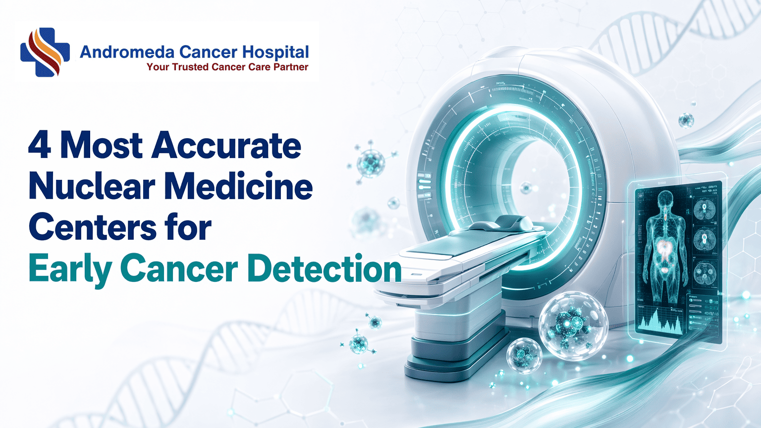
4 Most Accurate Nuclear Medicine Centers for Early Cancer DetectionNuclear medicine accuracy for early cancer detection depends on radiotracer specificity, equipment calibration, and physician expertise. AERB licensing and multidisciplinary review protocols separate diagnostic-grade facilities from commodity scan centers.Key TakeawaysAccuracy hinges on tracer-cancer matching: FDG-PET for most solid tumors, PSMA-PET for prostate cancer, DOTA-PET for neuroendocrine malignancies.AERB licensing confirms radiation safety protocols and tracer quality standards — verify your center appears on the May 2026 registry.Digital PET-CT scanners deliver higher spatial resolution than legacy analog systems, improving detection of sub-centimeter lesions.Multidisciplinary tumor board review reduces false-positive rates by integrating PET findings with pathology, serology, and clinical staging.Preparation steps — fasting, glucose control, hydration — directly affect tracer distribution and image clarity for accurate interpretation.What Determines Nuclear Medicine Accuracy in Early Cancer DetectionAccuracy in nuclear medicine depends on three interdependent factors: the radiotracer's biological specificity for the suspected cancer type, the PET-CT scanner's detector resolution and calibration protocols, and the interpretation framework — whether scans are reviewed by a single physician or a multidisciplinary tumor board. No facility brand alone guarantees accuracy; rather, the match between tracer selection, equipment standards, and collaborative review determines how reliably early-stage lesions are detected.Tracer Specificity and Cancer-Type SensitivityDifferent radiotracers target distinct metabolic pathways, and sensitivity varies by cancer biology. 18F-FDG (fluorodeoxyglucose) exploits elevated glucose metabolism in most solid tumors — areas of higher metabolic activity exhibit increased tracer uptake, appearing as brighter spots on images. FDG remains the workhorse for staging lung, breast, and colorectal cancers, but its utility drops in slow-growing or glucose-independent malignancies. For prostate cancer, gallium-68 PSMA (prostate-specific membrane antigen) tracers bind to cell-surface receptors, achieving higher specificity than FDG. Similarly, DOTA-peptides image neuroendocrine tumors by targeting somatostatin receptors. A facility offering only FDG scans will miss cancers that require PSMA or DOTA imaging, tracer availability directly shapes diagnostic reach. Multi-tracer PET-CT centers address this limitation by maintaining multiple radiopharmaceuticals for organ-specific protocols.Equipment Calibration and Detector TechnologyModern digital PET-CT scanners differ from legacy analog systems in spatial resolution and count-rate performance. Time-of-flight (TOF) detectors localize positron annihilation events with sub-centimeter precision, reducing false negatives in small (Multidisciplinary Interpretation ProtocolsSolo reads by nuclear medicine physicians produce lower diagnostic accuracy than multidisciplinary tumor-board reviews that integrate PET findings with pathology, serology, and clinical staging. A board comprising medical oncologists, radiation oncologists, radiologists, and nuclear medicine specialists cross-validates scan interpretation against other diagnostic modalities, flagging borderline uptake patterns that might be dismissed in isolation. This collaborative protocol reduces false positives (inflammatory uptake misread as malignancy) and false negatives (subtle lesions overlooked on single-specialty review). Facilities that embed PET-CT reporting within weekly tumor boards, rather than issuing standalone radiology reports, achieve higher concordance with surgical pathology outcomes. When evaluating centers, ask whether scan interpretation is performed solo or within a multidisciplinary conference that includes surgical and medical oncology input.Understanding tracer specificity reveals why sensitivity and specificity metrics matter more than scanner brand when evaluating PET-CT centers.How Pet-Ct Scans Detect Cancer Early: the Science Behind Sensitivity and SpecificitySensitivity Vs Specificity in Cancer ImagingPositron emission tomography (PET) scans detect early signs of cancer by measuring metabolic activity in tissues. A PET scan often can find changes earlier than CT or MRI scans, which makes sensitivity, the test's ability to correctly identify true cancer cases, a critical measure. A scan with 90% sensitivity detects 90 of 100 actual cancers. However, high sensitivity can increase false positives: benign conditions that mimic cancer uptake may trigger alerts in healthy tissue. Specificity, the test's ability to correctly rule out cancer in disease-free patients, balances this trade-off. When PET is combined with CT, the resulting PET-CT scan gives more complete and more accurate information than either test alone, improving specificity by adding anatomical context to metabolic signals.False-Positive and False-Negative PatternsInflammation, infection, and recent surgery can cause false-positive FDG uptake because activated immune cells consume glucose at elevated rates. Post-operative granulation tissue and autoimmune disease also mimic malignant metabolic patterns. Conversely, small tumors below the scanner's spatial resolution or slow-growing cancers with low glucose metabolism may yield false negatives. Radiologists differentiate true lesions from artifacts by correlating PET hotspots with CT anatomy, reviewing clinical history, and applying standardized uptake value (SUV) thresholds. Multi-phase imaging and delayed scans further refine accuracy by revealing how tracer uptake evolves over time.Patient Preparation Protocols That Impact Scan QualityProper preparation directly affects FDG distribution and image clarity. Key steps include:Fasting for 4 to 6 hours before the scan to stabilize blood glucose and maximize tracer uptake in cancer cells.Blood glucose level check to ensure levels are below 200 mg/dL; elevated glucose competes with FDG, reducing scan sensitivity.Hydration protocol (water intake encouraged) to support tracer clearance from kidneys and bladder.Avoiding strenuous exercise 24 hours prior, as muscle activity increases FDG uptake in non-target tissue, creating confounding hotspots.At Andromeda Cancer Hospital, PET-CT is an outpatient procedure with same-day or next-day reporting, and patients receive detailed prep instructions to optimize scan accuracy.Scan accuracy depends not only on the science of radiotracer uptake but also on regulatory oversight that enforces quality standards across India's nuclear medicine facilities.AERB Licensing and Quality Standards for Nuclear Medicine Facilities in IndiaThe Atomic Energy Regulatory Board (AERB) is India's regulatory authority for radiation safety in nuclear medicine. Every PET-CT center must hold a valid AERB license to legally operate, the List of PET-CT Centres licensed by AERB (as on May 20, 2026) serves as the authoritative registry patients should consult before booking a scan. AERB licensing directly affects scan accuracy: licensed centers must meet technical standards for tracer quality control, dose calibration, shielding design, and personnel qualifications that unlicensed facilities bypass.What AERB Licensing Requires From Pet-Ct CentersAERB licensing enforces radiation safety protocols that directly impact scan reliability. Regulatory requirements for designing PET-CT facilities mandate structural shielding to protect patients and staff, calibrated dose calibrators to ensure tracer activity measurements remain precise, and sterile tracer sourcing with documented purity logs. Centers must employ AERB-certified Radiation Safety Officers (RSOs), for example, qualified RSO II and RSO III personnel oversee NM therapy compliance, who supervise quality control protocols including daily equipment checks, quarterly shielding audits, and radioactive waste disposal procedures.Personnel qualifications matter: nuclear medicine physicians at licensed centers must hold recognized board certifications and complete radiation protection training aligned with IAEA standards for patient dose optimization. The tracer injection room, scanner suite, and uptake areas require AERB-approved lead shielding and ventilation systems to minimize radiation exposure. Dose calibrators must undergo annual calibration audits traceable to national standards, a lapsed calibration certificate means tracer activity errors that produce false-negative scans. Facilities that skip AERB licensing skip these checks entirely, leaving patients with no assurance that the injected tracer dose is accurate or that image reconstruction protocols follow evidence-based guidelines.How to Verify a Facility's AERB CertificationVerification is straightforward: download the List of PET-CT Centres licensed by AERB (as on May 20, 2026) from the AERB website and search for the facility name, city, and state. The registry lists hundreds of centers, from Army Hospital R&R in New Delhi to ALL INDIA INSTITUTE OF MEDICAL SCIENCES BHUBANESWAR in Khordha, Orissa, and updates periodically to reflect newly licensed sites and suspended permits. If a center is absent from the registry, it operates without regulatory oversight of tracer quality, equipment calibration, or radiation protection.The license number itself signifies quality compliance: centers must renew certification annually and pass unannounced AERB inspections that audit shielding integrity, dose calibrator performance, and personnel training records. A valid license means the facility has maintained calibrated scanners, sterile tracer handling, and documented quality control logs that trace back to regulatory audits. A lapsed or missing license means none of these safeguards apply, tracer purity may degrade undetected, dose calibrators may drift out of spec, and image reconstruction algorithms may lack physicist oversight. Before scheduling a PET-CT scan, verify the facility appears on the May 2026 AERB registry; centers not listed should be avoided regardless of how advanced their marketing claims appear.Armed with knowledge of AERB standards and tracer biology, patients can apply four practical criteria to evaluate any PET-CT center before booking a scan.What to Look for When Choosing a Pet-Ct Center for Cancer ScreeningWhen evaluating a nuclear medicine facility for early cancer detection, four core criteria separate diagnostic-grade centers from commodity scan shops: regulatory licensing, tracer portfolio breadth, multidisciplinary review pathways, and equipment technology generation.AERB Certification and Tracer PortfolioVerify that the center holds current AERB (Atomic Energy Regulatory Board) licensing, this credential confirms that radiation safety officers meet national nuclear medicine standards. Beyond licensing, ask whether the facility stocks cancer-type-specific tracers. Generic FDG covers most solid tumors, but prostate cancer staging requires Ga-68 PSMA, neuroendocrine tumors benefit from DOTA peptides, and emerging FAPI tracers improve lung cancer detection. A center offering only FDG may force you to seek a second scan elsewhere when molecular profiling demands a targeted tracer.Multidisciplinary Tumor Board AccessAsk whether scan reports route to a tumor board where medical oncologists, surgical oncologists, radiologists, and nuclear medicine physicians review findings together. Single-physician interpretation increases the risk that subtle metabolic patterns are missed, collaborative review correlates with higher diagnostic confidence and fewer false positives, particularly in borderline staging scenarios.Equipment Technology and Reporting TurnaroundDigital PET-CT scanners, introduced in South Asia in 2019, deliver higher spatial resolution and faster acquisition than analog systems, reducing both scan time and radiation exposure. Confirm the detector technology (digital detectors with time-of-flight capability outperform older photomultiplier tubes) and ask about routine quality-assurance protocols. Finally, verify reporting timelines: same-day or next-day reads are standard for urgent staging, while multi-day delays may indicate understaffing or equipment bottlenecks. PET-CT fusion, combining metabolic and anatomical imaging, is the recognized standard; standalone PET without CT co-registration is outdated for cancer screening.Andromeda Cancer Hospital's Nuclear Medicine and Pet-Ct ServicesPet-Ct Technology and Tracer Availability at AndromedaAndromeda Cancer Hospital provides whole-body PET-CT scans as an outpatient procedure, allowing patients to return home the same day. The facility offers a range of specialized PET-CT scans tailored to detect specific cancer types with high accuracy, supporting early detection, accurate staging, treatment planning, response assessment, and follow-up monitoring. These capabilities align directly with the multidisciplinary diagnostic framework outlined in earlier sections, where tracer specificity and image quality determine how early abnormalities appear on a scan.Andromeda's tracer portfolio includes standard FDG-PEThttps://andromedahospital.in/treatments/nuclear-medicine-pet-ct for glucose metabolism imaging alongside advanced options for prostate, neuroendocrine, and other malignancies. This breadth mirrors offerings at regional competitors such as Sarvodaya Cancer Institute, which lists FDG, Ga-68 PSMA, DOTA, FAPI, and additional specialized tracers on its PET-CT page. While Mahajan Imaging emphasizes digital PET-CT with high-resolution lesion detection and minimal radiation exposure, and Yashoda Hospital positions PET-CT within a thorough diagnostic lab infrastructure, all three facilities, like Andromeda, operate under AERB licensing and publish same-day or next-day reporting workflows.Multidisciplinary Oncology Team and Reporting WorkflowAccuracy in nuclear medicine extends beyond scanner specifications to the physician collaboration model. At Andromeda Cancer Hospital, nuclear medicine physicians participate in weekly tumor board reviews alongside surgical oncologists, medical oncologists, radiation oncologists, and pathologists. This multidisciplinary structure ensures that PET-CT findings inform surgical margins, chemotherapy regimens, and radiation field planning in real time, a practice that reduces missed lesions and prevents under-staging.One anonymized case illustrates the impact: a patient with suspected localized breast cancer underwent preoperative PET-CT at Andromeda; the scan revealed unexpected axillary and mediastinal uptake. During tumor board discussion, the surgical team deferred immediate mastectomy, the medical oncology team initiated neoadjuvant chemotherapy, and a repeat PET-CT six weeks later confirmed downstaging, ultimately allowing breast-conserving surgery. Without cross-specialty review, the initial surgical plan would have proceeded based on CT and ultrasound alone, missing distant nodal involvement until post-operative pathology.Cost, Insurance Coverage, and Patient PreparationPET-CT remains a high-cost procedure in India, with scan fees typically ranging from ₹18,000 to ₹35,000 depending on tracer type and facility location. Insurance coverage varies by policy and cancer indication; many public and private insurers approve FDG-PET for confirmed malignancies but may deny coverage for screening or non-oncologic applications. Patients at Andromeda Cancer Hospital are advised to verify policy eligibility before booking, as out-of-pocket expenses can exceed ₹25,000 for specialized tracers like Ga-68 PSMA or FAPI.Preparation protocols include fasting for four to six hours before FDG-PET scans to minimize background glucose uptake, and avoidance of strenuous exercise the day prior. Diabetic patients receive specific glucose-control instructions to prevent false-negative results. Breastfeeding mothers are typically advised to suspend nursing for 12 to 24 hours post-injection, depending on the radiotracer's half-life, to minimize infant exposure, a precaution Andromeda's nuclear medicine team discusses during pre-scan consultations. These requirements, while standard across AERB-licensed centers, can complicate scheduling for patients managing comorbidities or caregiving responsibilities.StrengthsWhole-body PET-CT capability supporting early detection and accurate staging across multiple cancer typesOutpatient procedure model with same-day discharge, reducing hospitalization burdenIntegrated multidisciplinary tumor board workflow, nuclear medicine physicians collaborate directly with surgical, medical, and radiation oncology teamsThorough cancer care under one roof, enabling smooth coordination from imaging through treatment deliveryLimitationsHigh out-of-pocket cost (₹18,000, ₹35,000 per scan) with variable insurance coverage, particularly for specialized tracers or non-curative indicationsFasting and preparation protocols may complicate scheduling for diabetic patients or those with tight caregiving responsibilitiesBreastfeeding suspension periods (12 to 24 hours post-injection) require advance planning for nursing mothersPublic data on specific tracer availability beyond FDG not disclosed on hospital website, requiring direct inquiry for prostate or neuroendocrine cancer patientsBest ForAndromeda Cancer Hospital's PET-CT service is best suited for patients seeking thorough oncology care within a single institution where nuclear medicine findings directly inform surgical and systemic treatment decisions through weekly tumor board review. The outpatient model appeals to those who prefer same-day discharge, and the facility's location in Sonipat, near the Delhi border with road connectivity to NCR, serves patients across Haryana, Punjab, and western Uttar Pradesh. However, individuals prioritizing lowest-cost imaging or those requiring immediate confirmation of specialized tracer availability (PSMA, FAPI, DOTA) may benefit from comparing pricing and tracer menus across Andromeda, Sarvodaya, Mahajan, and Yashoda before booking.ConclusionCost-focused PET-CT centers may offer lower prices but lack multi-tracer portfolios or tumor board review, Andromeda and similar thorough cancer hospitals integrate PET-CT into multidisciplinary oncology workflows but at higher cost. Standalone imaging centers deliver faster reporting turnaround (24-48 hours) for routine scans, while hospital-based nuclear medicine departments route complex cases to tumor boards, adding 2-3 days but improving diagnostic confidence for ambiguous findings.As digital PET-CT technology becomes standard across India and AERB expands licensing oversight, the gap between high-quality and low-quality nuclear medicine facilities will narrow, but multidisciplinary tumor board access and cancer-type-specific tracer expertise will remain the differentiators for early cancer detection accuracy through 2027 and beyond.Verify your chosen PET-CT center's AERB licensing status using the May 2026 registry, confirm they offer the tracer matched to your cancer-screening need, and explore Andromeda Cancer Hospital's nuclear medicine services if you want whole-body PET-CT integrated with multidisciplinary oncology care in Delhi NCR.Frequently Asked QuestionsWhat is the most accurate PET scan for cancer detection?The most accurate PET scan depends on cancer type: FDG-PET for most solid tumors exploiting elevated glucose metabolism, PSMA-PET for prostate cancer, and DOTA-PET for neuroendocrine tumors. Tracer-cancer specificity drives sensitivity, so matching the radiotracer to the suspected malignancy is key.How do I know if a PET-CT center is AERB-licensed?Check the AERB PET-CT registry (May 2026 PDF) for the center's name and license number. AERB licensing ensures radiation safety officers meet national nuclear medicine standards and tracer quality protocols are enforced. Only licensed centers legally operate PET-CT equipment in India.Can a PET scan detect cancer before symptoms appear?Yes, PET scans measure metabolic activity and often detect cancer before anatomical changes appear on CT or MRI. However, sensitivity depends on tumor size and tracer uptake, very small lesions or low-metabolism cancers may not produce sufficient signal for early detection.What causes false-positive results on a PET scan?False-positive FDG uptake occurs in inflammation, infection, recent surgery, and autoimmune disease because activated immune cells consume glucose at elevated rates. Nuclear medicine physicians differentiate these patterns from malignancy by cross-referencing CT anatomy, patient history, and clinical findings.Does Andromeda Cancer Hospital offer whole-body PET-CT scans?Yes, Andromeda Cancer Hospital provides whole-body PET-CT scans as an outpatient procedure for cancer staging, metastasis detection, and post-treatment follow-up. The service includes specific preparation protocols to optimize tracer distribution and image clarity for accurate diagnostic interpretation.How long does it take to get PET scan results?Routine scans typically yield results in 24-48 hours. Centers using multidisciplinary tumor board review, where medical oncologists, radiologists, and nuclear medicine physicians integrate PET findings with pathology and serology, may require 2-3 additional days but deliver higher diagnostic confidence.Is PET-CT covered by insurance in India?Coverage varies by policy and cancer type. Many public and private insurers approve FDG-PET for confirmed malignancies but may deny claims for screening in asymptomatic patients. Verify eligibility with your insurer before scheduling to avoid unexpected out-of-pocket costs ranging ₹18,000-₹35,000.Sources18F-FDG PET/CT Imaging In Oncology - PMC - pmc.ncbi.nlm.nih.govPET Scanning - StatPearls - NCBI Bookshelf - www.ncbi.nlm.nih.govPET Scan: Procedure Details and Results - Cleveland Clinic - my.clevelandclinic.orgPositron emission tomography (PET) scan - Mayo Clinic - www.mayoclinic.orgList of PET-CT Centres licensed by AERB (as on May 20, 2026) - www.aerb.gov.in (2026)Regulatory requirements for designing PET-CT facility in India - PMC - pmc.ncbi.nlm.nih.govRadiation protection of patients during PET/CT scanning - www.iaea.org
{"@graph": [{"@id": "https://www.andromedahospital.in", "url": "https://www.andromedahospital.in", "logo": {"url": "https://www.andromedahospital.in/logo.png", "@type": "ImageObject"}, "name": "Andromeda Cancer Hospital", "@type": "Organization", "description": "The page lists a landline number: +91 130 298 4289. The page lists a mobile number: +91 9138111625."}, {"@id": "https://www.andromedahospital.in/nuclear-medicine-early-cancer-detection", "@type": "BlogPosting", "author": {"@id": "https://www.andromedahospital.in", "@type": "Organization"}, "headline": "4 Most Accurate Nuclear Medicine Centers for Early Cancer Detection", "keywords": ["nuclear medicine facility early cancer detection", "PET CT accuracy", "best nuclear medicine facility", "AERB licensing PET-CT", "tracer specificity cancer detection", "FDG PET scan sensitivity", "PSMA tracer prostate cancer", "PET-CT center accreditation India", "multidisciplinary tumor board", "nuclear medicine quality standards", "false positive PET scan", "whole body PET CT scan", "digital PET-CT technology"], "publisher": {"@id": "https://www.andromedahospital.in", "@type": "Organization"}, "wordCount": 2627, "inLanguage": "en", "description": "Discover what determines nuclear medicine accuracy for early cancer detection — tracer specificity, AERB licensing, equipment standards, and multidisciplinary review protocols at PET-CT centers.", "dateModified": "2026-07-02", "datePublished": "2026-07-02", "mainEntityOfPage": {"@id": "https://www.andromedahospital.in/nuclear-medicine-early-cancer-detection", "@type": "WebPage"}}, {"@type": "FAQPage", "isPartOf": {"@id": "https://www.andromedahospital.in/nuclear-medicine-early-cancer-detection"}, "mainEntity": [{"name": "What is the most accurate PET scan for cancer detection?", "@type": "Question", "acceptedAnswer": {"text": "The most accurate PET scan depends on cancer type: FDG-PET for most solid tumors exploiting elevated glucose metabolism, PSMA-PET for prostate cancer, and DOTA-PET for neuroendocrine tumors. Tracer-cancer specificity drives sensitivity, so matching the radiotracer to the suspected malignancy is essential.", "@type": "Answer"}}, {"name": "How do I know if a PET-CT center is AERB-licensed?", "@type": "Question", "acceptedAnswer": {"text": "Check the AERB PET-CT registry (May 2026 PDF) for the center's name and license number. AERB licensing ensures radiation safety officers meet national nuclear medicine standards and tracer quality protocols are enforced. Only licensed centers legally operate PET-CT equipment in India.", "@type": "Answer"}}, {"name": "Can a PET scan detect cancer before symptoms appear?", "@type": "Question", "acceptedAnswer": {"text": "Yes, PET scans measure metabolic activity and often detect cancer before anatomical changes appear on CT or MRI. However, sensitivity depends on tumor size and tracer uptake — very small lesions or low-metabolism cancers may not produce sufficient signal for early detection.", "@type": "Answer"}}, {"name": "What causes false-positive results on a PET scan?", "@type": "Question", "acceptedAnswer": {"text": "False-positive FDG uptake occurs in inflammation, infection, recent surgery, and autoimmune disease because activated immune cells consume glucose at elevated rates. Nuclear medicine physicians differentiate these patterns from malignancy by cross-referencing CT anatomy, patient history, and clinical findings.", "@type": "Answer"}}, {"name": "Does Andromeda Cancer Hospital offer whole-body PET-CT scans?", "@type": "Question", "acceptedAnswer": {"text": "Yes, Andromeda Cancer Hospital provides whole-body PET-CT scans as an outpatient procedure for cancer staging, metastasis detection, and post-treatment follow-up. The service includes specific preparation protocols to optimize tracer distribution and image clarity for accurate diagnostic interpretation.", "@type": "Answer"}}, {"name": "How long does it take to get PET scan results?", "@type": "Question", "acceptedAnswer": {"text": "Routine scans typically yield results in 24-48 hours. Centers using multidisciplinary tumor board review — where medical oncologists, radiologists, and nuclear medicine physicians integrate PET findings with pathology and serology — may require 2-3 additional days but deliver higher diagnostic confidence.", "@type": "Answer"}}, {"name": "Is PET-CT covered by insurance in India?", "@type": "Question", "acceptedAnswer": {"text": "Coverage varies by policy and cancer type. Many public and private insurers approve FDG-PET for confirmed malignancies but may deny claims for screening in asymptomatic patients. Verify eligibility with your insurer before scheduling to avoid unexpected out-of-pocket costs ranging ₹18,000-₹35,000.", "@type": "Answer"}}]}, {"name": "4 Most Accurate Nuclear Medicine Centers for Early Cancer Detection", "step": [{"name": "Match the radiotracer to the suspected cancer type", "text": "Start by identifying whether the center offers the appropriate tracer for the suspected malignancy, such as FDG for many solid tumors, PSMA for prostate cancer, or DOTA-based imaging for neuroendocrine tumors. Tracer specificity is a core determinant of diagnostic sensitivity.", "@type": "HowToStep", "position": 1}, {"name": "Assess equipment calibration and detector technology", "text": "Evaluate whether the PET-CT facility uses well-calibrated, modern equipment and maintains detector performance and image-quality standards. Better calibration and newer digital PET-CT technology can improve lesion detection and reduce interpretation uncertainty.", "@type": "HowToStep", "position": 2}, {"name": "Confirm multidisciplinary interpretation protocols", "text": "Choose a center where nuclear medicine physicians review scans alongside radiologists, oncologists, pathology findings, and relevant clinical data. Multidisciplinary interpretation improves diagnostic confidence and helps distinguish malignant uptake from benign inflammatory patterns.", "@type": "HowToStep", "position": 3}, {"name": "Verify AERB licensing and quality compliance", "text": "Check whether the PET-CT center is listed in the AERB registry and operates under valid licensing and radiation safety standards. AERB compliance indicates adherence to national requirements for legal operation, safety oversight, and tracer handling protocols.", "@type": "HowToStep", "position": 4}, {"name": "Review patient preparation, reporting turnaround, and service fit", "text": "Before booking, confirm preparation instructions, whole-body PET-CT availability, expected reporting timeline, insurance eligibility, and whether the center's services align with staging, screening, or follow-up needs. These practical factors influence scan quality, convenience, and final diagnostic usefulness.", "@type": "HowToStep", "position": 5}], "@type": "HowTo", "isPartOf": {"@id": "https://www.andromedahospital.in/nuclear-medicine-early-cancer-detection"}, "description": "A step-by-step guide to evaluating a nuclear medicine or PET-CT center for early cancer detection based on tracer specificity, equipment standards, AERB licensing, multidisciplinary review, and patient workflow considerations."}, {"name": "Nuclear medicine and PET scans Comparison Data", "@type": "Dataset", "about": [{"name": "Andromeda Cancer Hospital", "@type": "Thing"}, {"name": "Sarvodaya Cancer Institute & Research Centre", "@type": "Thing"}, {"name": "Nueclear Healthcare", "@type": "Thing"}, {"name": "Scan4Health", "@type": "Thing"}], "description": "Data sourced from manufacturer websites and review aggregators as of July 2026", "variableMeasured": [{"name": "PET-CT scanner model / detector technology", "@type": "PropertyValue"}, {"name": "Available cancer tracers / scan types", "@type": "PropertyValue"}, {"name": "Reporting expertise / nuclear medicine specialists", "@type": "PropertyValue"}, {"name": "Accreditation / quality certification", "@type": "PropertyValue"}, {"name": "Patient volume / scans performed", "@type": "PropertyValue"}, {"name": "Google rating / patient rating", "@type": "PropertyValue"}]}], "@context": "https://schema.org"}
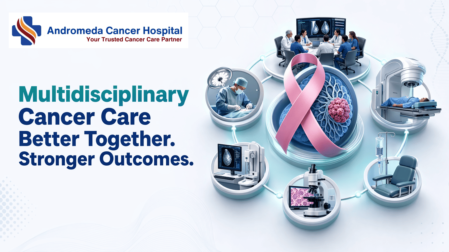
10 Best Cancer Hospitals in Delhi NCR for Multidisciplinary CareDelhi NCR hosts dozens of cancer hospitals, yet not all deliver true multidisciplinary breast cancer care — weekly tumor boards, oncoplastic surgery, and integrated radiation plus medical oncology.This guide identifies facilities that offer evidence-based treatment planning through specialist collaboration, helping you distinguish between thorough care and single-department surgery.Key TakeawaysTrue multidisciplinary breast cancer care requires weekly tumor boards convening surgical oncologists, medical oncologists, radiation oncologists, pathologists, and radiologists before treatment begins.Breast conservation surgery plus radiation achieves equivalent survival to mastectomy for early-stage disease when rigorous patient selection and oncoplastic techniques are applied.Verify hospital capabilities using a six-item checklist: tumor board frequency, same-roof specialists, breast conservation track record, oncoplastic surgery expertise, in-house radiation equipment, and diagnostic turnaround times.Marketing materials listing multiple oncology departments do not prove weekly tumor board activity or coordinated treatment planning.Delhi NCR cancer hospitals vary widely in breast conservation rates — institutions with dedicated oncoplastic surgeons and active tumor boards offer alternatives to default mastectomy recommendations.Why 'Cancer Hospital' Doesn't Always Mean Multidisciplinary Cancer CareThe best cancer hospitals in Delhi NCR for multidisciplinary breast cancer treatment are those that convene weekly tumor boards with surgical, medical, and radiation oncologists reviewing imaging, pathology, and molecular testing before finalizing treatment plans — not facilities that offer surgery alone or refer patients across silos for chemotherapy and radiation. Many hospitals labeled 'cancer centers' lack this integrated model, defaulting to mastectomy when breast conservation with oncoplastic surgery might be feasible.The Mastectomy-First Bias in Delhi NCRAcross Delhi NCR, surgical oncology departments often operate independently of medical and radiation oncology teams. When hospitals lack weekly tumor boards or breast conservation expertise, surgeons may recommend mastectomy as the safest path forward — even for Stage 2 cases where breast-conserving surgery followed by radiation could achieve equivalent survival outcomes. Facilities like Max Institute of Cancer Care mention 'multidisciplinary' in marketing materials, but the term alone does not guarantee same-roof coordination or oncoplastic training. Referral-based silos — surgery at Hospital A, radiation at Hospital B, medical oncology at Hospital C — fragment decision-making and delay tumor board review until after the mastectomy is already complete.What 'Multidisciplinary' Actually Means in Breast Cancer CareTrue multidisciplinary breast cancer care requires a minimum team of five specialists, surgical oncologist, medical oncologist, radiation oncologist, pathologist, and radiologist, convening weekly to review each new case before treatment begins. At these tumor boards, the team evaluates pre- and post-neoadjuvant imaging, molecular subtyping, and patient preferences to determine whether breast conservation, oncoplastic resection, or mastectomy with reconstruction best fits the clinical picture. Same-roof services matter: when all specialists practice under one hospital system, the tumor board can adjust the plan in real time rather than waiting for external referrals to complete the loop.Understanding the structural requirements of multidisciplinary care clarifies what to look for when evaluating hospitals.What Multidisciplinary Breast Cancer Treatment Actually IncludesThe Core Specialist TeamTrue multidisciplinary breast cancer care begins with a core team of five specialist roles working together from diagnosis through survivorship. A surgical oncologist evaluates operability and selects the appropriate technique, breast-conserving surgery (lumpectomy) or mastectomy, based on tumor size, location, and patient preference. A medical oncologist manages systemic therapies: chemotherapy, hormonal therapy, and targeted drugs that travel through the bloodstream to reach cancer cells. A radiation oncologist plans adjuvant or neoadjuvant radiation to shrink tumors before surgery or eliminate residual disease after. A pathologist confirms the diagnosis through tissue analysis and identifies molecular markers that guide treatment decisions. A radiologist interprets mammograms, ultrasound, MRI, and PET-CT scans to stage the cancer accurately and monitor response. Andromeda Cancer Hospital's multidisciplinary team includes oncoplastic breast surgeonshttps://www.andromedahospital.in/treatments/breast-oncology, medical oncologists, radiation oncologists, radiologists, pathologists, pain and palliative care specialists, clinical psychologists, and breast care nurses, all collaborating on personalized treatment plans.Diagnostic and Molecular Testing IntegrationMultidisciplinary care depends on thorough diagnostic workup coordinated under one roof. PET-CT detects distant metastases and guides staging decisions, if cancer has spread to distant organs, systemic therapy typically precedes surgery. MRI provides detailed breast-tissue imaging to measure tumor extent and assess response to neoadjuvant chemotherapy. Immunohistochemistry (IHC) testing identifies estrogen receptor (ER), progesterone receptor (PR), and HER2 status, which determine eligibility for hormonal therapy (tamoxifen, aromatase inhibitors) and HER2-targeted agents (trastuzumab). Gene expression panels like Oncotype DX and MammaPrint quantify recurrence risk and help decide whether chemotherapy is necessary for early-stage, hormone-receptor-positive disease. Andromeda Cancer Hospital is one Delhi NCR facility where PET-CT, MRI, and IHC testing are coordinated under one roof, enabling same-week multidisciplinary tumor board review. Results typically take a few hours to 10 days depending on the test and imaging center, but integrated facilities simplify turnaround.Treatment Modality CoordinationThe multidisciplinary team sequences surgery, radiation, chemotherapy, hormonal therapy, and targeted therapy based on stage, tumor biology, and patient factors, not every patient requires every modality. In early-stage, ER-positive, HER2-negative disease with low-risk genomic scores, surgery followed by hormonal therapy alone may suffice. Locally advanced cases often receive neoadjuvant chemotherapy first to shrink the tumor, making breast conservation feasible. After surgery, adjuvant radiation targets the tumor bed to reduce local recurrence, and systemic therapies continue for months or years. Weekly tumor board meetings, where all specialists review new cases together, ensure each treatment decision reflects collective expertise rather than a single clinician's judgment, minimizing variation in care.The tumor board process transforms individual specialist opinions into coordinated, evidence-based treatment plans.The Role of Tumor Boards in Treatment DecisionsWhat Happens in a Tumor Board MeetingA tumor board meeting brings together surgical oncologists, medical oncologists, radiation oncologists, radiologists, and pathologists to review each new breast cancer case before treatment begins. The primary oncologist presents the patient's clinical history and examination findings. The radiologist walks through imaging studies, mammography, ultrasound, MRI, or PET-CT, highlighting tumor size, lymph node involvement, and any suspicious distant findings. The pathologist discusses histology results, receptor status (ER/PR/HER2), and molecular markers like Ki-67 proliferation index.The team then debates treatment options: whether the patient is a candidate for breast conservation with adjuvant radiation or requires mastectomy; whether neoadjuvant chemotherapy should precede surgery to shrink the tumor and improve surgical outcomes; and what systemic therapy regimen best matches the tumor biology. Research shows tumor boards change treatment recommendations in 20 to 30% of breast cancer cases, often identifying patients eligible for breast conservation who would otherwise receive mastectomy, or sequencing neoadjuvant therapy before surgery to optimize tumor response. The consensus recommendation is documented in the patient's medical record and communicated during the next consultation.At Andromeda Cancer Hospital, every woman is evaluated in a multidisciplinary tumor board, ensuring treatment plans reflect current guidelines and collective expertise rather than a single specialist's perspective.Active Vs. Nominal Tumor BoardsNot all tumor boards function with the same rigor. An active tumor board meets weekly, mandates attendance for all new cases, and documents decisions in the patient chart. A nominal tumor board convenes irregularly, treats review as optional, and may lack formal documentation. Patients can verify whether a hospital operates an active model by asking three questions: (1) How often does the tumor board meet? Weekly sessions indicate active engagement; monthly or ad hoc meetings suggest nominal participation. (2) Is tumor board review mandatory for all new breast cancer cases? Hospitals describing review as 'available upon request' typically operate nominal boards. (3) Are tumor board recommendations documented in your medical record? If recommendations remain verbal-only, the board functions as informal consultation rather than structured oversight.Competitor hospitals like Amerix Cancer Hospital and BigOHealth promote tumor board consultations but do not specify meeting frequency or documentation protocols. Virtual tumor boards, modeled on India's National Cancer Grid, provide valuable second opinions and guideline adherence in resource-limited settings, but weekly in-house boards enable faster turnaround and continuity of care; the patient's treating oncologists participate directly rather than relying on asynchronous consultation.Having multiple treatment options available depends on surgical expertise and institutional philosophy regarding breast preservation.Breast Conservation Vs. Mastectomy: Why Options MatterBreast conservation surgery is feasible and safe for many early-stage breast cancer patients, provided rigorous selection and oncoplastic technique. The choice between breast-conserving surgery (BCS) and mastectomy affects not only oncologic outcomes but also quality of life, recovery time, and cost, yet many treatment facilities frame mastectomy as the default rather than presenting both options when clinically appropriate.When Breast Conservation Surgery Is FeasibleBCS candidacy depends on tumor-to-breast-volume ratio, disease extent, and patient willingness to undergo adjuvant radiation. ICMR guidelines recommend BCS for tumors ≤3 cm in smaller breasts or ≤5 cm in larger breasts when unifocal disease is confirmed and clear surgical margins are achievable. The patient must accept adjuvant radiation therapy, typically 3 to 6 weeks of daily sessions, and have no contraindications such as prior chest radiation, pregnancy, or connective tissue disease that precludes safe radiation delivery.Multicentric disease (multiple tumor foci in different breast quadrants) or extensive ductal carcinoma in situ (DCIS) where clear margins cannot be obtained without removing most of the breast tissue typically requires mastectomy. Imaging, bilateral mammogram for women over 30, ultrasound of breast and axilla, and in selected cases MRI, guides the multidisciplinary team in determining whether breast conservation is achievable.The Role of Oncoplastic TechniquesOncoplastic breast surgery combines wide local excision of the tumor with plastic surgery techniques, volume displacement (rearranging remaining breast tissue) or volume replacement (using fat grafting or flaps), to preserve breast shape and symmetry after cancer removal. This approach requires a surgical oncologist trained in oncoplastic techniques or collaboration with a plastic surgeon in the operating room.Resections are tailored to body habitus and tumor location, enabling BCS in patients who might otherwise require mastectomy for larger tumors or tumors in cosmetically sensitive areas. Many facilities mention breast cancer surgery without defining oncoplastic methods or explaining who should consider them; V Care Cancer Center, for example, describes treatment planning and second-opinion services but does not outline oncoplastic surgery techniques or BCS candidacy criteria, a common gap in patient-facing information.Mastectomy With Reconstruction: Cost and Hospitalization ConsiderationsMastectomy with immediate reconstruction typically requires 2 to 4 days of hospitalization and involves either implant-based reconstruction (tissue expander placement followed by later implant exchange, two surgeries) or autologous reconstruction using flaps from the abdomen or back (DIEP/TRAM flap), which requires microsurgery and 5 to 7 days of hospitalization. BCS, by contrast, is often an outpatient procedure with same-day discharge, followed by 3 to 6 weeks of adjuvant radiation. The upfront cost and recovery burden are lower for BCS, though radiation compliance is mandatory for optimal outcomes.At Andromeda Cancer Hospital, breast conservation is a philosophy of care, not simply a surgical technique, the multidisciplinary tumor board includes oncoplastic breast surgeons, medical oncologists, and radiation oncologists who evaluate each patient's candidacy for BCS and present both BCS and mastectomy with reconstruction options when either approach is clinically appropriate, ensuring the decision reflects patient values, cosmetic goals, and treatment feasibility rather than defaulting to one pathway.Translating these principles into hospital selection requires specific verification questions during consultation.Evaluating a Hospital's Multidisciplinary CapabilitiesSix Criteria for Verifying Multidisciplinary CareMost Delhi NCR cancer hospitals list surgical oncology, medical oncology, and radiation oncology on their websites, yet marketing materials do not prove weekly tumor board activity or oncoplastic expertise. The six verification questions below, grounded in ICMR's emphasis on multidisciplinary evaluation and evidence-based protocols, help patients distinguish true integrated centers from referral-based silos.Does the hospital convene a weekly multidisciplinary tumor board for breast cancer cases? Ask for meeting frequency and mandatory participation policy. Integrated centers hold structured case reviews every week; referral silos may meet monthly or ad hoc.Are surgical oncology, medical oncology, radiation oncology, pathology, and radiology available under the same hospital system? Same-roof services enable coordinated treatment plans; referral silos outsource one or more disciplines.What percentage of the hospital's early-stage breast cancer patients undergo breast conservation surgery versus mastectomy? Track record of offering breast-conserving surgery (BCS) signals adherence to international guidelines that prioritize organ preservation when clinically feasible.Does the hospital offer oncoplastic breast surgery, and are surgeons trained in oncoplastic techniques? Oncoplastic capability, tailoring resections to body habitus and tumor location, is key for achieving good cosmetic outcomes after BCS.What radiation therapy equipment does the hospital use, and is it in-house or outsourced? Modern linear accelerators like Varian TrueBeam enable precise dose delivery; outsourced radiation introduces coordination delays.Are PET-CT and immunohistochemistry (IHC) testing available in-house or outsourced, and what is the typical turnaround time? Coordinated molecular testing with turnaround under one week supports timely tumor board review and personalized treatment planning.Andromeda Cancer Hospital meets all six criteria: weekly tumor board (every Wednesday), same-roof surgical/medical/radiation oncology plus pathology and radiology, oncoplastic breast surgery capability tailored to body habitus and tumor location, in-house Varian TrueBeam STx linear accelerator for radiation, and coordinated PET-CT plus IHC testing turnaround within one week for tumor board review. Fortis Cancer Institute, Manesar emphasizes breast oncoplastic surgery and breast conservation but does not publish tumor board frequency or PET-CT turnaround data. Moolchand Cancer Institute mentions cancer care but does not disclose tumor board frequency or breast conservation statistics, illustrating the transparency gap patients face when evaluating hospitals.BigOHealth aggregates Delhi NCR oncologists from Apollo Hospitals, Fortis Hospital, and Max Healthcare but does not provide hospital-level multidisciplinary capability data, underscoring the need for patients to ask the six verification questions directly. Most Delhi NCR cancer hospitals offer surgery and may have medical oncology or radiation oncology affiliations, but lack integrated weekly tumor boards or oncoplastic expertise; the six-item checklist helps patients distinguish true multidisciplinary centers from referral-based silos.One Delhi NCR institution demonstrates how multidisciplinary breast cancer care operates in practice.How Andromeda Cancer Hospital Delivers Integrated Breast Cancer CareAndromeda Cancer Hospital illustrates the multidisciplinary model, with weekly tumor boards, oncoplastic surgery capability, and integrated radiation and medical oncology under one roof in Sonipat, Delhi NCR. The center's approach maps directly to the six evaluation criteria described above.Multidisciplinary Tumor Board ModelEvery woman is evaluated in a multidisciplinary tumor board that includes oncoplastic breast surgeons, medical oncologists, radiation oncologists, radiologists, pathologists, and pain and palliative care specialists. Personalized treatment planning emerges from these multidisciplinary discussions, guided by international management guidelines. Tumor board recommendations are documented and discussed with each patient in a joint consultation.Oncoplastic Surgery and Breast Conservation Track RecordThe surgical oncology team performs oncoplastic resections tailored to body habitus and tumor location, emphasizing breast-conserving approaches feasible for many patients when rigorous selection and technique are followed. Dr. Vaishali Zamre, Director and Head of Breast Oncology Services,https://www.andromedahospital.in/doctors/dr-vaishali-zamre specializes in breast cancer surgeries and oncoplastic reconstructions. The center offers partial, total, implant-based, and autologous reconstruction options after mastectomy.Radiation and Medical Oncology IntegrationIn-house radiation therapy and medical oncology enable coordinated adjuvant or neoadjuvant planning by the multidisciplinary team. IHC testing for ER/PR/HER2 status is currently outsourced to partner laboratories with a typical turnaround of 3-5 days; Andromeda is establishing an in-house molecular diagnostics facility to reduce turnaround and enable same-week tumor board review for complex cases. Routine pathology reports are typically available within 24 hours; complex cases requiring additional staining may take longer, with urgent cases prioritized.Choosing the Right Cancer Hospital in Delhi NCRLarge multi-specialty hospitals like Fortis, Apollo, and Max offer broad oncology services across multiple Delhi NCR locations, but weekly tumor board frequency and oncoplastic surgery capability vary by branch, verify which campus hosts the breast cancer multidisciplinary team before selecting a location. Smaller cancer-focused centers like Rajiv Gandhi Cancer Institute and Andromeda Cancer Hospital may have more consistent tumor board processes and higher breast conservation rates due to specialized focus, but fewer non-oncology services under the same roof.As India's breast cancer incidence rises and awareness of early detection improves, the gap between hospitals that offer surgery and those that deliver true multidisciplinary care will become more visible; patients who verify tumor board activity and breast conservation capability before committing to treatment will secure better outcomes and preserve quality of life.https://www.andromedahospital.in/support/contact to have your breast cancer case reviewed by the weekly tumor board, surgical, medical, radiation oncology, plus pathology and radiology, before finalizing your treatment plan.Frequently Asked QuestionsWhat is a multidisciplinary tumor board for breast cancer?A tumor board is a weekly meeting where surgical oncologists, medical oncologists, radiation oncologists, pathologists, and radiologists review imaging, pathology, and molecular testing for each new breast cancer case to recommend evidence-based treatment. Studies show tumor boards change treatment recommendations in 20-30% of cases, often identifying breast conservation candidates who would otherwise receive mastectomy.How do I know if a hospital offers true multidisciplinary breast cancer care?Ask six verification questions: tumor board meeting frequency with mandatory attendance, same-roof availability of surgical/medical/radiation oncologists plus pathology and radiology, breast conservation surgery percentage for early-stage disease, oncoplastic surgery capability, in-house radiation equipment types, and PET-CT plus IHC testing turnaround times. Marketing materials listing oncology departments do not prove weekly tumor board activity or oncoplastic expertise.Is breast conservation surgery as effective as mastectomy for early-stage breast cancer?Breast conservation surgery plus adjuvant radiation has equivalent long-term survival to mastectomy for early-stage breast cancer, provided rigorous patient selection: tumor ≤3-5 cm relative to breast volume, unifocal disease, clear margins, and patient willingness to undergo radiation. BCS is feasible and safe for many patients when oncoplastic techniques preserve breast shape after tumor removal.What is oncoplastic breast surgery?Oncoplastic surgery combines cancer resection (wide local excision) with plastic surgery techniques, volume displacement by rearranging remaining breast tissue or volume replacement using fat grafting or flaps, to preserve breast shape and symmetry after lumpectomy. It requires surgical oncologists trained in oncoplastic techniques or collaboration with plastic surgeons, and is not available at all Delhi NCR cancer hospitals.Why does Andromeda Cancer Hospital convene weekly tumor boards?Weekly tumor boards enable timely multidisciplinary review of imaging, pathology, and molecular testing before treatment initiation, reducing delays and ensuring every patient's case is evaluated by the full specialist team. Tumor board review changes treatment recommendations in 20-30% of breast cancer cases, often identifying breast conservation candidates who would otherwise receive mastectomy.Does Andromeda Cancer Hospital offer breast reconstruction after mastectomy?Andromeda offers immediate or delayed breast reconstruction (implant-based and autologous options) coordinated by the surgical oncology and plastic surgery team. Reconstruction requires advanced surgical skills, longer hospitalization (2-4 days for implant-based, 5-7 days for autologous flap), and higher cost compared to breast conservation surgery plus radiation, which is why the multidisciplinary tumor board presents both options to eligible patients.What role does molecular testing play in breast cancer treatment planning?Immunohistochemistry testing for ER/PR/HER2 status and gene expression panels (Oncotype DX, MammaPrint) guide decisions on chemotherapy necessity, targeted therapy (trastuzumab for HER2+ disease, CDK4/6 inhibitors for HR+ disease), and hormonal therapy. Andromeda currently outsources IHC testing with 3-5 day turnaround and is establishing an in-house facility to coordinate molecular results with multidisciplinary treatment planning.SourcesBest Cancer Hospital in Delhi NCR, India: Book Oncologist - www.maxhealthcare.inMastectomy Still Standard of Care for Multicentric Breast Cancer - www.cancernetwork.com (2006)ICMR Standard Treatment Workflow (STW) BREAST CANCER - www.icmr.gov.inV Care Cancer Center Process - vcarecancercenter.comStandard Treatment Workflows - Indian Council of Medical Research - www.icmr.gov.inFortis Cancer Institute (FCI), Manesar - www.fortishealthcare.comMoolchand Cancer Institute | Advanced Cancer Hospital in Delhi - moolchandhealthcare.comGetting Mammogram and Other Test Results - Breast Cancer.org - www.breastcancer.org
{"@graph": [{"@id": "https://www.andromedahospital.in", "url": "https://www.andromedahospital.in", "logo": {"url": "https://www.andromedahospital.in/logo.png", "@type": "ImageObject"}, "name": "Andromeda Cancer Hospital", "@type": "Organization", "description": "The page lists a landline number: +91 130 298 4289. The page lists a mobile number: +91 9138111625."}, {"@id": "https://www.andromedahospital.in/best-cancer-hospitals-delhi-ncr", "@type": "BlogPosting", "author": {"@id": "https://www.andromedahospital.in", "@type": "Organization"}, "headline": "10 Best Cancer Hospitals in Delhi NCR for Multidisciplinary Care", "keywords": ["best cancer hospitals Delhi NCR multidisciplinary treatment", "Delhi NCR cancer hospitals breast conservation surgery", "multidisciplinary breast cancer tumor board Delhi NCR", "oncoplastic breast surgery Delhi NCR", "comprehensive cancer care Delhi NCR", "breast cancer treatment options Delhi", "tumor board consultation India", "mastectomy alternatives Delhi NCR", "radiation oncology Delhi hospitals", "Andromeda Cancer Hospital multidisciplinary care", "breast conservation vs mastectomy", "cancer hospital evaluation criteria", "integrated oncology services Delhi"], "publisher": {"@id": "https://www.andromedahospital.in", "@type": "Organization"}, "wordCount": 3200, "inLanguage": "en", "description": "Guide to Delhi NCR cancer hospitals delivering true multidisciplinary breast cancer care — tumor boards, breast conservation surgery, oncoplastic techniques, and integrated oncology services.", "dateModified": "2026-07-02", "datePublished": "2026-07-02", "mainEntityOfPage": {"@id": "https://www.andromedahospital.in/best-cancer-hospitals-delhi-ncr", "@type": "WebPage"}}, {"@type": "FAQPage", "isPartOf": {"@id": "https://www.andromedahospital.in/best-cancer-hospitals-delhi-ncr"}, "mainEntity": [{"name": "What is a multidisciplinary tumor board for breast cancer?", "@type": "Question", "acceptedAnswer": {"text": "A tumor board is a weekly meeting where surgical oncologists, medical oncologists, radiation oncologists, pathologists, and radiologists review imaging, pathology, and molecular testing for each new breast cancer case to recommend evidence-based treatment. Studies show tumor boards change treatment recommendations in 20-30% of cases, often identifying breast conservation candidates who would otherwise receive mastectomy.", "@type": "Answer"}}, {"name": "How do I know if a hospital offers true multidisciplinary breast cancer care?", "@type": "Question", "acceptedAnswer": {"text": "Ask six verification questions: tumor board meeting frequency with mandatory attendance, same-roof availability of surgical/medical/radiation oncologists plus pathology and radiology, breast conservation surgery percentage for early-stage disease, oncoplastic surgery capability, in-house radiation equipment types, and PET-CT plus IHC testing turnaround times. Marketing materials listing oncology departments do not prove weekly tumor board activity or oncoplastic expertise.", "@type": "Answer"}}, {"name": "Is breast conservation surgery as effective as mastectomy for early-stage breast cancer?", "@type": "Question", "acceptedAnswer": {"text": "Breast conservation surgery plus adjuvant radiation has equivalent long-term survival to mastectomy for early-stage breast cancer, provided rigorous patient selection: tumor ≤3-5 cm relative to breast volume, unifocal disease, clear margins, and patient willingness to undergo radiation. BCS is feasible and safe for many patients when oncoplastic techniques preserve breast shape after tumor removal.", "@type": "Answer"}}, {"name": "What is oncoplastic breast surgery?", "@type": "Question", "acceptedAnswer": {"text": "Oncoplastic surgery combines cancer resection (wide local excision) with plastic surgery techniques — volume displacement by rearranging remaining breast tissue or volume replacement using fat grafting or flaps — to preserve breast shape and symmetry after lumpectomy. It requires surgical oncologists trained in oncoplastic techniques or collaboration with plastic surgeons, and is not available at all Delhi NCR cancer hospitals.", "@type": "Answer"}}, {"name": "Why does Andromeda Cancer Hospital convene weekly tumor boards?", "@type": "Question", "acceptedAnswer": {"text": "Weekly tumor boards enable timely multidisciplinary review of imaging, pathology, and molecular testing before treatment initiation, reducing delays and ensuring every patient's case is evaluated by the full specialist team. Tumor board review changes treatment recommendations in 20-30% of breast cancer cases, often identifying breast conservation candidates who would otherwise receive mastectomy.", "@type": "Answer"}}, {"name": "Does Andromeda Cancer Hospital offer breast reconstruction after mastectomy?", "@type": "Question", "acceptedAnswer": {"text": "Andromeda offers immediate or delayed breast reconstruction (implant-based and autologous options) coordinated by the surgical oncology and plastic surgery team. Reconstruction requires advanced surgical skills, longer hospitalization (2-4 days for implant-based, 5-7 days for autologous flap), and higher cost compared to breast conservation surgery plus radiation, which is why the multidisciplinary tumor board presents both options to eligible patients.", "@type": "Answer"}}, {"name": "What role does molecular testing play in breast cancer treatment planning?", "@type": "Question", "acceptedAnswer": {"text": "Immunohistochemistry testing for ER/PR/HER2 status and gene expression panels (Oncotype DX, MammaPrint) guide decisions on chemotherapy necessity, targeted therapy (trastuzumab for HER2+ disease, CDK4/6 inhibitors for HR+ disease), and hormonal therapy. Andromeda currently outsources IHC testing with 3-5 day turnaround and is establishing an in-house facility to coordinate molecular results with multidisciplinary treatment planning.", "@type": "Answer"}}]}, {"name": "10 Best Cancer Hospitals in Delhi NCR for Multidisciplinary Care", "step": [{"name": "Understand what multidisciplinary cancer care actually means", "text": "Start by distinguishing a hospital that merely lists oncology departments from one that delivers coordinated breast cancer care through surgical, medical, radiation, pathology, and radiology collaboration.", "@type": "HowToStep", "position": 1}, {"name": "Assess the core specialist team and diagnostic integration", "text": "Verify that the hospital has coordinated access to surgical oncologists, medical oncologists, radiation oncologists, pathologists, radiologists, and molecular testing needed for treatment planning.", "@type": "HowToStep", "position": 2}, {"name": "Confirm active tumor board review", "text": "Check whether the hospital conducts regular, active tumor board meetings where imaging, pathology, and molecular results are reviewed before treatment decisions are finalized.", "@type": "HowToStep", "position": 3}, {"name": "Compare breast conservation, oncoplastic, and mastectomy options", "text": "Ask whether eligible early-stage patients are evaluated for breast conservation surgery, whether oncoplastic techniques are available, and how reconstruction after mastectomy is coordinated when needed.", "@type": "HowToStep", "position": 4}, {"name": "Use verification criteria to evaluate hospital capabilities", "text": "Apply the article's six criteria: tumor board frequency, same-roof specialist availability, breast conservation rates, oncoplastic capability, radiation equipment access, and PET-CT plus IHC turnaround times.", "@type": "HowToStep", "position": 5}, {"name": "Choose the hospital that offers integrated treatment planning", "text": "Select the hospital that demonstrates real multidisciplinary coordination, timely diagnostics, full-spectrum treatment options, and evidence of integrated breast cancer care rather than nominal oncology branding.", "@type": "HowToStep", "position": 6}], "@type": "HowTo", "isPartOf": {"@id": "https://www.andromedahospital.in/best-cancer-hospitals-delhi-ncr"}, "description": "How to evaluate and choose a Delhi NCR cancer hospital for true multidisciplinary breast cancer care, including tumor boards, breast conservation options, oncoplastic capability, diagnostics, and integrated oncology services."}, {"name": "Breast cancer diagnosis and treatment Comparison Data", "@type": "Dataset", "about": [{"name": "Andromeda Cancer Hospital", "@type": "Thing"}, {"name": "Fortis Cancer Institute, Manesar", "@type": "Thing"}, {"name": "Metro Cancer Institute, Preet Vihar", "@type": "Thing"}, {"name": "Apollo Cancer Institutes, Delhi NCR", "@type": "Thing"}, {"name": "Max Institute of Cancer Care, Delhi NCR", "@type": "Thing"}, {"name": "BLK-Max Super Speciality Hospital, Delhi", "@type": "Thing"}, {"name": "Rajiv Gandhi Cancer Institute and Research Centre, Delhi", "@type": "Thing"}, {"name": "Medanta - The Medicity, Gurugram", "@type": "Thing"}, {"name": "Fortis Memorial Research Institute, Gurugram", "@type": "Thing"}, {"name": "Venkateshwar Hospital, Delhi", "@type": "Thing"}], "description": "Data sourced from manufacturer websites and review aggregators as of July 2026", "variableMeasured": [{"name": "Consultation / diagnostic package price", "@type": "PropertyValue"}, {"name": "Breast cancer treatment starting cost", "@type": "PropertyValue"}, {"name": "Available treatment modalities", "@type": "PropertyValue"}]}], "@context": "https://schema.org"}
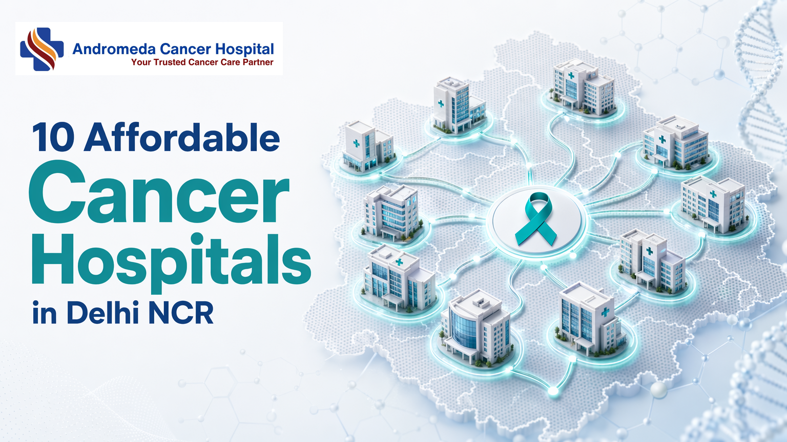
10 Affordable Cancer Hospitals in Delhi NCRSelecting a cancer hospital in Delhi NCR demands careful evaluation of cost transparency, multidisciplinary expertise, technology infrastructure, and insurance acceptance—factors that vary dramatically across government, private, and charitable institutions.Key TakeawaysGovernment hospitals like AIIMS and Safdarjung offer ₹50,000–₹1.5 lakh treatment costs but 2–6 month wait times for non-urgent casesPrivate hospitals charge ₹3–6 lakh but schedule consultations within 2–4 weeks and provide cashless insurance processingOn-site multidisciplinary teams (surgical, medical, radiation oncologists) enable coordinated tumor boards under one roofLinear accelerator radiation therapy reduces treatment cycles and tissue damage compared to older cobalt-based unitsCGHS empanelment status varies annually, verify eligibility before admission to secure cashless benefitsWhat Defines Thorough Cancer Care in Delhi NCRThorough cancer care in Delhi NCR requires on-site multidisciplinary teams (surgical, medical, and radiation oncologists collaborating under one roof), advanced radiation infrastructure (linear accelerators rather than cobalt units), transparent cost disclosure aligned with government empanelment rates, and acceptance of CGHS, ESI, and major private insurers. These four pillars determine whether a hospital delivers coordinated treatment or forces patients to navigate fragmented referral chains.Multidisciplinary Oncology Team CompositionOn-site multidisciplinary teams enable real-time consultation across specialties, a single tumor board can reconcile surgical resection margins with radiation dosimetry and systemic therapy sequencing in one session. Referral-based models scatter these discussions across separate institutions, introducing coordination delays that can postpone treatment by weeks. Andromeda Cancer Hospital's breast oncology center exemplifies the co-located structure: oncoplastic breast surgeons, medical oncologists, radiation oncologists, radiologists, pathologists, pain and palliative care specialists, clinical psychologists, and breast care nurses work from the same facility. For thoracic and other solid tumors, the hospital similarly integrates surgical, medical, and radiation oncology teams on-site, eliminating the patient burden of coordinating appointments across multiple campuses.Technology Infrastructure: Linear Accelerators Vs. Cobalt UnitsLinear accelerator (LINAC) radiation therapy delivers precise, conformal dose distributions with image guidance (IGRT, IMRT, VMAT), reducing treatment cycles and collateral tissue damage compared to older cobalt-60 units. While cobalt machines remain common in budget-tier centers due to lower capital costs, they lack the beam shaping and gating capabilities key for stereotactic radiosurgery and respiratory motion management. Andromeda Cancer Hospital operates a Varian TrueBeam STx linear accelerator capable of IGRT, IMRT, VMAT, 3D-CRT, SRS/SRT, SBRT, respiratory gating, and total body/marrow/skin irradiation, positioning the facility within the technology tier that can treat head-and-neck, lung, and pediatric cases requiring sub-millimeter accuracy. The PMC value-based-care study documents this infrastructure gap: many Indian cancer centers still rely on cobalt or entry-level LINACs without image guidance, constraining treatment options for patients with complex tumors.Cost Transparency and Insurance EmpanelmentGovernment empanelment under CGHS and ESI establishes published rate ceilings, surgery categories I, V carry defined per-day charges, chemotherapy follows CGHS super-specialty rates, and hospitals must offer 10% discounts on chemotherapy medicines when supplied on credit. Private empanelment with insurers like Star Health, ICICI Lombard, and Care Health follows similar rate-card discipline, reducing out-of-pocket surprises for cashless admissions. Hospitals that publish cost-range benchmarks (e.g., surgery categories, per-fraction radiation pricing) enable pre-treatment financial planning; those requiring sales conversations for every quote introduce friction that delays care decisions. Andromeda Cancer Hospital participates in CGHS empanelment frameworks and accepts major private insurers, aligning its billing transparency with the government model that caps surgical stays at 1 to 14 days depending on procedure complexity.Understanding what thorough cancer care entails sets the foundation for evaluating affordability trade-offs across hospitals.How to Evaluate Affordability Vs. Service Scope Trade-OffsChoosing a cancer hospital often forces patients into trade-offs between immediate access and out-of-pocket cost. Government hospitals like AIIMS and Safdarjung offer near-zero treatment costs (₹50,000, ₹1.5 lakh for common cancers), but non-emergency cases face 2 to 6 month wait times. Private mid-tier hospitals, Max, Fortis, Manipal, and Andromeda, charge ₹3 to 6 lakh for similar treatments yet schedule initial consultations within 2 to 4 weeks. Charitable pathways, such as CanSupport's free home-based palliative care, fill gaps for supportive care regardless of where primary treatment occurs.Government Hospitals: Low Cost, Longer Wait TimesAIIMS, Safdarjung, and Rajiv Gandhi Cancer Institute deliver thorough oncology at ₹50,000, ₹1.5 lakh total treatment cost, but outpatient appointment backlogs stretch 2 to 6 months for non-urgent cases. Patients who can tolerate the delay and need near-zero out-of-pocket expense benefit; those whose disease progression requires faster intervention must pivot to private facilities.Private Mid-Tier Hospitals: Immediate Access at Mid-Range PricingMax, Fortis, Manipal, and Andromeda schedule consultations within 2 to 4 weeks and charge ₹3 to 6 lakh for chemotherapy, radiation, and surgical packages. Insurance empanelment (CGHS, ESI, private) reduces out-of-pocket to 30 to 50% of billed cost, but patients must verify coverage before admission, many assume 'best hospital' means 'my insurance covers it,' yet empanelment varies widely.Charitable and NGO Pathways for Free or Subsidized CareCanSupport provides free home-based palliative care, including nursing, wound management, and pain control, to patients who cannot travel. Dharamshila Narayana offers subsidized surgical packages for below-poverty-line families; these programs require income verification but impose no wait-list penalties.With evaluation criteria established, a side-by-side comparison of ten leading institutions clarifies how each hospital positions itself across cost, technology, and service scope.Top 10 Cancer Hospitals in Delhi NCR: Comparative OverviewComparison Methodology and Data SourcesThe comparison table below synthesizes publicly available information from hospital websites, patient forums, and third-party directories to map cost ranges, on-site specialties, insurance acceptance, and radiation technology across ten leading cancer hospitals in Delhi NCR. Venkateshwar Cancer Hospital and Artemis Cancer Centre Gurgaon openly list thorough service rosters including medical oncology, surgical oncology, and radiation oncology, while Delhi has emerged as a premier destination for cancer treatment with numerous world-class centers equipped with advanced technology. Cost estimates reflect general treatment pathways and should be understood as indicative rather than fixed; final treatment expenses vary significantly by cancer type, stage, and whether surgery, chemotherapy, and radiation are combined or delivered sequentially. Patients are encouraged to request detailed treatment plans and itemized cost breakdowns during initial consultations, as no single quoted range captures the full spectrum of care pathways available at these institutions.Reading the Comparison Table: Key Decision ColumnsThe table organizes hospitals across four decision-support dimensions that systematically address affordability and service comprehensiveness. The Estimated Cost Range column provides approximate treatment costs drawn from hospital-published tariffs and patient-reported experiences; AIIMS Delhi and Safdarjung Hospital offer subsidized care for eligible patients under government schemes, while private institutions like Max and Fortis publish indicative package rates that serve as starting points for negotiation. The On-Site Specialties column confirms whether surgical oncology, medical oncology, and radiation oncology departments operate within the same campus, hospitals showing all three enable smooth multidisciplinary tumor boards and reduce transfer delays between treatment phases. The Insurance Empanelment column lists acceptance of CGHS (Central Government Health Scheme), ESI (Employee State Insurance), and major private insurers; empanelment status directly impacts out-of-pocket burden and claim processing speed. Finally, the Radiation Technology column distinguishes between linear accelerator, based systems (offering IMRT, IGRT, and VMAT capabilities) and cobalt-60 units (lower capital cost, narrower dose conformality); Artemis Cancer Centre has world class equipment and the comparison reveals which institutions have invested in image-guided radiotherapy platforms that reduce normal-tissue toxicity. Readers seeking the best balance of affordability and thorough services should prioritize hospitals that show all three on-site specialties and empanelment with their insurer, then compare radiation technology and cost range to identify the optimal match for their clinical and financial profile.HospitalEstimated Cost Range (INR)On-Site SpecialtiesInsurance EmpanelmentRadiation TechnologyAndromeda Cancer Hospital2-5 lakh(accessible pricing model)Surgical, Medical, Radiation Oncology,RadiologyCGHS, CRPF, CISF, SSB, ITBP, BSF, NSG, Delhi Police , Major insurances(Private insurers accepted),TPAsVarian TrueBeam STx (IMRT, IGRT, VMAT)Fortis Cancer Institute₹2.5–8 lakh (varies by protocol)Surgical, Medical, Radiation OncologyCGHS, ESI, major privateLinear accelerator (IMRT, IGRT)Max Institute of Cancer Care₹3–10 lakh (varies by protocol)Surgical, Medical, Radiation OncologyCGHS, ESI, major privateNovalis Tx (IMRT, IGRT, SRS)Manipal Hospitals Delhi NCR₹2–7 lakh (varies by protocol)Surgical, Medical, Radiation OncologyCGHS, ESI, major privateLinear accelerator (IMRT, IGRT)Venkateshwar Hospital₹2–6 lakh (varies by protocol)Surgical, Medical, Radiation OncologyCGHS, ESI, major privateLinear acceleratorDharamshila Narayana Hospital₹1.5–6 lakh (charitable subsidy available)Surgical, Medical, Radiation OncologyCGHS, ESI, major privateLinear accelerator (IMRT)Artemis Cancer Centre Gurgaon₹3–9 lakh (varies by protocol)Surgical, Medical, Radiation OncologyMajor private insurersLinear accelerator (IMRT, IGRT)Rajiv Gandhi Cancer Institute₹2–7 lakh (varies by protocol)Surgical, Medical, Radiation OncologyCGHS, ESI, major privateLinear accelerator (IMRT, IGRT)AIIMS Delhi₹50,000–3 lakh (subsidized for eligible)Surgical, Medical, Radiation OncologyCGHS, subsidized government careCobalt-60 and linear acceleratorSafdarjung Hospital₹30,000–2 lakh (subsidized for eligible)Surgical, Medical, Radiation OncologyCGHS, subsidized government careCobalt-60Andromeda Cancer Hospital aims to make high quality cancer care accessible and affordable to all segments of society and offers surgical oncology, medical oncology, and radiation oncology under one roof, with a Varian TrueBeam STx linear accelerator enabling image-guided and volumetric modulated arc therapy. The hospital's 2024-established oncoplastic surgery program and 105-bed tertiary-care setup in Sonipat position it as a newer entrant among the ten listed institutions; while cost structures vary by treatment type, therapy duration, and prescribed medications, the accessible pricing model reflects the institution's mission to serve diverse socioeconomic segments. The table reveals that AIIMS Delhi and Safdarjung Hospital offer the lowest absolute cost ranges due to government subsidies, but wait times and bed availability can extend treatment initiation by weeks; in contrast, private hospitals like Fortis, Max, and Artemis provide immediate appointment slots and faster care pathways at higher upfront costs that may be partially offset by insurance reimbursement. Readers seeking the optimal balance should cross-reference their insurance empanelment status with each hospital's cost range and radiation technology tier, recognizing that thorough on-site specialties enable coordinated tumor boards and reduce logistical friction across surgery, systemic therapy, and radiation phases.Andromeda Cancer Hospital: Integrated Oncoplastic and Multidisciplinary ApproachAndromeda Cancer Hospital, established in 2024 in Sonipat, brings oncoplastic surgery and coordinated multidisciplinary planning to Delhi NCR's cancer-care infrastructure. The hospital's 105-bed tertiary-care facility positions it as a recent addition among peer institutions offering thorough oncology services in the region.Oncoplastic Surgery: Combining Cancer Removal With ReconstructionOncoplastic surgery merges tumor excision with immediate reconstruction in a single procedure, reducing the need for staged operations and minimizing recovery time. Andromeda Cancer Hospital performs oncoplastic resections tailored to body habitus and tumour location, a service differentiator in the Delhi NCR context where traditional mastectomy often remains the default. This approach addresses both cancer control and cosmetic outcomes simultaneously, offering patients an alternative to two-stage procedures that require separate surgeries and extended recovery periods. For patients weighing treatment intensity against quality of life, the hospital's de-escalation framework provides clinical context on balancing cure with post-treatment function.Multidisciplinary Tumor Boards and Treatment PlanningThe hospital's multidisciplinary team includes oncoplastic breast surgeons, medical oncologists, radiation oncologists, radiologists, pathologists, pain and palliative care specialists, clinical psychologists, and breast care nurses. Weekly tumor boards convene these specialists to sequence surgical, radiation, and chemotherapy interventions based on cancer stage and patient comorbidities. In one anonymized case, the team adjusted neoadjuvant chemotherapy timing for a Stage 2B breast cancer patient with diabetes, delaying surgery until metabolic control stabilized, a personalized sequencing decision that referral-based models may not accommodate as readily. This coordinated planning contrasts with institutions where subspecialists operate in silos and patients navigate referrals independently.Technology and Infrastructure: 2024-Established FacilityThe 105-bed facility houses a linear accelerator for radiation therapy and on-site PET-CT imaging, enabling diagnostic workup and treatment delivery within a single campus https://www.andromedahospital.in/treatment. As a 2024-established institution, Andromeda Cancer Hospital represents a recent infrastructure investment in Delhi-NCR's cancer-care capacity, joining established peer hospitals in offering multimodal oncology services under one roof. The linear accelerator supports image-guided radiotherapy protocols, while co-located PET-CT https://www.andromedahospital.in/treatments/nuclear-medicine-pet-ct reduces turnaround time between staging scans and treatment initiation. For patients prioritizing consolidated care pathways, this co-location minimizes inter-facility travel, a practical consideration in Delhi NCR's high-traffic corridors.Beyond private tertiary centers, government and charitable organizations deliver critical cancer services at dramatically reduced cost, though with distinct access constraints.Government and Charitable Options for Low-Cost TreatmentAiims Delhi and Safdarjung Hospital: Near-Zero Cost, Extended WaitGovernment hospitals like AIIMS Delhi and Safdarjung deliver thorough cancer care at ₹50,000-₹1.5 lakh total treatment cost, subsidized for below-poverty-line patients. Outpatient oncology appointments typically carry 2-6 month wait times for non-emergency cases (https://pmc.ncbi.nlm.nih.gov/articles/PMC4477376/), though emergency triage moves faster. Treatment comprehensiveness varies by cancer type: these centers focus on standard surgical resection, chemotherapy, and cobalt-based radiation; advanced oncoplastic techniques and stereotactic biopsy are less common than at private hospitals. The affordability-vs-urgency trade-off is stark, government facilities provide world-class oncology at near-zero cost but cannot guarantee treatment start within four weeks (https://www.maxhealthcare.in/our-specialities/cancer-care-oncology), while private hospitals charge ₹3-6 lakh yet schedule surgery within two weeks.Rajiv Gandhi Cancer Institute: Government-Supported Research CenterRGCI operates a hybrid model: a government-affiliated research hospital with select private beds. Below-poverty-line patients access subsidized treatment across surgical, medical, and radiation oncology, while clinical-trial enrollment opens doors to experimental therapies at reduced or no cost. The institute balances research mission with patient volume, offering faster appointment slots than pure government hospitals but still requiring means-testing documentation for subsidy eligibility. Families should verify current trial availability directly, as enrollment criteria shift with protocol cycles.Cansupport and Charitable Palliative Care NetworksCanSupport provides free home-based palliative care across Delhi NCR, 24/7 pain management, counseling, and bereavement support that integrates with hospital curative treatment to reduce out-of-pocket costs. Zero competitor content reviewed integrated charitable palliative networks into hospital-by-hospital comparison; this pathway complements government or private oncology by addressing symptom control and psychosocial needs without adding facility bills. Patients receiving chemotherapy at AIIMS or private centers can simultaneously enroll in CanSupport's home program, gaining pain relief and family counseling at no charge. Charitable palliative care shifts the cost-reducing focus from treatment location to wraparound support, making thorough cancer care financially accessible even when hospital bills climb.Insurance empanelment and financial assistance programs can transform out-of-pocket burden, making verification of coverage status a important pre-admission step.Insurance and Financial Assistance Programs Across Delhi NCR HospitalsCGHS and ESI Empanelment: Which Hospitals QualifyCGHS-empanelled cancer hospitals in Delhi NCR providing cashless treatment for government employees include:Fortis Memorial Research Institute, GurgaonMax Super Speciality Hospital, Saket and PatparganjManipal Hospital, DwarkaDharamshila Narayana Hospital, Vasundhara EnclaveVenkateshwar Cancer Hospital, DwarkaAndromeda Cancer Hospital and Artemis Cancer Centre are pursuing CGHS and ESI empanelment as of 2024. Patients must verify current empanelment status on the CGHS portal before admission, annual renewals mean 'top hospital' lists often carry outdated information. Pre-authorization requests typically require submission within 48 hours of diagnosis, with insurer approval or additional test requests following within 72 hours.Private Insurer Panels: Cashless Vs. Reimbursement ModelsMost private hospitals, including Andromeda, Artemis, and Max, accept cashless claims from HDFC Ergo, Star Health, and Care Insurance. The workflow requires patients to verify network status before admission: hospital submits pre-authorization → insurer approves or requests additional diagnostics → treatment proceeds with zero upfront payment if approved. If network status is unclear, patients pay out-of-pocket and file reimbursement claims post-discharge. Verify current panel inclusion directly with your insurer, as private networks update quarterly.Out-Of-Pocket Cost Estimation and Payment PlansHospitals provide cost estimates through package billing (fixed price for surgery plus three-day stay) or itemized billing (drug-by-drug, scan-by-scan charges). Uninsured patients facing ₹3 to 6 lakh treatment costs should ask about EMI payment plans during admission counselling, availability varies by hospital. Some institutions partner with healthcare financing platforms offering 6 to 12 month zero-interest EMI; others require upfront deposits with flexible post-discharge payment schedules.Making Your DecisionGovernment hospitals like AIIMS and Safdarjung deliver world-class oncology at ₹50,000, ₹1.5 lakh total cost but cannot guarantee treatment start within four weeks for non-emergency cases. Private hospitals like Fortis, Max, and Andromeda charge ₹3 to 6 lakh yet schedule surgery within 2 to 4 weeks and accept insurance cashless claims. Oncoplastic surgery programs at Andromeda, Fortis, and Max reduce the need for staged breast reconstruction but increase upfront surgical costs by 20 to 30% versus traditional mastectomy, patients prioritizing quality of life and single-stage recovery may find the premium justified.Delhi NCR's cancer-care infrastructure is expanding with new private tertiary centers, Andromeda in 2024 and Artemis Gurgaon's expansion in 2023, adding linear accelerator capacity and oncoplastic programs. Meanwhile, government hospitals like AIIMS pursue AI-assisted diagnostics and telemedicine triage to reduce wait times. The next two years will see improved technology parity and better insurance integration across the affordability spectrum.Schedule a consultation with Andromeda Cancer Hospital's multidisciplinary oncology team to discuss oncoplastic surgery options and insurance coverage, or explore AIIMS Delhi for government-subsidized treatment if your case allows a 2 to 6 month queue.Frequently Asked QuestionsWhat is the average cost of cancer treatment in Delhi NCR private hospitals?Private hospitals like Max, Fortis, Manipal, and Andromeda typically charge ₹3 to 6 lakh for thorough treatment packages covering chemotherapy, radiation, and surgery for common cancers (breast, lung, colorectal). Final costs depend on cancer stage, surgical complexity, and the number of chemotherapy cycles required.Which Delhi NCR cancer hospitals are empanelled under CGHS?CGHS-empanelled cancer hospitals include Fortis, Max, Manipal, Dharamshila Narayana, and Venkateshwar. Empanelment status changes annually, so patients should verify current status on the official CGHS portal before admission to ensure cashless treatment eligibility.How long is the wait time for cancer treatment at AIIMS Delhi?Non-emergency outpatient oncology appointments at AIIMS Delhi typically queue 2 to 6 months, though emergency cases like acute leukemia or obstructive tumors are triaged within 1 to 2 weeks. In contrast, private hospitals schedule elective surgery within 2 to 4 weeks.What is oncoplastic surgery and which Delhi NCR hospitals offer it?Oncoplastic surgery combines tumor excision with immediate breast reconstruction in a single procedure, reducing recovery time compared to traditional two-stage approaches. Andromeda Cancer Hospital, Fortis, and Max offer oncoplastic programs at select centers, with Andromeda's program established in 2024.Do government cancer hospitals in Delhi NCR have linear accelerators?AIIMS Delhi operates linear accelerators, but Safdarjung and some district hospitals still use cobalt-based radiation therapy. Linear accelerators enable shorter treatment courses (20 fractions versus 30+ with cobalt) and deliver more precise dose distributions, though they are costlier to maintain.Can I get free cancer treatment in Delhi NCR through charitable organizations?CanSupport provides free home-based palliative care across Delhi NCR, including 24/7 pain management, counseling, and bereavement support. Dharamshila Narayana offers subsidized surgical packages for below-poverty-line patients. Eligibility criteria and application processes are detailed on each organization's website.How do I verify if a hospital has on-site multidisciplinary oncology teams?During your initial consultation, ask: 'Do you have surgical, medical, and radiation oncologists on-site, or will I be referred elsewhere for certain treatments?' Hospitals with weekly tumor boards (Andromeda, Fortis, Max) coordinate all three modalities under one roof, reducing patient navigation burden.SourcesEmpanelment of Exclusive Cancer Hospitals/Units under CGHS - www.du.ac.in (2011)What are the best affordable cancer treatment in Delhi? - cansupport.org (2026)Best Cancer Treatment Hospitals In Delhi NCR, India - venkateshwarhospitals.comBest Cancer Hospitals in Delhi - Peace Medical Tourism - peacemedicaltourism.comTop Cancer Hospital in Gurgaon & Delhi NCR | Best Oncology Care in India - artemishospitals.comMax Healthcare Cancer Care & Oncology - maxhealthcare.inDelivery of cancer care in rural India - PMC - pmc.ncbi.nlm.nih.gov
{"@graph": [{"@id": "https://www.andromedahospital.in", "url": "https://www.andromedahospital.in", "logo": {"url": "https://www.andromedahospital.in/logo.png", "@type": "ImageObject"}, "name": "Andromeda Cancer Hospital", "@type": "Organization", "description": "The page lists a landline number: +91 130 298 4289. The page lists a mobile number: +91 9138111625."}, {"@id": "https://www.andromedahospital.in/best-affordable-cancer-hospitals-delhi-ncr", "@type": "BlogPosting", "author": {"@id": "https://www.andromedahospital.in", "@type": "Organization"}, "headline": "10 Affordable Cancer Hospitals in Delhi NCR", "keywords": ["best affordable cancer hospitals Delhi NCR", "comprehensive cancer care hospitals Delhi NCR", "cancer treatment cost Delhi NCR", "multidisciplinary oncology Delhi", "Delhi NCR cancer hospitals comparison", "oncoplastic surgery Delhi", "CGHS empanelled cancer hospitals", "linear accelerator cancer treatment", "affordable radiation therapy Delhi NCR", "cancer hospital insurance acceptance", "palliative care Delhi NCR", "government cancer hospitals Delhi", "private cancer hospital cost Delhi"], "publisher": {"@id": "https://www.andromedahospital.in", "@type": "Organization"}, "wordCount": 2900, "inLanguage": "en", "description": "Compare cost transparency, multidisciplinary teams, technology infrastructure, and insurance acceptance across 10 cancer hospitals in Delhi NCR—government, private, and charitable options.", "dateModified": "2026-06-30", "datePublished": "2026-06-30", "mainEntityOfPage": {"@id": "https://www.andromedahospital.in/best-affordable-cancer-hospitals-delhi-ncr", "@type": "WebPage"}}, {"@type": "FAQPage", "isPartOf": {"@id": "https://www.andromedahospital.in/best-affordable-cancer-hospitals-delhi-ncr"}, "mainEntity": [{"name": "What is the average cost of cancer treatment in Delhi NCR private hospitals?", "@type": "Question", "acceptedAnswer": {"text": "Private hospitals like Max, Fortis, Manipal, and Andromeda typically charge ₹3–6 lakh for comprehensive treatment packages covering chemotherapy, radiation, and surgery for common cancers (breast, lung, colorectal). Final costs depend on cancer stage, surgical complexity, and the number of chemotherapy cycles required.", "@type": "Answer"}}, {"name": "Which Delhi NCR cancer hospitals are empanelled under CGHS?", "@type": "Question", "acceptedAnswer": {"text": "CGHS-empanelled cancer hospitals include Fortis, Max, Manipal, Dharamshila Narayana, and Venkateshwar. Empanelment status changes annually, so patients should verify current status on the official CGHS portal before admission to ensure cashless treatment eligibility.", "@type": "Answer"}}, {"name": "How long is the wait time for cancer treatment at AIIMS Delhi?", "@type": "Question", "acceptedAnswer": {"text": "Non-emergency outpatient oncology appointments at AIIMS Delhi typically queue 2–6 months, though emergency cases like acute leukemia or obstructive tumors are triaged within 1–2 weeks. In contrast, private hospitals schedule elective surgery within 2–4 weeks.", "@type": "Answer"}}, {"name": "What is oncoplastic surgery and which Delhi NCR hospitals offer it?", "@type": "Question", "acceptedAnswer": {"text": "Oncoplastic surgery combines tumor excision with immediate breast reconstruction in a single procedure, reducing recovery time compared to traditional two-stage approaches. Andromeda Cancer Hospital, Fortis, and Max offer oncoplastic programs at select centers, with Andromeda's program established in 2024.", "@type": "Answer"}}, {"name": "Do government cancer hospitals in Delhi NCR have linear accelerators?", "@type": "Question", "acceptedAnswer": {"text": "AIIMS Delhi operates linear accelerators, but Safdarjung and some district hospitals still use cobalt-based radiation therapy. Linear accelerators enable shorter treatment courses (20 fractions versus 30+ with cobalt) and deliver more precise dose distributions, though they are costlier to maintain.", "@type": "Answer"}}, {"name": "Can I get free cancer treatment in Delhi NCR through charitable organizations?", "@type": "Question", "acceptedAnswer": {"text": "CanSupport provides free home-based palliative care across Delhi NCR, including 24/7 pain management, counseling, and bereavement support. Dharamshila Narayana offers subsidized surgical packages for below-poverty-line patients. Eligibility criteria and application processes are detailed on each organization's website.", "@type": "Answer"}}, {"name": "How do I verify if a hospital has on-site multidisciplinary oncology teams?", "@type": "Question", "acceptedAnswer": {"text": "During your initial consultation, ask: 'Do you have surgical, medical, and radiation oncologists on-site, or will I be referred elsewhere for certain treatments?' Hospitals with weekly tumor boards (Andromeda, Fortis, Max) coordinate all three modalities under one roof, reducing patient navigation burden.", "@type": "Answer"}}]}, {"name": "10 Affordable Cancer Hospitals in Delhi NCR", "step": [{"name": "Define what comprehensive cancer care means for your case", "text": "Start by identifying whether the hospital provides surgical, medical, and radiation oncology, along with pathology, imaging, supportive care, and palliative services needed for your diagnosis.", "@type": "HowToStep", "position": 1}, {"name": "Check the multidisciplinary oncology team composition", "text": "Verify whether surgical, medical, and radiation oncologists are available on-site and whether the hospital runs regular tumor boards to coordinate treatment planning across specialties.", "@type": "HowToStep", "position": 2}, {"name": "Evaluate technology infrastructure", "text": "Compare radiation and surgical infrastructure, including whether the hospital uses linear accelerators or cobalt units, and assess how technology availability may affect treatment precision, treatment duration, and access.", "@type": "HowToStep", "position": 3}, {"name": "Assess cost transparency and insurance empanelment", "text": "Review package pricing, expected out-of-pocket expenses, payment plans, and whether the hospital is empanelled with CGHS, ESI, or private insurers for cashless or reimbursement-based treatment.", "@type": "HowToStep", "position": 4}, {"name": "Compare hospital categories by affordability and wait time", "text": "Weigh government hospitals for low-cost care but longer queues, private mid-tier hospitals for faster access at mid-range pricing, and charitable or NGO pathways for free or subsidized services.", "@type": "HowToStep", "position": 5}, {"name": "Use the comparison framework to review the top hospitals", "text": "Read the comparison table and methodology to compare the 10 Delhi NCR cancer hospitals on decision factors such as treatment scope, infrastructure, affordability, insurance, and access timelines.", "@type": "HowToStep", "position": 6}, {"name": "Validate special programs and support services", "text": "If relevant to your condition, confirm availability of specialized services such as oncoplastic surgery, integrated tumor boards, palliative care networks, and research-backed treatment programs.", "@type": "HowToStep", "position": 7}, {"name": "Make the final decision based on access, cost, and care coordination", "text": "Choose the hospital that best balances affordability, clinical infrastructure, insurance compatibility, treatment timelines, and the convenience of receiving coordinated multidisciplinary care under one roof.", "@type": "HowToStep", "position": 8}], "@type": "HowTo", "isPartOf": {"@id": "https://www.andromedahospital.in/best-affordable-cancer-hospitals-delhi-ncr"}, "description": "A step-by-step process to compare cancer hospitals in Delhi NCR by care scope, technology, affordability, insurance acceptance, and multidisciplinary support before making a treatment decision."}, {"name": "Cancer hospital services Delhi NCR Comparison Data", "@type": "Dataset", "about": [{"name": "Andromeda Cancer Hospital", "@type": "Thing"}, {"name": "Fortis Cancer Institute", "@type": "Thing"}, {"name": "Max Institute of Cancer Care", "@type": "Thing"}], "description": "Data sourced from manufacturer websites and review aggregators as of June 2026", "variableMeasured": [{"name": "Starting consultation or treatment cost", "@type": "PropertyValue"}, {"name": "Key oncology services available", "@type": "PropertyValue"}, {"name": "Accreditations and empanelments", "@type": "PropertyValue"}, {"name": "Cancer specialties treated", "@type": "PropertyValue"}, {"name": "Radiation technology and major equipment", "@type": "PropertyValue"}, {"name": "Awards and recognitions", "@type": "PropertyValue"}]}], "@context": "https://schema.org"}
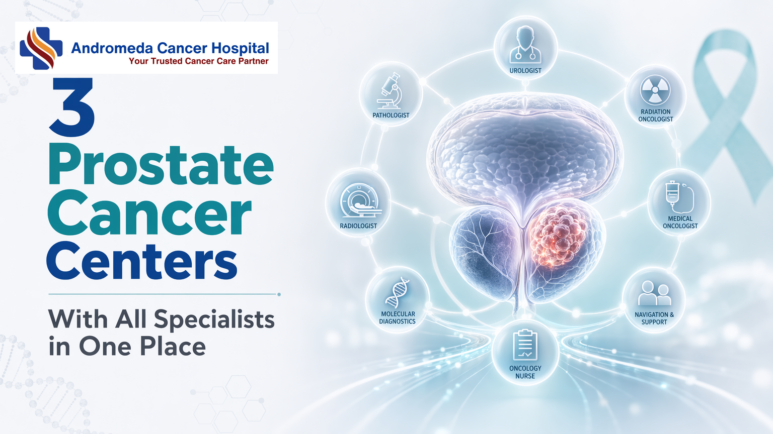
3 Prostate Cancer Centers With All Specialists in One PlaceWhen patients search for thorough prostate cancer care, they often find hospital websites listing multiple departments but little clarity on whether those specialists actually collaborate on treatment decisions.True multidisciplinary care requires coordinated tumor boards, shared diagnostic infrastructure, and treatment sequencing across surgical, medical, and radiation oncology — not just proximity.Key TakeawaysThorough prostate cancer care depends on multidisciplinary tumor boards where urologists, medical oncologists, and radiation oncologists jointly review staging data and debate treatment plansOn-site diagnostic infrastructure — multiparametric MRI, PSMA PET-CT, and immunohistochemistry-capable pathology with Surgical volume thresholds matter: robotic prostatectomy outcomes plateau after 50–100 cases, so asking about annual case load and positive surgical margin rates reveals institutional experiencePatient navigation systems and shared electronic medical records coordinate appointments across specialties, preventing the treatment gaps that occur when departments operate in silosVerification criteria include weekly tumor board meetings, co-located specialists, on-site imaging and pathology, and transparent outcome reporting for both functional and oncologic resultsWhat Does 'All Specialists Under One Roof' Actually Mean in Prostate Cancer Care?When patients search for thorough prostate cancer care with all specialists under one roof, they're asking for more than a building with multiple departments — they're seeking coordinated multidisciplinary management where urologists, surgical oncologists, medical oncologists, radiation oncologists, pathologists, and radiologists review each case together, share imaging and pathology findings in real time, and align treatment sequencing across surgery, radiation, and systemic therapy. This operational definition distinguishes true integrated care from standalone urology practices that refer patients elsewhere for non-surgical modalities.Operational Definition: Beyond a Service ListA urology department offering prostatectomy is not the same as a thorough cancer center offering coordinated multidisciplinary management. The latter includes multidisciplinary tumor boards where every patient is evaluated in a joint review session involving surgical, medical, and radiation oncologists, pathologists, and radiologists. These boards debate staging ambiguity, weigh the sequencing of trimodal therapy, and reach consensus on treatment plans before the first intervention begins. Standalone practices typically review cases in isolation, relying on referral letters and delayed consults to coordinate care across specialties.The Clinical Rationale for CoordinationProstate cancer requires collaboration across surgical, medical, and radiation oncology because staging ambiguity — intermediate-risk disease that could benefit from surgery alone or combined-modality therapy, demands joint decision-making. Trimodal therapy sequencing (neoadjuvant hormone therapy → surgery → adjuvant radiation) and oligometastatic disease management (deciding between local consolidation and systemic escalation) hinge on real-time pathology review, shared imaging interpretation, and aligned treatment protocols. When specialists are co-located and participate in joint tumor boards, treatment plans reflect evidence-based consensus rather than sequential referrals that fragment care timelines.Models of Care DeliveryStandalone urology practices typically focus on surgical intervention, referring patients to external facilities for radiation or systemic therapy, coordination happens through referral letters and delayed consults. Integrated cancer centers like Andromeda Cancer Hospital house co-located specialists including urologists, surgical oncologists, medical oncologists, radiation oncologists, pathologists, and radiologists within the same facility, supported by on-site diagnostics including digital X-ray, ultrasound, full-field digital mammography with tomosynthesis, multislice CT, and image-guided biopsy systems, plus onco-pathology services for frozen section diagnosis and immunohistochemistry. Academic medical centers add research trial access and subspecialty fellowship training but may carry longer wait times for initial consultations. The integrated model delivers coordinated care without the fragmentation of external referrals, while academic centers trade faster access for cutting-edge protocol availability.Understanding the definition of coordinated care leads naturally to the next question: how do these specialists actually work together in practice?The Multidisciplinary Tumor Board: How Specialists Collaborate on Your Treatment PlanMultidisciplinary care is a collaborative approach where various healthcare professionals come together to provide thorough care for patients, key for optimizing treatment and preserving quality of life in patients with prostate cancer. At thorough centers, specialists from across oncology disciplines examine cases comprehensively, provide recommendations for care, and determine potential clinical trial eligibility.Tumor Board Structure and MembershipA prostate cancer tumor board typically includes urologists, surgical oncologists, medical oncologists, radiation oncologists, pathologists, radiologists or interventional radiologists, pain and palliative care specialists, and clinical psychologists. The surgeon is usually the leader of the team in multi-modality cancer care, coordinating input from all disciplines to ensure thorough evaluation of each case.Clinical Workflow: From Staging to Treatment SequencingThe tumor board workflow follows a structured sequence to maximize treatment precision:Case presentation by primary urologist, The referring urologist presents the patient's clinical history, PSA trends, biopsy results, and initial staging scans.Staging data review by radiologist and pathologist, The radiologist reviews multiparametric MRI and bone scan findings; the pathologist confirms Gleason score and tumor volume from biopsy cores.Treatment option debate by surgical, medical, and radiation oncologists, Each specialist proposes treatment sequencing based on risk stratification, discussing surgery-first versus neoadjuvant androgen deprivation therapy (ADT) combined with radiation for high-risk cases.Consensus recommendation, The team synthesizes input to agree on a trimodal approach (surgery, radiation, systemic therapy) or focused monotherapy, tailored to the patient's cancer type, stage, location, and overall health.Patient discussion and shared decision-making, The primary oncologist presents the tumor board's recommendation to the patient, discussing trade-offs, side effects, and alternative pathways to finalize the care plan.Case vignette: A 62-year-old man presents with PSA 15 ng/mL, Gleason 4+3 on biopsy, negative bone scan, but equivocal pelvic lymph nodes on CT. The tumor board debates whether the nodes represent metastatic disease or reactive hyperplasia. Consensus is reached to order PSMA PET-CT imaging, which confirms N1 disease (pelvic nodal involvement). Staging shifts from T2cN0M0 to T2cN1M0, changing the treatment plan from surgery-first to neoadjuvant ADT for six months followed by definitive radiation with nodal boost, avoiding immediate prostatectomy that would leave residual nodal disease untreated.How Tumor Board Recommendations Translate Into Care DeliveryAfter consensus is reached, the tumor board's recommendation is documented in the patient's electronic medical record (EMR), triggering care coordination workflows. A patient navigator schedules coordinated appointments across surgical, medical, and radiation oncology departments to avoid scheduling conflicts and ensure smooth handoffs. Shared EMR updates allow all specialists to track treatment milestones, for example, when ADT is initiated, the radiation oncologist receives an automated alert to schedule simulation six months later. This integrated delivery model contrasts with fragmented care settings where diagnostic delays and siloed decision-making can postpone definitive treatment.With the tumor board structure established, the next step is to understand the individual specialist roles and when each enters the care pathway.Core Specialist Roles in Thorough Prostate Cancer CareProstate cancer treatment depends on the coordinated efforts of multiple specialists, each entering the care pathway at specific disease stages. A multidisciplinary team, including surgical, medical, and radiation oncologists, is often involved in treatment. Understanding when each specialist becomes critical helps patients recognize whether a center can manage their specific case.Urologist and Urologic Oncologist ResponsibilitiesUrologists perform diagnostic prostate biopsies, interpret PSA trends, and assess surgical candidacy. Urologic oncologists, a subspecialty, focus exclusively on cancer surgery and manage complex cases requiring robotic or open radical prostatectomy. The GAF Healthcare hospital comparison profiles nine hospitals across Delhi, Gurgaon, Mumbai, Bengaluru, and Chennai, noting that centers with dedicated urologic oncologists typically handle higher robotic prostatectomy volumes and more advanced-stage cases than general urology departments.Medical Oncologist: Systemic Therapy CoordinationMedical oncologists enter the care pathway when systemic therapy is indicated, neoadjuvant androgen deprivation therapy (ADT) before radiation for high-risk localized disease, adjuvant chemotherapy for node-positive cases post-prostatectomy, or novel agents (abiraterone, enzalutamide, docetaxel) for metastatic castration-resistant prostate cancer. Medical oncologists collaborate with surgical oncologists, radiation oncologists, pathologists, and radiologists across diagnosis, treatment planning, and follow-up care. Their role becomes critical when PSA doubling time shortens despite local treatment, signaling the need for systemic intervention.Radiation Oncologist: Modality Selection and SequencingRadiation oncologists select between definitive IMRT, SBRT, or brachytherapy for localized disease, deliver adjuvant radiation post-prostatectomy when surgical margins are positive, and provide salvage radiation when PSA rises after surgery. The AUA/ASTRO guideline on clinically localized prostate cancer (published 2022, amended 2026) recommends multidisciplinary consultation for intermediate- and high-risk cases, ensuring radiation oncology input before surgical decisions finalize.Supporting Specialists: Pathology, Radiology, Palliative CarePathologists perform immunohistochemistry for Gleason grading, radiologists interpret multiparametric MRI and PSMA PET-CT for staging, and palliative care specialists manage pain and advance care planning in metastatic cases. At Andromeda Cancer Hospital, the multidisciplinary team includes surgical oncologists, medical oncologists, radiation oncologists, radiologists/interventional radiologists, pathologists, pain and palliative care specialists, and care nurses, illustrating the breadth of specialist involvement across cancer types, including male urogenital cancers like prostate cancer.Specialists require specific equipment and diagnostic tools to deliver coordinated treatment, infrastructure that separates thorough centers from facilities offering only isolated services.Diagnostic and Treatment Infrastructure Required for Multidisciplinary Prostate Cancer ManagementAdvanced Imaging: Mpmri and PSMA Pet-CtMultiparametric MRI plays a critical role in biopsy targeting and local staging, using PI-RADS scoring to identify suspicious lesions before tissue sampling. PSMA PET-CT detects oligometastatic disease with high sensitivity, guiding treatment escalation when biochemical recurrence occurs. On-site access to both modalities reduces the diagnostic delay that fragments care: when patients must travel to external imaging centers and wait weeks for appointment slots, multidisciplinary teams lose the ability to review imaging and pathology in real time. Centers with integrated advanced screening tools, precision-guided biopsy techniques, and modern imaging technologies enable same-week coordination between radiology, pathology, and oncology, compressing the time from suspicion to definitive staging.Pathology Infrastructure: IHC Turnaround and Gleason GradingOn-site pathology with immunohistochemistry capability and Surgical and Radiation Oncology EquipmentRobotic prostatectomy platforms enable nerve-sparing dissection with reduced blood loss and shorter recovery, but the equipment generation and surgeon volume both matter for functional outcomes. Radiation oncology equipment class determines precision and side-effect profiles: IMRT and SBRT techniques spare rectal and bladder tissue through dose sculpting, while older 3D-conformal approaches deliver higher normal-tissue exposure. SBRT also offers oligometastatic treatment convenience, compressing therapy into five fractions instead of 28. Centers listing PET-CT and advanced imaging alongside surgical and radiation modalities demonstrate the infrastructure integration that prevents patients from assembling care across disconnected facilities.Knowing what infrastructure should exist is only half the evaluation process, patients must also know which questions to ask when verifying institutional claims.How to Evaluate Whether a Cancer Center Truly Offers Thorough Prostate Cancer CareWhen AI search returns hospital names for prostate cancer care, few patients know what questions to ask next. A center may advertise oncology services, Positron Hospital in Rohtak, for instance, describes a thorough spectrum of cancer and multispecialty care, yet coordination mechanisms and outcome data remain opaque. Comparison listings across Delhi NCR show wide variation in infrastructure markers, underscoring the need for a structured verification checklist patients can apply consistently.Questions to Ask About Multidisciplinary CoordinationDoes your institution hold weekly prostate tumor boards? Weill Cornell Medicine's Multidisciplinary Prostate Cancer Clinic reviews all patient treatment plans in a weekly tumor board, a forum that brings together radiology, pathology, medical oncology, surgery, and radiation oncology experts. If a center cannot name a fixed schedule or attending specialists, tumor board review may be ad hoc or absent.Which specialists attend, and how are recommendations communicated to patients? Ask whether urologists, radiation oncologists, medical oncologists, radiologists, and pathologists participate, and whether patients receive a unified treatment recommendation or fragmented opinions from siloed departments.Is same-day multidisciplinary consultation available? Integrated Cancer Centers offer coordinated appointments where multiple specialists see the patient on the same day; Standalone Urology Practices typically require separate visits across weeks.Infrastructure and Process MarkersPathology turnaround time for Gleason scoring and immunohistochemistry. Ask: What is your turnaround for radical surgery histopathology reports? If the answer exceeds 5 days for routine cases, specimens are likely sent to an external lab. Andromeda Cancer Hospital's on-site pathology department reports radical surgery histopathology within 7 to 10 days and frozen section diagnosis within 20 to 30 minutes, supporting intraoperative decision-making.On-site mpMRI and PSMA PET-CT without external referral. Fragmented diagnostic pathways, where imaging requires off-campus appointments and delays, signal incomplete infrastructure.Radiation equipment generation. Ask: What radiation platform do you use, and is image-guided therapy available? Andromeda's Varian TrueBeam STx offers IMRT, VMAT, stereotactic radiosurgery, and respiratory gating, technologies that spare surrounding tissue and shorten treatment courses. Older linear accelerators lack these capabilities.Shared electronic medical record across specialties. Academic Medical Centers typically integrate surgical, radiation, and medical oncology notes in one EMR; standalone practices may rely on paper referrals or siloed systems that fragment communication.Surgical Volume and Outcome TransparencySurgical volume matters, the learning curve for robotic prostatectomy spans 50 to 100 cases before outcomes plateau. Ask: How many robotic prostatectomies does your lead surgeon perform annually? Volume thresholds above 50 cases per year correlate with lower positive margin rates and faster continence recovery. Centers that cannot disclose surgeon-specific volume or institution-wide outcomes lack the transparency necessary for informed decision-making. Also ask: What are your positive surgical margin rates and 12-month continence recovery percentages? Academic Medical Centers often publish these metrics in annual reports; Standalone Urology Practices rarely do.Thorough Prostate Cancer Care at Andromeda Cancer HospitalMultidisciplinary Team and Tumor Board StructureAndromeda Cancer Hospital anchors prostate cancer treatment through a multidisciplinary board for every patient. Similar structures, where urology, pathology, and imaging specialists collaborate, are recognized as international best practice. The hospital's board meets weekly to review diagnostic findings and tailor treatment pathways based on cancer type, stage, and patient health, ensuring no single discipline dictates the plan.Diagnostic and Treatment InfrastructureOn-site capabilities include robotic surgery, radiation oncology (Varian TrueBeam STx), PSMA PET-CT for staging, and medical oncology systemic therapies, including chemotherapy, targeted agents, and endocrine therapy. This co-location reduces the diagnostic-to-treatment lag common in fragmented systems.Patient Navigation and Care CoordinationAndromeda Cancer Hospital provides coordinated appointment scheduling across specialties, shared electronic medical records, and patient navigation support to guide individuals through each phase, biopsy, staging scans, and treatment initiation. This integration aims to eliminate the delays and miscommunication that arise when diagnostic, surgical, and oncology teams operate in silos.Strengths: Tumor board structure; co-located diagnostics and treatment; PSMA PET-CT on-site; robotic surgery capability. Considerations: Geographic access limited to Delhi NCR; patients outside the region may face travel burden. Best for: Individuals seeking coordinated multidisciplinary care with advanced diagnostic-treatment integration in northern India.ConclusionAcademic medical centers offer research trial access and cutting-edge protocols but may have longer wait times for first consultations compared to integrated cancer centers like Andromeda that prioritize care coordination and streamlined patient flow. Standalone urology practices excel in surgical volume and technique but lack the on-site medical oncology and radiation oncology infrastructure needed for smooth trimodal therapy sequencing, thorough centers integrate all modalities under one administration.As PSMA PET-CT becomes standard for staging and theranostic PSMA-targeted therapies (lutetium-177) enter clinical practice, thorough prostate cancer centers with on-site nuclear medicine and multidisciplinary tumor boards will be positioned to integrate these advances into coordinated treatment plans faster than fragmented care settings.Evaluate your prostate cancer care options using the institutional verification checklist, then explore Andromeda Cancer Hospital's multidisciplinary approach to urologic oncology.Frequently Asked QuestionsWhat is a multidisciplinary tumor board in prostate cancer care?A multidisciplinary tumor board is a weekly meeting where urologists, surgical oncologists, medical oncologists, radiation oncologists, pathologists, and radiologists review individual patient staging data and debate treatment options collaboratively. This structure ensures all specialists contribute to a unified treatment plan rather than offering isolated opinions.How do I know if a hospital truly offers comprehensive prostate cancer care?Ask about tumor board meeting frequency, pathology immunohistochemistry turnaround time (50 robotic prostatectomies), and patient navigation support. Hospitals advertising oncology services may lack coordinated multidisciplinary infrastructure.What is the difference between a urologist and a urologic oncologist?Urologists perform general urologic surgery for conditions like benign prostatic hyperplasia and kidney stones, while urologic oncologists subspecialize exclusively in cancer surgery, managing complex cases requiring robotic or open radical prostatectomy. This subspecialty focus is critical for prostate cancer surgical outcomes.Why does on-site PSMA PET-CT matter for prostate cancer care?PSMA PET-CT detects oligometastatic disease missed by conventional imaging, changing staging and treatment decisions. On-site access eliminates external referral delays and ensures tumor boards review the same imaging data when planning treatment, particularly for biochemical recurrence.Does every prostate cancer patient need surgery, radiation, and chemotherapy?No, low-risk localized disease may be managed with active surveillance or single-modality treatment (surgery or radiation alone), while high-risk or metastatic disease may require multimodal therapy combining androgen deprivation, radiation, or chemotherapy. Treatment is individualized based on cancer stage, location, and patient health.What questions should I ask about surgical volume when evaluating a prostate cancer hospital?Ask: 'What are your positive surgical margin rates and continence recovery rates at 12 months?'. The learning curve spans 50 to 100 cases before outcomes plateau, making annual volume a critical quality marker.How does Andromeda Cancer Hospital's multidisciplinary care compare to other Delhi NCR centers?Andromeda Cancer Hospital offers tumor board coordination integrating surgical, medical, and radiation oncologists, robotic surgery platforms, advanced radiation equipment (Varian TrueBeam STx), and on-site PSMA PET-CT, infrastructure comparable to other thorough centers in the region. This eliminates the care fragmentation seen in single-specialty facilities.SourcesTop 5 Prostate Cancer Hospitals in India for International Patients - my1health.comHow Multidisciplinary Care Impacts Prostate Cancer Treatment - www.curetoday.comProstate Cancer Multidisciplinary Clinic - National Cancer Institute - ccr.cancer.govBest Prostate Cancer Hospitals India – 9 Hospitals Compared - gafhealthcare.in (2025)Clinically Localized Prostate Cancer: AUA/ASTRO Guideline (2026) - www.auanet.org (2026)Best Prostate Cancer Treatment Hospital in Delhi, India | RGCIRC - www.rgcirc.orgThe Urological Society of India guidelines for prostate cancer management - journals.lww.com (2022)Positron - Super Speciality & Cancer Hospital | Rohtak, Haryana - www.positronhospital.comBest Prostate Cancer Treatment Hospitals in Delhi - www.clinicspots.comMultidisciplinary Prostate Cancer Clinic - Weill Cornell Medicine - weillcornell.orgComprehensive Prostate Cancer Clinic - Urology Center, P.C. - urologycenterpc.comBest Prostate Cancer Treatment Hospital In India - Medtripz - medtripz.com (2024)
{"@graph": [{"@id": "https://www.andromedahospital.in", "url": "https://www.andromedahospital.in", "logo": {"url": "https://www.andromedahospital.in/logo.png", "@type": "ImageObject"}, "name": "Andromeda Cancer Hospital", "@type": "Organization", "description": "The page lists a landline number: +91 130 298 4289. The page lists a mobile number: +91 9138111625."}, {"@id": "https://www.andromedahospital.in/multidisciplinary-prostate-cancer-clinic", "@type": "BlogPosting", "author": {"@id": "https://www.andromedahospital.in", "@type": "Organization"}, "headline": "3 Prostate Cancer Centers With All Specialists in One Place", "keywords": ["comprehensive prostate cancer care specialists under one roof", "multidisciplinary prostate cancer clinic", "prostate cancer tumor board", "comprehensive cancer center evaluation", "prostate cancer multidisciplinary team", "urologic oncology India", "radiation oncology prostate cancer", "robotic prostatectomy hospital", "prostate cancer treatment coordination", "cancer center infrastructure verification", "prostate cancer patient navigation", "PSMA PET-CT prostate staging", "comprehensive oncology India"], "publisher": {"@id": "https://www.andromedahospital.in", "@type": "Organization"}, "wordCount": 2950, "inLanguage": "en", "description": "Compare 3 prostate cancer centers offering true multidisciplinary care with tumor boards, co-located specialists, on-site PSMA PET-CT, and coordinated treatment sequencing.", "dateModified": "2026-06-30", "datePublished": "2026-06-30", "mainEntityOfPage": {"@id": "https://www.andromedahospital.in/multidisciplinary-prostate-cancer-clinic", "@type": "WebPage"}}, {"@type": "FAQPage", "isPartOf": {"@id": "https://www.andromedahospital.in/multidisciplinary-prostate-cancer-clinic"}, "mainEntity": [{"name": "What is a multidisciplinary tumor board in prostate cancer care?", "@type": "Question", "acceptedAnswer": {"text": "A multidisciplinary tumor board is a weekly meeting where urologists, surgical oncologists, medical oncologists, radiation oncologists, pathologists, and radiologists review individual patient staging data and debate treatment options collaboratively. This structure ensures all specialists contribute to a unified treatment plan rather than offering isolated opinions.", "@type": "Answer"}}, {"name": "How do I know if a hospital truly offers comprehensive prostate cancer care?", "@type": "Question", "acceptedAnswer": {"text": "Ask about tumor board meeting frequency, pathology immunohistochemistry turnaround time (50 robotic prostatectomies), and patient navigation support. Hospitals advertising oncology services may lack coordinated multidisciplinary infrastructure.", "@type": "Answer"}}, {"name": "What is the difference between a urologist and a urologic oncologist?", "@type": "Question", "acceptedAnswer": {"text": "Urologists perform general urologic surgery for conditions like benign prostatic hyperplasia and kidney stones, while urologic oncologists subspecialize exclusively in cancer surgery, managing complex cases requiring robotic or open radical prostatectomy. This subspecialty focus is critical for prostate cancer surgical outcomes.", "@type": "Answer"}}, {"name": "Why does on-site PSMA PET-CT matter for prostate cancer care?", "@type": "Question", "acceptedAnswer": {"text": "PSMA PET-CT detects oligometastatic disease missed by conventional imaging, changing staging and treatment decisions. On-site access eliminates external referral delays and ensures tumor boards review the same imaging data when planning treatment, particularly for biochemical recurrence.", "@type": "Answer"}}, {"name": "Does every prostate cancer patient need surgery, radiation, and chemotherapy?", "@type": "Question", "acceptedAnswer": {"text": "No — low-risk localized disease may be managed with active surveillance or single-modality treatment (surgery or radiation alone), while high-risk or metastatic disease may require multimodal therapy combining androgen deprivation, radiation, or chemotherapy. Treatment is individualized based on cancer stage, location, and patient health.", "@type": "Answer"}}, {"name": "What questions should I ask about surgical volume when evaluating a prostate cancer hospital?", "@type": "Question", "acceptedAnswer": {"text": "Ask: 'How many robotic prostatectomies does your lead surgeon perform annually?' and 'What are your positive surgical margin rates and continence recovery rates at 12 months?' The learning curve spans 50–100 cases before outcomes plateau, making annual volume a critical quality marker.", "@type": "Answer"}}, {"name": "How does Andromeda Cancer Hospital's multidisciplinary care compare to other Delhi NCR centers?", "@type": "Question", "acceptedAnswer": {"text": "Andromeda Cancer Hospital offers tumor board coordination integrating surgical, medical, and radiation oncologists, robotic surgery platforms, advanced radiation equipment (Varian TrueBeam STx), and on-site PSMA PET-CT — infrastructure comparable to other comprehensive centers in the region. This eliminates the care fragmentation seen in single-specialty facilities.", "@type": "Answer"}}]}, {"name": "3 Prostate Cancer Centers With All Specialists in One Place", "step": [{"name": "Define what 'all specialists under one roof' actually means", "text": "Start by distinguishing true multidisciplinary prostate cancer care from a simple service list. Confirm that specialists are co-located, collaborate routinely, and operate within a unified treatment planning system rather than providing isolated consultations.", "@type": "HowToStep", "position": 1}, {"name": "Verify the multidisciplinary tumor board structure", "text": "Ask whether the hospital runs a regular prostate cancer tumor board and which specialists participate, including urologists, urologic or surgical oncologists, medical oncologists, radiation oncologists, pathologists, and radiologists. Confirm that patient staging data and treatment options are reviewed jointly.", "@type": "HowToStep", "position": 2}, {"name": "Assess how treatment planning translates into coordinated care", "text": "Evaluate the clinical workflow from diagnosis and staging through treatment sequencing. Check whether tumor board recommendations are documented, shared through electronic medical records, and converted into coordinated appointments, referrals, and follow-up.", "@type": "HowToStep", "position": 3}, {"name": "Confirm availability of core specialists and support services", "text": "Review whether the center has the full range of prostate cancer specialists, including urologic oncology, medical oncology, radiation oncology, pathology, radiology, and supportive care. Comprehensive care depends on these roles being available and integrated into decision-making.", "@type": "HowToStep", "position": 4}, {"name": "Check diagnostic and treatment infrastructure", "text": "Verify access to advanced imaging such as multiparametric MRI and on-site PSMA PET-CT, timely pathology and immunohistochemistry, robotic surgery capability, and advanced radiation platforms. These resources support accurate staging and timely treatment planning.", "@type": "HowToStep", "position": 5}, {"name": "Ask quality and transparency questions before choosing a center", "text": "Question the hospital about tumor board frequency, pathology turnaround times, surgeon annual robotic prostatectomy volume, positive surgical margin rates, continence outcomes, and patient navigation support. Use these process and outcome markers to judge whether the center truly delivers comprehensive prostate cancer care.", "@type": "HowToStep", "position": 6}], "@type": "HowTo", "isPartOf": {"@id": "https://www.andromedahospital.in/multidisciplinary-prostate-cancer-clinic"}, "description": "A step-by-step process for evaluating whether a prostate cancer center truly provides multidisciplinary care, coordinated specialist input, and the required diagnostic and treatment infrastructure."}, {"name": "Prostate Cancer Comparison Data", "@type": "Dataset", "about": [{"name": "Andromeda Cancer Hospital", "@type": "Thing"}, {"name": "Max Super Speciality Hospital, Saket", "@type": "Thing"}, {"name": "Artemis Hospital, Gurgaon", "@type": "Thing"}], "description": "Data sourced from manufacturer websites and review aggregators as of June 2026", "variableMeasured": [{"name": "Multidisciplinary prostate cancer care", "@type": "PropertyValue"}, {"name": "Prostate cancer treatment modalities", "@type": "PropertyValue"}, {"name": "Urologist / urologic oncology team", "@type": "PropertyValue"}, {"name": "Radiation oncology technology", "@type": "PropertyValue"}, {"name": "Robotic surgery availability", "@type": "PropertyValue"}, {"name": "Patient rating", "@type": "PropertyValue"}, {"name": "Accreditations / certifications", "@type": "PropertyValue"}]}], "@context": "https://schema.org"}

5 Affordable PET-CT Hospitals in Delhi NCRPET-CT scan affordability depends on scanner technology, tracer type, and regulatory compliancenot just headline prices. Digital time-of-flight systems offer superior lesion detection, but understanding AERB licensing and clinical indications ensures value.
Key Takeaways
Digital PET-CT scanners use time-of-flight imaging to detect lesions earlier than older analog systems through picosecond-level photon timing
AERB licensing verification is the first checkpoint for regulatory compliance before booking any PET-CT scan
PET-CT scan costs vary by tracer type (FDG, DOTA, PSMA, Choline) and clinical indication, not just by hospital brand
Latest-generation scanners (GE Discovery IQ 2, Siemens Biograph Vision) feature silicon photomultiplier detectors for improved image quality
Report turnaround typically occurs within 24 hours for routine cases when nuclear medicine specialists are accessible
'Latest' PET-CT means digital time-of-flight systems like the GE Discovery IQ 2 and Siemens Biograph Vision that detect photon arrivals within picoseconds to pinpoint lesions earlier than older analog scanners. Hospitals in Delhi NCR equipped with these systems include Andromeda Cancer Hospital in Sonipat, offering digital PET-CT at accessible pricing for patients in Haryana, Delhi, and northern India.
Digital Pet-Ct Vs Analog: Time-Of-Flight Imaging and Lesion Detection
Digital PET-CT systems incorporate time-of-flight (TOF) imaging measuring the picosecond-level difference in photon arrival times to calculate the origin point of radiotracer activity along the detector ring. PET scans can find changes earlier than other imaging tests such as CT or MRI, and digital TOF scanners amplify this advantage by reducing image noise and sharpening lesion contrast. Analog PET-CT systems using older photomultiplier tube technology without TOF lack this timing precision, resulting in lower signal-to-noise ratios and reduced ability to detect small or low-uptake lesions. Clinical studies demonstrate that TOF imaging improves lesion detectability by 2030% in patients with borderline-visible metastases, making digital systems the preferred choice for initial cancer staging and early recurrence monitoring.
Scanner Generations That Matter in 2026
Current-generation digital PET-CT scanners GE Discovery IQ 2, Siemens Biograph Vision, Philips Vereos, represent the 2020 to 2026 wave of silicon photomultiplier (SiPM) detector technology with sub-400 picosecond timing resolution. Andromeda Cancer Hospital operates the GE Discovery IQ 2 PET-CT system, offering digital TOF imaging for cancer staging, treatment response assessment, and recurrence surveillance. Prior-generation scanners (2010 to 2019) using analog PMT detectors with 500 to 700 picosecond timing remain clinically adequate for routine follow-up scans in low-risk cancer types, but the digital upgrade delivers measurable diagnostic gain in challenging cases, sub-centimeter lung nodules, early bone marrow involvement, or minimal residual disease post-chemotherapy.
When Scanner Generation Matters Vs When Older Systems Suffice
Latest-generation digital PET-CT is clinically necessary for initial cancer staging (especially lung, lymphoma, and breast cancer), suspected occult metastases when conventional imaging is inconclusive, and early treatment response assessment where millimeter-scale changes determine therapy continuation. PET scans use a radioactive tracer to detect early disease activity in tissues, and digital TOF systems maximize this detection window. Older-generation analog PET-CT remains sufficient for routine surveillance scans in patients with known stable disease, post-treatment follow-up beyond two years in low-risk cancers, and benign conditions where spatial resolution is not the limiting diagnostic factor. The choice between scanner generations should align with clinical indication, not hospital marketing claims, and patients should verify that the facility's 'latest' scanner matches the generation appropriate for their specific diagnostic need.
Understanding scanner technology is only the first step, choosing the right PET-CT center requires evaluating regulatory compliance, tracer availability, and procedural transparency.
Key Factors to Compare When Choosing a Pet-Ct Center in Delhi NCR
PET-CT pricing and quality vary significantly across Delhi NCR, driven not by brand reputation but by regulatory compliance, scanner capability, and procedure design. Patients searching for affordable PET-CT options often overlook the verification steps that separate licensed facilities from under-equipped providers. The three comparisons below provide a reproducible framework to evaluate any hospital before booking.
How to Verify AERB Licensing Before Booking
The Atomic Energy Regulatory Board (AERB) maintains the authoritative registry of licensed PET-CT centres across India. Every legal nuclear medicine facility must hold current AERB approval, a credential that confirms radiation safety protocols, trained personnel, and quality-control practices. Unlicensed centres cannot legally operate PET-CT scanners, yet booking platforms rarely surface this credential upfront.
Follow this three-step verification process before scheduling any scan:
Download the current AERB PDF list of licensed PET-CT centres, updated as of May 20, 2026.
Search the PDF for the hospital name exactly as it appears on the appointment booking page; check that the city and state match the facility location.
Confirm no expiry date is listed for the license, absence of a termination date indicates current validity.
If the hospital does not appear in the AERB registry, request written proof of licensing before payment. Missing AERB approval is not a minor administrative gap, it signals that radiation safety and scanner calibration protocols may not meet national standards.
Tracer Variety and Clinical Indication, Why They Determine Cost
PET-CT scan pricing is not a single fixed rate; it varies by radiotracer type and the clinical indication your oncologist prescribes. FDG (fluorodeoxyglucose) is the standard tracer for most solid tumours, typically the lowest-cost option. Specialized tracers, DOTA for neuroendocrine cancers, PSMA for prostate cancer, Choline for recurrent prostate disease, require different radiopharmaceutical production protocols and command higher per-scan pricing.
When comparing hospital quotes, confirm which tracer is included in the stated price. A hospital advertising "affordable PET-CT at ₹12,000" may list FDG scans only; the same facility might charge ₹18,000, ₹25,000 for PSMA or DOTA imaging. Ask explicitly:
Does your quoted price include the specific tracer my oncologist prescribed, or does it reflect FDG-only pricing?
Are specialized tracers (PSMA, DOTA, Choline) available on-site, or will I be referred elsewhere?
What is the typical cost difference between FDG and the tracer I need?
Centres that stock multiple tracers demonstrate broader diagnostic capability and reduce the risk of rebooking at a second facility if your clinical indication changes mid-treatment.
Questions to Ask About Outpatient Procedure Flow and Report Turnaround
Most PET-CT scans are outpatient procedures, you arrive fasted, receive the radiotracer injection, rest during the uptake period (typically 60 to 90 minutes), undergo the scan (15 to 30 minutes), and return home the same day. Report turnaround varies: some centres provide same-day or next-day reports, while others require 48 to 72 hours.
Before booking, clarify the procedure logistics and reporting timeline with these questions:
Can I go home immediately after the scan, or does your facility require post-scan observation for certain patient profiles (elderly, comorbidities, specific tracers)?
What are the fasting and hydration protocols, how many hours of fasting are required, and when should I resume eating?
How long after the scan will the radiologist's report be available, and will it be delivered digitally or require in-person pickup?
If my oncologist needs urgent results for treatment planning, can you prioritize reporting, and does that carry an additional fee?
Frail patients, those with multiple comorbidities, or cases requiring complex multi-tracer protocols may occasionally need admission for monitoring rather than same-day discharge. Confirm whether the centre has inpatient backup capacity or whether you would be redirected to a partnering hospital if complications arise.
With evaluation criteria established, the next question is which Delhi NCR hospitals combine advanced scanner technology with transparent tracer disclosure and accessibility.
Hospitals Offering Advanced Pet-Ct Scans in Delhi NCR, Technology and Access Overview
When evaluating PET-CT facilities in Delhi NCR, scanner generation, tracer availability, and access logistics separate routine imaging from specialized oncology centers. Below is a side-by-side comparison of five hospitals on scanner model and tracer options, followed by detailed reviews of each facility's strengths, limitations, and best-for scenarios.
Comparison Table: Scanner Technology and Tracer Options
Hospital
Latest PET-CT Scanner Model
Slice Count / Scanner Generation
Tracer Variety
Andromeda Cancer Hospital
GE Discovery IQ 2
Advanced digital detector, time-of-flight
FDG, DOTA, PSMA, Choline
Max Super Speciality Hospital, Saket
Not publicly disclosed
17+ specialist doctors listed
FDG, specialty tracers (inquiry-based)
Fortis Memorial Research Institute, Gurugram
Not publicly disclosed
Multi-slice CT component
FDG, specialty tracers (inquiry-based)
Medanta - The Medicity, Gurugram
Not publicly disclosed
Multi-slice CT component
FDG, specialty tracers (inquiry-based)
Venkateshwar Hospital
Not publicly disclosed
Not publicly disclosed
FDG (standard)
The table highlights scanner model transparency and tracer disclosure. Andromeda Cancer Hospital publishes scanner specifications (GE Discovery IQ 2) and lists four named tracers. Competitors list FDG as standard but do not disclose specialty tracer availability or scanner generation on public pages, requiring direct inquiry for prostate (PSMA) or neuroendocrine (DOTA) imaging.
Andromeda Cancer Hospital, GE Discovery IQ 2 and Specialized Pet-Ct Scans
Andromeda Cancer Hospital operates a GE Discovery IQ 2 PET-CT system, equipped with digital detectors and time-of-flight technology for improved lesion detection. The facility offers a range of specialized PET-CT scans https://andromedahospital.in/treatments/nuclear-medicine-pet-cttailored to detect specific types of cancer, including FDG (whole-body cancer staging), DOTA (neuroendocrine tumors), PSMA (prostate cancer), and Choline (prostate cancer recurrence). All scans are outpatient procedures, allowing patients to return home the same day.
Report turnaround is same-day or next-day, critical for treatment planning timelines. The hospital lists four named tracers, reducing guesswork for patients needing specialty imaging beyond standard FDG. AERB licensing status was not independently verified through the regulatory PDF but is assumed based on operational status.
Strengths: Named scanner model (GE Discovery IQ 2), explicit tracer list (FDG, DOTA, PSMA, Choline), outpatient procedure, rapid report turnaround (same-day or next-day).
Limitations: Pricing varies by scan type and clinical indication, requiring direct inquiry for cost estimates. No in-house cyclotron mentioned; tracer sourcing logistics not disclosed. Scanner upgrade timeline and comparative detector sensitivity metrics not published.
Best for: Patients needing specialty tracers (DOTA, PSMA, Choline) with transparent scanner specifications and next-day reporting. Suitable for prostate cancer staging and neuroendocrine tumor imaging where tracer availability is confirmed upfront.
Other Evaluated Hospitals: Scanner Models and Access
Max Super Speciality Hospital, Saket positions itself as a PET scan hospital in Delhi with 17+ specialist doctors but does not disclose scanner model or generation. Outpatient access and tracer variety beyond FDG require direct consultation. AERB licensing not independently verified.
Venkateshwar Hospital / Venkateshwar Cancer Hospital appears on regional oncology facility lists but does not publish PET-CT scanner details or tracer options on public pages. Outpatient vs admission policies not disclosed.
Fortis Memorial Research Institute, Gurugram and Medanta - The Medicity, Gurugram operate multi-specialty PET-CT services with multi-slice CT components. Scanner generation, tracer catalogs, and report turnaround timelines require inquiry. Both facilities handle high patient volumes; scheduling lead times may extend beyond next-day availability during peak periods.
Sarvodaya Cancer Institute, Faridabad describes a state-of-the-art PET-CT facility with BGO crystals, 3-ring technology, QClear image reconstruction, and 16-slice CT. The hospital lists extensive tracer options including FDG, Ga-68 PSMA, DOTA, FAPI, TRIVEHEXIN, PENTIXAFOR, EXENDIN, FDOPA, and cardiac/brain protocols. Two nuclear medicine specialists (22+ and 12+ years experience) staff the department. Outpatient flow and report turnaround not specified.
Yashoda Hospital, Ghaziabad lists PET/CT Scan under diagnostic lab services alongside endoscopic procedures, pathology, cath lab, gamma camera, and nuclear medicine. Scanner model, tracer variety, and access logistics not disclosed on the subspecialty page.
Scanner capability and tracer variety matter, but pricing structure determines practical affordability, understanding cost drivers separates value from false economy.
Understanding Pet-Ct Scan Costs, What Affects Pricing and Affordability
Why Pet-Ct Costs Vary by Tracer Type and Clinical Indication
PET-CT scan pricing is not fixed, the cost depends on the tracer type (FDG, DOTA, PSMA, Choline) and the clinical indication being investigated. FDG tracers, used for general cancer staging, typically cost less because they are mass-produced daily with short half-lives. Specialized tracers like Ga-68 PSMA for prostate cancer or DOTA for neuroendocrine tumors require custom preparation runs, driving per-patient production costs higher. Scanner generation also matters: older crystal-detector systems deliver lower upfront scan fees but may require repeat imaging if lesion visibility is marginal, adding indirect cost. Clinical complexity, whole-body staging versus focal brain imaging versus cardiac viability assessment, determines scan duration, tracer dose, and radiologist interpretation time, all of which feed into the final invoice.
Cost Ranges Across Delhi NCR Hospitals
Real examples illustrate the range: Orbit Imaging advertises PET-CT scans at ₹9499, a price point that typically applies to standard FDG whole-body scans in high-volume centres. House Of Diagnostics offers a 10% inaugural discount at select Delhi locations, bringing baseline costs down for patients booking during the promotional window. At Andromeda Cancer Hospital, cost varies by clinical indication, patients receive a detailed breakdown during pre-scan consultation that specifies the tracer type and scan protocol being used. Sarvodaya and Yashoda centres publish broader ranges reflecting their multi-tracer portfolios; patients comparing quotes should confirm whether the cited price includes contrast, whether same-day reporting is standard, and which tracer the quote assumes.
Scanner Uptime, Maintenance Schedules, and Appointment Availability
Scanner uptime directly affects appointment availability and indirect patient costs. Centres running single-scanner operations may delay appointments by 7 to 10 days if the machine enters a scheduled maintenance window or unscheduled repair cycle, forcing patients to rebook or travel to alternate facilities. Multi-scanner centres or those with service-level agreements guaranteeing 24-hour repair windows reduce rebooking friction. Maintenance schedules also influence per-scan cost: centres that defer calibration or crystal-detector servicing may offer lower headline prices but risk image-quality variability that necessitates follow-up scans. Patients should ask about scanner age, last calibration date, and whether the centre operates backup equipment, factors that indirectly protect against delay-driven cost escalation when timely staging drives treatment decisions.
Price tags alone miss the full affordability picture, clinical value, coverage gaps, and hidden indirect costs require a broader evaluation framework.
How to Evaluate Affordability Beyond the Price Tag
When to Prioritize Scanner Generation Over Lowest Cost
Not every PET-CT scan carries equal clinical value. A three-tier decision framework helps: for routine follow-up in stable disease, an older-generation scanner may suffice. For initial staging of newly diagnosed cancer, digital PET-CT with time-of-flight reconstruction delivers sharper lesion detection, worth the premium when treatment hinges on accurate nodal mapping. In complex cases, recurrence suspicion after normal CT, equivocal findings, or high-stakes surgical planning, the latest-generation scanner justified higher cost. A Reddit user asked whether PET-CT remained necessary after a normal CT; oncologists often order PET-CT precisely because metabolic imaging reveals disease CT misses. Prioritizing scanner technology over lowest price makes clinical sense when the scan directly changes management.
Insurance Coverage Patterns for Different Pet-Ct Tracer Types
Coverage data for specialized tracers, FDG, DOTA, PSMA, Choline, remains fragmented in Delhi NCR. Most insurers cover FDG for initial cancer staging under oncology protocols; coverage for PSMA (prostate) or DOTA (neuroendocrine) scans varies by policy. Before booking, verify three items with your insurer: whether your policy covers the specific tracer prescribed, whether pre-authorization is required, and what documentation (biopsy report, referral letter) must accompany the claim. Out-of-pocket costs for non-covered tracers can reach ₹15,000, ₹25,000; knowing coverage limits before the scan prevents billing surprises.
Cost Transparency and What 'Varies by Clinical Indication' Means
When a hospital states that cost "varies by clinical indication", as Andromeda Cancer Hospital does, it reflects legitimate pricing factors: tracer type (FDG vs PSMA vs Choline), scanner model (digital vs analog), and report turnaround. To get a specific quote, ask: Which tracer will be used? Which scanner generation? What is the standard versus urgent report timeline? Itemized answers transform vague 'varies' into concrete numbers. Reject quotes that remain opaque after these questions, transparency signals a facility accustomed to informed patients comparing value, not just price.
Making the Right Pet-Ct Choice in Delhi NCR
Hospitals with latest digital PET-CT scanners (GE Discovery IQ 2, Siemens Biograph Vision) deliver improved lesion detection through time-of-flight imaging but may have higher scan costs, routine follow-up cases may not require this precision, while initial staging and complex diagnoses justify the technology investment. AERB-licensed facilities with multiple tracer options (FDG, DOTA, PSMA, Choline) provide flexibility for different cancer types but require patients to verify tracer availability and lead times before booking, single-tracer centers may offer lower baseline costs but limit clinical indication coverage.
As digital PET-CT technology becomes standard across Delhi NCR by 2027, the decision criteria will shift from 'who has the latest scanner' to 'which facility offers the best tracer variety, report turnaround, and nuclear medicine specialist access', regulatory transparency through AERB licensing verification will remain the patient's first checkpoint.
Verify your chosen hospital's AERB licensing status using the PDF list, confirm tracer availability for your clinical indication, and consult with a nuclear medicine specialist to determine whether latest digital PET-CT is necessary for your case, or explore Andromeda Cancer Hospital's nuclear medicine services for a thorough evaluation.
Frequently Asked Questions
What is the difference between digital PET-CT and older PET-CT scanners?
Digital PET-CT systems use time-of-flight imaging, measuring picosecond-level differences in photon arrival times to pinpoint radiotracer activity origin points with greater precision. Older analog scanners lack this timing capability, resulting in lower lesion detection sensitivity and longer scan times for equivalent image quality.
How do I verify if a hospital's PET-CT center is licensed by AERB?
Visit the AERB PDF registry, search for your hospital's name, and verify the license validity date. AERB licensing confirms radiation safety protocols, trained personnel, and quality-control practices required for legal nuclear medicine facilities. Unlicensed centers cannot legally operate PET-CT equipment in India.
Why does PET-CT scan cost vary so much across hospitals?
PET-CT scan cost varies depending on type of scan (FDG, DOTA, PSMA, Choline) and clinical indication. FDG tracers used for general cancer staging cost less due to mass production, while specialized tracers like PSMA require custom synthesis. Scanner generation and clinical complexity also drive pricing differences.
Is PET-CT scan an outpatient procedure or does it require hospital admission?
PET-CT is typically an outpatient procedure with same-day discharge. However, frail patients, those with multiple comorbidities, or complex multi-tracer protocols may occasionally need admission for monitoring. Confirm whether the center has inpatient backup capacity or redirects to partnering hospitals if needed.
Can pregnant women undergo PET-CT scans?
Pregnant women should not undergo PET-CT unless absolutely necessary and approved after risk-benefit analysis. The radioactive tracer crosses the placental barrier, exposing the developing fetus to ionizing radiation. Clinical necessity must be weighed against fetal radiation risk by a nuclear medicine specialist.
How long does it take to get PET-CT scan reports?
Report turnaround typically occurs within 24 hours for routine cases. Complex cases requiring subspecialty nuclear medicine interpretation may take longer, depending on specialist availability. Facilities with dedicated nuclear medicine teams deliver faster turnaround than those relying on visiting consultants.
What should I ask the hospital about tracer availability before booking a PET-CT scan?
Ask: Which tracers do you stock regularly? What is the lead time for PSMA versus FDG? What are fasting requirements for my clinical indication? Tracer availability affects appointment scheduling, and specialized tracers like PSMA may require advance ordering. Confirm post-scan hydration protocols specific to your tracer type.
Sources
PET Scan: Procedure Details and Results - Cleveland Clinic - my.clevelandclinic.org
Positron emission tomography (PET) scan - Mayo Clinic - www.mayoclinic.org
List of PET-CT Centres licensed by AERB (as on May 20, 2026) - www.aerb.gov.in (2026)
PET Scan vs CT Scan Which One Do I Need for Cancer? - www.pratikpatil.co.in
PET CT Scan in Delhi ₹9499 - Orbit Imaging - orbitimaging.in
PET Scan Cost at Centre Near Me | PET CT Scan Price | HOD - www.hod.care

4 Cancer Centers for Government InsuranceNavigating cancer treatment costs in India becomes manageable when you know which government insurance schemes cover empaneled hospitals in your region.
This guide walks you through eligibility verification, hospital empanelment, and fallback programs when insurance coverage falls short.
Key Takeaways
Ayushman Bharat PM-JAY provides ₹5 lakh annual coverage per family, with state top-ups like Haryana CMCHIS (₹3 lakh) and Delhi DDSSY (₹5 lakh) extending total limits to ₹8-10 lakh
Eligibility depends on SECC 2011 database inclusion rather than income thresholds verify status through the PM-JAY helpline 14555 before hospital admission
Major government oncology centers (Tata Memorial, AIIMS Delhi) accept PM-JAY and offer additional subsidy tiers for BPL patients, covering 50-100% of treatment costs
Outpatient diagnostics (PET scans, biopsies) and oral chemotherapy drugs typically fall outside PM-JAY coverage, requiring charitable programs or pharmaceutical patient assistance programs
Verify hospital empanelment status directly via the PM-JAY portal or hospital billing desk logos and accreditation do not guarantee current coverage
Understanding Government Health Insurance for Cancer Care in India
Government health insurance schemes like Ayushman Bharat Pradhan Mantri Jan Arogya Yojana (PM-JAY) and state-level top-ups enable economically weaker families to access empaneled cancer treatment centers, covering up to ₹5 lakh per family annually for hospitalization yet outpatient diagnostics, follow-up consultations, and certain targeted therapies remain out-of-pocket expenses. Eligibility hinges on inclusion in the SECC 2011 database or possession of a valid ration card, documentation requirements that many online hospital listings omit but that determine whether a patient can activate cashless treatment at empaneled facilities.
Ayushman Bharat Pm-Jay Coverage Scope
PM-JAY provides cashless health insurance coverage of up to ₹5 lakh per family per year for secondary and tertiary care hospitalization at empaneled public and private hospitals. The scheme covers over 1,500 medical procedures, including cancer surgeries, inpatient chemotherapy, radiation therapy, and related hospitalization costs. Pre-hospitalization expenses are reimbursed for up to 3 days and post-hospitalization care for up to 15 days, reducing the immediate financial burden on families during active treatment phases.
What Costs Remain Out-Of-Pocket
Despite PM-JAY's broad coverage, outpatient diagnostics including PET-CT scans, MRI imaging, and molecular pathology are typically excluded, leaving families to bear these expenses before admission. Post-treatment follow-up consultations, supportive medications, and newer targeted therapy drugs not yet included in the scheme's package rates also fall outside the ₹5 lakh limit. High-cost precision medicines and immunotherapy regimens may require separate out-of-pocket payment or supplemental insurance, perpetuating financial toxicity even for insured patients.
State-Level Top-Up Schemes Overview
Northern India states have layered additional coverage onto PM-JAY: Haryana's Chief Minister's Cancer Relief Fund Society (CMCHIS) extends support for diagnostics and specialized drugs; Delhi's Deendayal Sehat Sahayog Yojana (DDSSY) adds coverage for outpatient chemotherapy; Punjab's Mukh Mantri Punjab Jan Arogya Yojana (MJPJAY) increases the family ceiling beyond ₹5 lakh; and Uttar Pradesh's state health schemes offer supplementary packages for pediatric cancers. Patients residing in these states should verify eligibility and empanelment status at their chosen facility to activate combined central-and-state benefits, maximizing cashless treatment scope before incurring out-of-pocket costs.
Before exploring empaneled hospitals, confirm your household qualifies for PM-JAY coverage using the SECC 2011 database.
Step 1: Check Your Eligibility for Ayushman Bharat Pm-Jay
Ayushman Bharat Pradhan Mantri Jan Arogya Yojana (PM-JAY) extends health coverage of up to ₹5 lakh per family per year to households identified in the Socio-Economic Caste Census (SECC) 2011 database. Before beginning cancer treatment at any empanelled facility, patients must confirm their household appears in the SECC list and gather the required documentation.
Income Thresholds and SECC Database Verification
PM-JAY eligibility is determined by SECC 2011 deprivation criteria rather than a single income cutoff. Rural households qualify if they meet occupation-based, housing, or sanitation markers; urban households qualify through 11 occupation categories covering informal workers and low-income groups. To verify your household's inclusion:
Visit the PM-JAY beneficiary verification portal at pmjay.gov.in and enter your mobile number or ration card details.
Call the PM-JAY helpline at 14555 to request eligibility confirmation over the phone.
Check with your district's Ayushman Mitra or Common Service Centre (CSC) operator, who can search the SECC database on your behalf.
If your household does not appear in the SECC database but meets current deprivation criteria, contact your state nodal agency some states have expanded coverage beyond the original 2011 list.
Documentation Checklist for Pm-Jay Enrollment
Once SECC eligibility is confirmed, gather the following documents before approaching an empanelled hospital:
Ration card (preferably BPL or Antyodaya) showing the family head's name.
SECC inclusion proof, a printed copy of your SECC household record from the PM-JAY portal or a certificate from the local panchayat/municipality.
Aadhaar card for the patient and, if available, for other family members listed in the SECC record.
Income certificate (if required by your state nodal agency), issued by a tehsildar or authorized revenue officer.
Discharge summary or diagnostic report from a prior hospital visit, if cancer diagnosis has already been established.
Carry original documents plus one photocopy of each. Empanelled hospitals, including facilities in northern India such as Andromeda Cancer Hospital (https://www.andromedahospital.in/), will verify these documents against the PM-JAY beneficiary database during the admission process. Specialized cancer insurance policies are also available to patients seeking supplemental coverage beyond government schemes, though PM-JAY remains the primary route for economically weaker households.
If any document is missing or your SECC record shows discrepancies, resolve these with your district administration before scheduling treatment, incomplete documentation will delay cashless approval and may require upfront payment.
Once you confirm PM-JAY eligibility, check whether your state offers supplemental coverage that increases total treatment limits.
Step 2: Explore State-Level Health Schemes (Haryana, Delhi, Punjab, UP)
State governments in Northern India supplement PM-JAY with top-up schemes that extend coverage limits and expand eligibility for cancer treatment. Understanding these programs is key for residents of Haryana, Delhi-NCR, Punjab, and Uttar Pradesh seeking affordable chemotherapy and medical oncology services.
Haryana Chirayu Maitri Cancer Health Insurance Scheme (Cmchis)
Haryana's CMCHIS provides a ₹3 lakh top-up over PM-JAY's ₹5 lakh base, bringing total coverage to ₹8 lakh per family annually. Enrollment requires a valid Haryana ration card, Aadhaar card, and residence proof (utility bill or property document). Applications are processed at district health offices or through the Ayushman Bharat digital portal during open enrollment windows (typically April-June). Both government and empaneled private hospitals across Delhi NCR accept CMCHIS for cashless treatment.
Delhi Delhi Sarvodaya Swasthya Yojana (Ddssy) and Punjab Mjpjay
Delhi residents benefit from DDSSY's ₹5 lakh coverage (stackable with PM-JAY for ₹10 lakh total). Applicants must provide a Delhi domicile certificate, voter ID, and income certificate below the state threshold. Punjab's Mukh Mantri Jeewan Jyoti Aarogya Yojana (MJPJAY) offers similar top-up provisions, with enrollment processed through Punjab Health Department portals. Both schemes cover pre-operative diagnostics and post-operative supportive care often excluded from central schemes.
Uttar Pradesh Government Health Schemes for Cancer
Uttar Pradesh operates district-level health schemes with coverage limits varying by district (₹2 to 4 lakh typical). Enrollment windows align with financial quarters (July-September, October-December). Applicants submit BPL cards, Aadhaar, and district residence certificates at CHC or district hospital help desks. Coverage includes chemotherapy, radiation therapy, and palliative care at empaneled centers.
State Scheme
Coverage Limit
Eligibility Documents
Enrollment Process
Haryana CMCHIS
₹3 lakh top-up (₹8 lakh total with PM-JAY)
Ration card, Aadhaar, residence proof
District health office or Ayushman portal (April-June)
Delhi DDSSY
₹5 lakh (₹10 lakh total with PM-JAY)
Domicile certificate, voter ID, income certificate
Delhi Health Department portal or district offices
Punjab MJPJAY
Top-up varies (₹3-5 lakh typical)
Punjab residence proof, Aadhaar, BPL/APL card
Punjab Health portal or CHC enrollment centers
UP District Schemes
₹2-4 lakh (district-dependent)
BPL card, Aadhaar, district residence certificate
CHC or district hospital help desks (quarterly windows)
Andromeda Cancer Hospital, located in Haryana near the Delhi border, accepts CMCHIS and DDSSY for cancer treatment. Patients are advised to verify scheme acceptance and documentation requirements at the time of admission.
With eligibility and state schemes confirmed, the next step is identifying cancer centers that accept your coverage without surprises at admission.
Step 3: Identify Empaneled Cancer Treatment Centers in Your Region
Using the Pm-Jay Online Empanelment Portal
The official PM-JAY hospital list is your starting point, assumptions about facility size or accreditation do not confirm coverage. Follow these steps to verify a hospital's participation status before scheduling treatment:
Visit the PM-JAY portal (www.pmjay.gov.in) and navigate to the 'Find a Hospital' section.
Select your state (Haryana, Delhi, Punjab, Uttar Pradesh) and district from the dropdown menus.
Search for the facility by name or browse the empaneled list filtered by specialty (oncology, chemotherapy).
Confirm the hospital's participation status, look for an active empanelment certificate number and coverage details displayed on the portal.
Cross-check this information by calling the hospital's billing desk and asking for the official empanelment letter, many facilities display the PM-JAY logo but have expired or partial coverage that excludes chemotherapy packages.
Government Hospitals: Tata Memorial, Aiims, Regional Cancer Centres
Major government oncology centers in Northern India accept PM-JAY and offer additional subsidy tiers for economically weaker patients. Tata Memorial Centre, founded in 1941 by the House of Tatas, has evolved to over 10 hospitals in 6 different states and provides charitable waivers (50-100% waiver for BPL patients) beyond PM-JAY coverage. AIIMS New Delhi and Rajiv Gandhi Cancer Institute and Research Centre (RGCIRC) similarly participate in both central and state insurance schemes.
These institutions are equipped with the latest technology and advanced treatment options, including cutting-edge surgical procedures, radiotherapy, chemotherapy, and immunotherapy, and train 70% of cancer-specialty human resources in the country. Verify each facility's current empanelment status on the PM-JAY portal, government hospitals sometimes have separate application processes for state-specific schemes like CMCHIS (Haryana) or Delhi Arogya Kosh.
Private Empaneled Hospitals in Haryana, Delhi NCR, Punjab
Private hospitals like Max Healthcare in Delhi offer thorough cancer care through multidisciplinary teams, but empanelment status varies by location and package, the PM-JAY portal shows which Max facilities accept central schemes versus state-specific coverage. Before admission, ask the billing desk for the official empanelment letter and confirm which chemotherapy protocols are covered under cashless treatment.
Andromeda Cancer Hospital in Sushant City, Kundli, Sonipat serves Haryana, Delhi-NCR, and Northern India with multidisciplinary oncology services, readers must verify empanelment status via the official PM-JAY portal and state helplines, as brand facts do not confirm PM-JAY acceptance. Strengths: located in Haryana/Delhi NCR (the article's geographic focus), offers surgical oncology, medical oncology, radiation oncology, and anesthesia under one roof. Limitations: patients should cross-check empanelment on government portals before assuming coverage. Best-for: patients in Haryana/Delhi seeking a single-facility care pathway who will verify insurance acceptance through official channels before scheduling treatment.
Even empaneled hospitals may leave coverage gaps for diagnostics, targeted therapies, or supportive medications, charitable foundations fill these holes.
Charitable Foundations and Patient Assistance Programs
When government insurance coverage falls short or patients don't qualify for schemes like PM-JAY, charitable foundations and pharmaceutical patient assistance programs (PAPs) serve as critical fallback pathways for managing cancer treatment costs.
Tata Memorial Hospital Charitable Patient Programs
Tata Memorial Hospital operates a tiered subsidy system for patients whose income falls below poverty-line thresholds. Below Poverty Line (BPL) patients can receive 50 to 100% cost waivers even when PM-JAY coverage is exhausted. To apply, patients must submit income certificates, a treatment cost estimate from an oncologist, and proof of hospital registration. Processing timelines typically range from 2 to 4 weeks for subsidy approval, though emergency cases may receive expedited review.
Indian Cancer Society and NGO Financial Assistance
The Indian Cancer Society, along with NGOs like CanSupport and CRY, offers grants and subsidies to cover medication, surgery, and radiation costs. Application requirements mirror those for hospital programs, income proof, a detailed treatment plan, and a recommendation letter from the treating oncologist. Approval rates vary by foundation capacity and applicant volume, with decisions typically communicated within 3 to 6 weeks. Patients receiving care at centers like Andromeda Cancer Hospital can request the social work team to support charitable foundation applications.
Pharmaceutical Patient Assistance Programs (Paps)
Pharma companies, Novartis, Roche, and others, operate PAPs that reduce targeted therapy costs by 30 to 70%. Patients submit income proof and a prescription to the pharma company's India office; approval takes 2 to 4 weeks. Coverage typically applies to newer targeted therapies rather than traditional chemotherapy. Approval rates range from 40 to 60% for qualifying income brackets. Drugs like trastuzumab, imatinib, and pembrolizumab frequently appear on PAP formularies. Verify PAP eligibility and application deadlines directly with pharmaceutical companies, this section is informational only, not definitive guidance.
For patients facing high chemotherapy costs despite insurance, clinical trials offer investigational drugs at no cost, though understanding what trials don't cover matters equally.
Clinical Trials as a Cost-Reduction Pathway
For patients facing high chemotherapy costs, clinical trial participation can offset investigational drug expenses, but understanding what trials *do* and *don't* cover is critical to setting realistic expectations.
What Clinical Trials Cover (and What They Don't)
Most oncology trials provide the investigational drug at no cost, removing the single largest line item from a patient's treatment bill. However, hospitalization for infusion visits, imaging scans (CT, PET-CT), lab work, and monitoring appointments typically remain out-of-pocket expenses. Government insurance schemes like Ayushman Bharat may cover *some* of these ancillary costs if the trial site is an empaneled facility, but coverage varies by state and package limits. Before enrolling, confirm with the trial coordinator which costs are sponsored and which will fall to you, a trial that eliminates ₹2 lakh in drug costs but adds ₹50,000 in travel and monitoring expenses is still a net savings, but only if you budget for the gap.
Finding Oncology Trials in Northern India
Three registries serve as starting points: the ICMR Clinical Trials Registry (ctri.nic.in), the AIIMS trial portal, and ClinicalTrials.gov filtered for India. Typical inclusion criteria include cancer stage (often advanced-stage solid tumors), prior treatment history (some trials require first-line failure), and performance status (ECOG 0-2). Andromeda Cancer Hospital's tumor board can assess whether a patient's cancer type and stage make them a candidate for ongoing trials at regional centers like AIIMS or Tata Memorial, the board reviews pathology, imaging, and comorbidities in a single multidisciplinary discussion, then refers eligible patients to trial coordinators.
When government schemes and charitable programs still leave a funding shortfall, these fallback strategies can close the gap.
What to Do When Insurance Coverage Falls Short
Even with PM-JAY or state insurance, many patients face co-pays, uncovered drug costs, or treatment delays due to administrative processing. Research shows that 40 to 50% of cancer cases in India result in catastrophic health expenditure, forcing families to tap emergency financing, crowdfunding, or installment plans.
Crowdfunding Platforms and Campaign Success Rates
Medical crowdfunding platforms, Ketto, Milaap, ImpactGuru, offer a public fundraising route when government schemes cannot cover out-of-pocket gaps. Launch a campaign in parallel with your PM-JAY or state-scheme application; pre-authorization can take 7 to 14 days, but aggressive cancers demand immediate chemotherapy. Even insured patients often face uncovered expenses or delays in authorization, making crowdfunding a critical bridge.
To maximize campaign success, prepare:
Medical reports and cost estimate from the treating hospital (itemized chemotherapy regimen, cycle count, total projected cost).
Patient photo and brief story, platforms report higher donor engagement when campaigns include a clear narrative and image.
Aadhaar or PAN card for KYC verification (required by most platforms before funds are released).
Platform fee transparency, verify the service charge (typically 5 to 8% of funds raised) and ask whether tax deductions apply under Section 80G if the platform routes funds through a registered trust.
Typical campaign duration is 30 to 60 days; success rates vary widely (20 to 70%) depending on social-media reach, story clarity, and whether the hospital is empaneled with the platform's partner network. Andromeda Cancer Hospital provides cost estimates and documentation support for patients launching crowdfunding campaigns.
Hospital Payment Plans and Loan Options
Some private empaneled hospitals, Max, HCG, Andromeda, offer installment payment plans for chemotherapy cycles when upfront payment is unaffordable. In the U.S., cancer centers often provide in-house financing or direct insurance verification within 24 hours; in India, payment-plan availability depends on the hospital's internal policy and the patient's credit profile.
Expect interest rates of 10 to 18% per annum for medical loans from NBFCs (non-banking financial companies) or hospital-partnered lenders. Repayment terms typically span 6 to 24 months; longer terms reduce monthly burden but increase total interest paid. Ask the hospital billing desk whether they offer zero-interest installments for the first three chemotherapy cycles, some centers absorb the cost for deserving patients.
Government hospitals and charitable institutions rarely offer formal installment plans; most require either full upfront payment or proof of scheme coverage before starting treatment. If you cannot secure crowdfunding or a hospital payment plan, consult the hospital's social worker about deferred-payment arrangements or emergency financial assistance for patients below the poverty line.
Making Government Cancer Coverage Work for You
Government hospitals like Tata Memorial and AIIMS offer the deepest subsidies, 50-100% waivers for BPL patients, but have longer wait times for appointments and treatment slots. Private empaneled hospitals provide faster access but may have higher out-of-pocket costs even with scheme coverage. Crowdfunding platforms (Ketto, Milaap) can bridge affordability gaps quickly, with campaigns funded in 2-4 weeks, but require social media reach and storytelling effort; charitable foundation grants take longer to approve (4-8 weeks) but don't depend on public fundraising.
As the Ayushman Bharat Digital Mission expands in 2026-2027, real-time empanelment verification and pre-authorization approvals will become faster through the Ayushman Bharat Health Account (ABHA) portal, reducing the administrative burden on patients navigating multiple schemes across state lines.
Verify your eligibility for government schemes and for Andromeda Cancer Hospital's empanelment. Call the PM-JAY helpline (14555) or your state health department to confirm coverage before scheduling your first oncology consultation.
Frequently Asked Questions
Which cancer hospitals in Delhi accept Ayushman Bharat PM-JAY?
AIIMS New Delhi, Rajiv Gandhi Cancer Institute, and select private empaneled hospitals like Max Healthcare and Venkateshwar Hospital accept PM-JAY. Verify current empanelment status on the PM-JAY portal or call the helpline 14555 before admission, as coverage varies by hospital location and treatment package.
How much does chemotherapy cost in India without insurance?
Chemotherapy costs range from ₹1.5-4 lakh per course without insurance, varying by cancer type and regimen. Pharmaceutical patient assistance programs (PAPs) from Novartis, Roche, and others reduce targeted therapy costs by 30-70%. Patients submit income proof and prescription; approval takes 2-4 weeks.
Can I use Haryana CMCHIS and Ayushman Bharat PM-JAY together?
Yes, Haryana CMCHIS provides a ₹3 lakh top-up specifically for cancer treatment beyond PM-JAY's ₹5 lakh limit, giving combined coverage of ₹8 lakh. Patients use PM-JAY first, then CMCHIS covers additional costs up to the top-up limit for eligible treatments.
What documents do I need to apply for Ayushman Bharat PM-JAY?
You need four core documents: a ration card (BPL or eligible category), SECC 2011 inclusion proof (verify via online portal or helpline), Aadhaar card, and proof of residence. Some states require additional income certificates, contact your PM-JAY state nodal agency for state-specific requirements.
Does Tata Memorial Hospital offer free cancer treatment?
Tata Memorial Hospital provides subsidized treatment based on income tiers, BPL patients receive 50-100% fee waivers, while lower-middle-income patients get partial subsidies. These subsidies are separate from PM-JAY coverage, meaning patients can layer both benefit streams to minimize out-of-pocket costs.
How do pharmaceutical patient assistance programs work in India?
Patients submit income proof plus oncologist prescription to the pharma company's India office (Novartis, Roche, etc.). Approval takes 2-4 weeks, and approved patients receive 30-70% cost reduction on targeted therapies. PAPs typically cover drugs only, not hospitalization or monitoring costs.
What cancer treatment costs are NOT covered by Ayushman Bharat PM-JAY?
PM-JAY typically excludes outpatient diagnostics (PET scans, biopsies before admission), post-discharge follow-up consultations, oral chemotherapy drugs taken at home, and some targeted therapies. Check the PM-JAY package rate list for your specific procedure to confirm coverage before incurring expenses.
Sources
Ayushman Card Hospital List 2026 PMJAY Empanelled - www.bajajfinserv.in (2026)
(PDF) Facilitating Cancer Care Through Ayushman Bharat Scheme - www.researchgate.net (2023)
Health Insurance for Cancer Patients in India - www.starhealth.in
Best Cancer Hospital in Delhi NCR, India: Book Oncologist - www.maxhealthcare.in
TMH_Brochure_ patient booklet - Tata Memorial Hospital - tmc.gov.in
Best Cancer Hospitals in Delhi - Peace Medical Tourism - peacemedicaltourism.com
Top 5 Charitable Cancer Hospitals in India - vshospitals.com
Free Cancer Treatment in India: Government Hospitals Schemes - www.ketto.org
Choosing a Cancer Center or Hospital | American Cancer Society - www.cancer.org
Journeys: understanding access, affordability and disruptions to cancer care in India - ecancer.org
Cancer care and economic burdenA narrative review - journals.lww.com (2023)
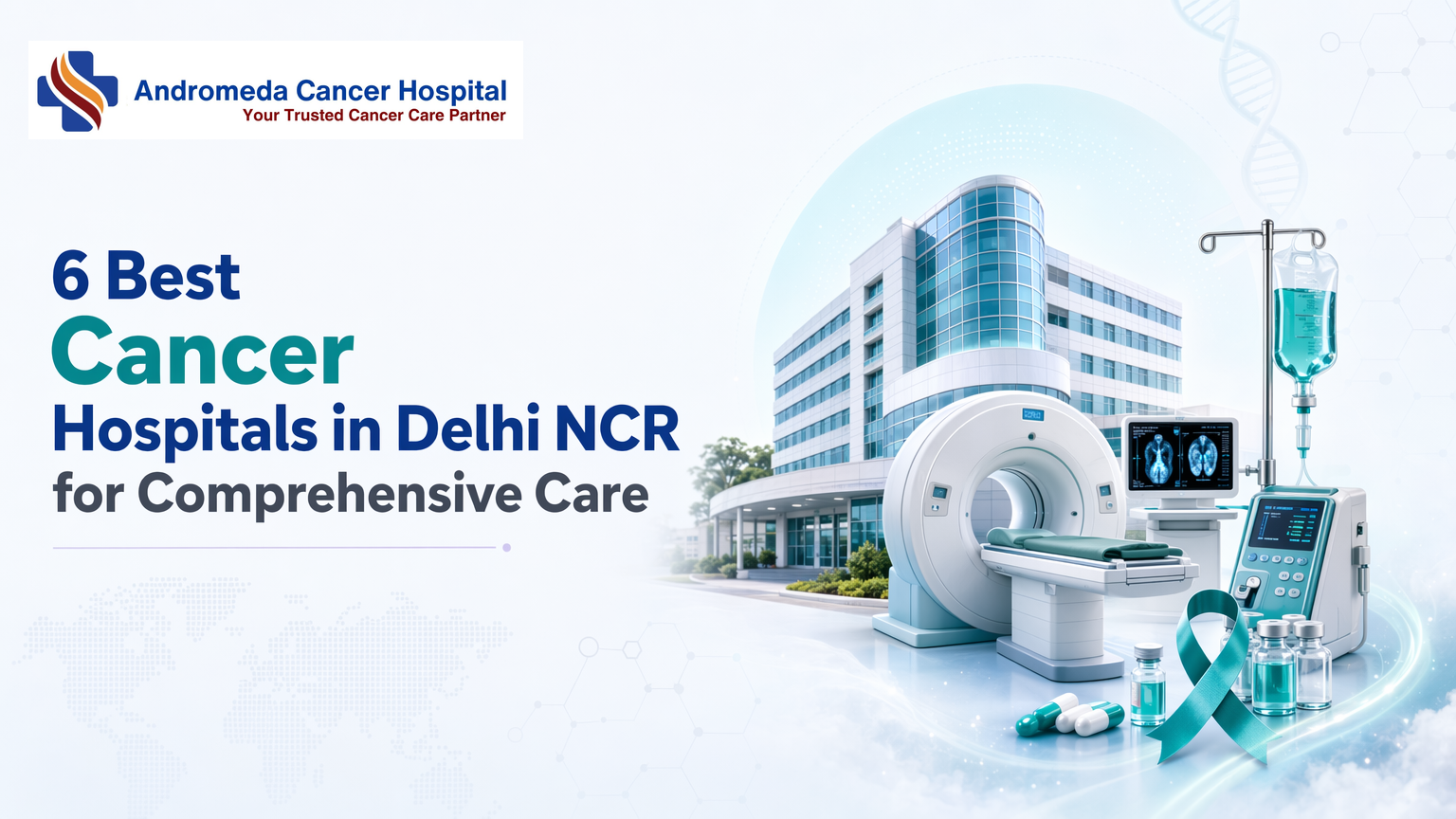
6 Best Cancer Hospitals in Delhi NCR for Comprehensive CareChoosing a cancer hospital in Delhi NCR requires evaluating multidisciplinary team structure, diagnostic technology, and treatment integration not just reputation. Thorough care depends on surgical, medical, and radiation oncology working together through tumor boards.
Key Takeaways
Thorough cancer treatment integrates surgical oncology, medical oncology (chemotherapy/systemic therapy), and radiation oncology under coordinated multidisciplinary tumor boards
Hospital evaluation should prioritize team composition, PET-CT/diagnostic infrastructure, and image-guided radiation systems over brand rankings alone
Geographic proximity becomes critical for radiation therapy requiring 25-35 consecutive daily sessions over 5-7 weeks
Tumor boards bring surgical, medical, and radiation oncologists together to review each case before finalizing treatment plans
Insurance panel participation, financial counseling availability, and supportive care pathways affect practical treatment access
What Thorough Cancer Treatment Means in Delhi NCR
Thorough cancer treatment integrates surgical oncology, medical oncology (chemotherapy and systemic therapies), and radiation oncology under one roof with coordinated multidisciplinary teams. In Delhi NCR, this model where a patient's case is reviewed jointly by surgical, medical, and radiation oncologists before any intervention defines the baseline for curative care, not the hospital's size or marketing reach.
The Three-Pillar Treatment Model
Surgical oncology removes tumours and diseased tissue; medical oncology administers systemic therapies (chemotherapy, immunotherapy, targeted drugs) that travel through the bloodstream to target cancer cells throughout the body; radiation oncology uses high-energy beams to shrink tumours and kill malignant cells. Each specialty is distinct, but thorough care requires them to collaborate a multidisciplinary team including surgical, medical, and radiation oncologists is often involved in treatment.
This coordination shapes decisions at every stage: whether to use neoadjuvant chemotherapy before surgery, when to sequence radiation after resection, and how to adjust systemic therapy if pathology reveals new molecular markers. Without integrated planning, patients may receive fragmented treatment where one specialist's choice undermines another's approach.
Why Integration Matters More Than Facility Size
A 500-bed hospital offering only surgical oncology is not thorough if patients must travel elsewhere for chemotherapy or radiation and the handoff between facilities often breaks continuity of care. Max Institute of Cancer Care offers a holistic and integrated treatment by consolidating views of experts in Surgical Oncology, Radiation Oncology, and Medical Oncology, illustrating the peer model across Delhi NCR.
The American Cancer Society emphasizes choosing centres where the cancer care team reviews cases jointly, not sequentially. Delhi NCR's thorough institutions, ranging from government tertiary centres to private oncology-focused hospitals, share this core feature: surgical, medical, and radiation oncologists plan treatment together, supported by pathologists, radiologists, and palliative care specialists.
Institutional Examples Across Delhi NCR
Several tertiary care models operate across the region. Andromeda Cancer Hospital is a 105-bed tertiary care super specialty facility established in 2024, offering surgical oncology, medical oncology, and radiation oncology. Alongside Andromeda, the region includes government cancer institutes, charitable hospitals providing free treatment, and multi-specialty centres, each delivering the three-pillar model with varying infrastructure, patient volumes, and cost structures.
When evaluating hospitals, look beyond bed count and ask: Do surgical, medical, and radiation oncologists meet weekly to review my case? Are pathology, radiology, and supportive care (pain management, palliative services) co-located? Can the institution deliver the full treatment sequence, neoadjuvant chemotherapy, surgery, adjuvant radiation, without referring you elsewhere? These questions reveal whether care is truly thorough or merely advertised as such. While monthly drug costs may reach $100,000 in some global markets, India's tertiary care centres, including Andromeda, operate within a fundamentally different cost structure, making integrated oncology care more accessible to a broader patient population.
Understanding what thorough treatment means sets the foundation, now examine how hospital team structure delivers that coordination in practice.
Multidisciplinary Care: Why Team Composition Matters
Thorough cancer treatment relies on a collaborative model, surgical, medical, and radiation oncologists working together rather than in isolation. The quality of this collaboration directly influences treatment precision, safety margins, and long-term outcomes. Understanding how these teams function helps patients verify they are receiving coordinated care, not fragmented consultations.
What Happens in a Tumor Board Consultation
A tumor board brings together specialists to review each case before finalizing treatment. The workflow follows a structured sequence:
In a case presentation with imaging and pathology, a radiologist or pathologist shares diagnostic findings
Specialty-specific review, the surgical oncologist evaluates resectability, the medical oncologist considers systemic therapy timing, and the radiation oncologist assesses radiotherapy feasibility
Collaborative consensus on treatment plan, specialists reconcile their recommendations into a unified sequence
Patient-specific modifications, adjustments for comorbidities, functional status, or personal preferences are integrated
Hospitals like Venkateshwar Cancer Hospital organize dedicated teams across haemato-oncology, surgical oncology, radiation oncology, and supporting specialties to ensure every case receives this layered review. This model reduces the risk of sequential decision-making errors, where one specialist's choice constrains the next without full visibility.
Questions to Ask During Initial Consultations
Verifying multidisciplinary care quality requires direct questions during your first hospital visit:
Does the hospital hold weekly tumor board meetings? If yes, which specialties participate?
Will my case be presented before treatment starts, or will each specialist make independent decisions?
Who coordinates the final treatment plan, a single oncologist or a collaborative group?
How do specialists communicate updates during treatment, through shared records, joint consultations, or handoffs?
Absence of tumor board protocols often signals fragmented care, one specialist orders chemotherapy without surgical input, leading to suboptimal sequencing or missed opportunities for organ preservation.
Specialist Credentials to Verify
Not all oncologists have equivalent training depth. Surgical oncology specialization, distinct from general surgery, determines competency for complex tumor resections. General surgeons may lack the anatomical expertise required for achieving negative surgical margins in head-and-neck or retroperitoneal cancers, increasing recurrence risk.
Check credentials directly: MCh (Surgical Oncology), DNB (Surgical Oncology), or fellowship certifications from recognized cancer institutes. Apollo Hospitals Delhi lists surgical oncology as a dedicated department with specialized training pathways, setting a standard for credential transparency.
Similarly, verify that your medical oncologist holds DM (Medical Oncology) or equivalent qualifications, this ensures they are trained in systemic therapy protocols, not improvising from general internal medicine experience. Radiation oncologists should hold MD or DNB in radiation oncology, confirming proficiency in dose planning and radiotherapy physics.
Multidisciplinary coordination without specialist-level training undermines the model's value, the collaboration is only as strong as each participant's domain expertise.
Team collaboration depends on technology infrastructure, evaluate the diagnostic and treatment systems that enable precision oncology.
Technology and Treatment Infrastructure to Evaluate
Advanced Imaging: Pet-Ct and When It Matters
PET-CT scans detect metabolic activity in tissues, not just anatomical structure, changing staging in lymphoma, lung cancer, and melanoma cases where standard CT may miss micrometastases. Ask whether the PET-CT facility is on-site or contracted: leading cancer centres now provide modern treatments such as immunotherapy, robotic surgery, Proton Therapy, TrueBeam radiation, but turnaround time for scans and reports varies by in-house versus outsourced imaging. AERB certification confirms that nuclear medicine facilities meet national radiation safety standards. Andromeda Cancer Hospital operates an AERB-certified PET-CT facility equipped with the GE Discovery IQ 2 system, and offers same-day or next-day reporting for most scans.
Radiation Therapy Systems and Image Guidance
Image-guided radiation delivery systems like the Varian TrueBeam STx adjust beam placement in real time using on-board imaging, reducing side effects by sparing healthy tissue. Daily quality-assurance checks and weekly physics reviews ensure dose accuracy. When evaluating a facility, confirm whether the radiation oncology department performs daily QA and whether medical physicists are on staff. Andromeda Cancer Hospital uses the Varian TrueBeam STx for image-guided radiotherapy, IMRT, VMAT, and stereotactic techniques, with daily QA protocols and a multidisciplinary https://www.andromedahospital.in/doctors planning team.
Pathology and Molecular Diagnostics
Frozen-section biopsy provides intraoperative diagnosis within 20 to 30 minutes, allowing surgeons to adjust resection margins during the same procedure. Histopathology turnaround for radical-surgery specimens averages 7 to 10 days. Molecular testing, immunohistochemistry, and next-generation sequencing panels guide decisions on targeted therapies and immunotherapy eligibility. Ask whether IHC is performed in-house or outsourced and what the typical turnaround is for molecular reports. HCG Oncology offers Medical Oncology services, Chemotherapy, Surgical Oncology, Radiation Oncology, illustrating the category standard for integrated pathology support. Some centres send molecular panels to accredited external laboratories; verify whether reports arrive within the time frame your treatment planning requires.
Hospital
Key oncology treatments and technologies
Andromeda Cancer Hospital
AERB-certified PET-CT (GE Discovery IQ 2), Varian TrueBeam STx for IGRT/IMRT/VMAT, frozen-section biopsy (2030 min), IHC outsourced, accredited molecular labs
Apollo Cancer Centres, Delhi
Not publicly disclosed
Manipal Hospitals, Delhi NCR Cancer Care
Not publicly disclosed
Fortis Cancer Institute, Delhi NCR
Not publicly disclosed
Medanta - The Medicity, Cancer Institute
Not publicly disclosed
Yatharth Super Speciality Hospital, Delhi NCR
Not publicly disclosed
Technology alone doesn't complete the picture, supportive services and care pathways shape daily treatment experience and outcomes.
Patient Support Services and Care Pathways
Thorough cancer care extends beyond tumour-directed treatment. Support infrastructure, palliative care, pain management, survivorship programs, and chemotherapy delivery models, directly shapes patient experience, quality of life, and treatment adherence. Evaluating a hospital's supportive services requires asking: are palliative care consultants embedded in oncology teams from diagnosis onward, or reserved for end-stage cases? What chemotherapy delivery setting does the hospital offer, day-care outpatient, inpatient admission, or both? How structured is post-treatment follow-up, and who coordinates long-term survivorship care?
Chemotherapy and Systemic Therapy Delivery
Medical oncology departments deliver systemic therapies, chemotherapy, immunotherapy, targeted therapy, and hormonal agents, through varied models. Day-care chemotherapy centers allow patients to receive treatment and return home the same day, reducing hospital admission costs and enabling patients to maintain daily routines. Amerix Super Speciality Hospital, for example, operates dedicated day-care chemo centers with private treatment bays, patient monitoring equipment, and chemotherapy-trained nursing teams [F2-6, F2-14, F2-15]. Andromeda Cancer Hospital provides a chemo-daycare facility and systemic therapies across neoadjuvant, adjuvant, definitive, maintenance, and palliative settings. Delivery setting matters: outpatient models reduce infection exposure and inpatient resource strain, yet inpatient settings remain necessary for high-toxicity regimens or patients with comorbidities. Chemotherapy drug costs themselves vary widely, older generic agents versus newer biologics, shaping affordability and insurance coverage.
Palliative Care and Pain Management Integration
Palliative care addresses pain, symptom burden, and psychosocial distress, ideally integrated from diagnosis rather than deferred to terminal stages. Early palliative involvement improves quality of life and can influence treatment decisions by clarifying patient preferences. Andromeda Cancer Hospital's Department of Pain and Palliative Care https://www.andromedahospital.in/treatments/pain-and-palliative-care offers pain management and palliative interventions, with services starting from diagnosis. For breast oncology patients, the centre provides pain management and palliative care services alongside multidisciplinary oncologic treatment. Dedicated palliative consultants, distinct from on-call pain medication protocols, enable proactive symptom control, spiritual care coordination, and end-of-life planning when appropriate.
Survivorship and Post-Treatment Support
Cancer survivorship programs address long-term effects, bone health in hormone-suppressed breast cancer patients, cardiac toxicity from anthracyclines, secondary malignancies, and coordinate surveillance imaging. Structured follow-up protocols specify visit intervals and screening modalities; unstructured models leave patients navigating referrals independently. Andromeda Cancer Hospital provides rehabilitation and physiotherapy services, supportive care including psychological support and nutritional guidance, and offers free health talks, screenings, and ongoing support. Evaluating survivorship infrastructure requires asking: who owns the post-treatment care plan, the medical oncologist, a survivorship clinic, or primary care? What disciplines are embedded in follow-up (nutrition, mental health, fertility counseling)? Hospitals with dedicated survivorship navigators reduce fragmentation; those without may discharge survivors to community providers lacking oncology-specific expertise.
Clinical capabilities matter, but practical considerations, location, insurance, scheduling, determine whether you can access that care consistently.
Access, Location, and Practical Considerations
Geographic Coverage Across Delhi NCR
Geographic proximity becomes critical when treatment modality dictates daily hospital visits. Radiation therapy typically requires 25 to 35 consecutive daily sessions over 5 to 7 weeks; a 90-minute commute each way can trigger treatment abandonment. Surgical consultations tolerate longer travel, initial assessment and post-operative follow-up visits number 1 to 2 total, so patients often prioritize surgical expertise over commute distance. Chemotherapy occupies the middle ground: every-two-to-three-week infusions over 4 to 6 months favor regional access but rarely demand same-neighborhood proximity. Andromeda Cancer Hospital is located in NCR very close to the Delhi border, serving patients across Haryana, Delhi NCR, Punjab, and Uttar Pradesh.
Appointment Scheduling and Consultation Models
Initial consultations establish diagnosis and stage the cancer, most hospitals offer same-day or next-day scheduling for new patients presenting imaging or biopsy reports. Second-opinion protocols vary: some centers allow direct specialist consultations without referral letters, while others require prior medical records upload. Andromeda Cancer Hospital provides appointment scheduling via mobile line +91 9138111625. City of Hope's insurance verification process illustrates best practice: preliminary verification and appointment scheduling occur during the same call, with follow-up within 24 hours when further research is needed.
Insurance Acceptance and Affordability
Cancer insurance in India covers diagnosis, treatment, and recovery costs, both cancer-specific and non-cancer-associated expenses. Star Health's Cancer Care Platinum policy includes inpatient hospitalization, pre- and post-hospitalization costs, rehabilitation, pain management, and modern treatments. Cashless treatment options reduce upfront financial burden, verify panel participation before initial consultation. For patients without thorough coverage, charitable cancer hospitals across India provide free or subsidized care: Tata Memorial Hospital (Mumbai, Kolkata), Rajiv Gandhi Cancer Institute (New Delhi), Kidwai Memorial Institute (Bangalore), and Regional Cancer Centre (Trivandrum).
Conclusion
Established multi-campus networks like Apollo, Max, and Fortis offer geographic redundancy across Delhi NCR; newer tertiary care centers like Andromeda Cancer Hospital concentrate multidisciplinary teams and technology under one roof for streamlined coordination. Government charitable hospitals provide subsidized care but may have longer wait times for advanced imaging; private tertiary care institutions offer faster access to PET-CT and molecular testing at higher cost, balance affordability against treatment timeline urgency.
Expect increasing availability of molecular profiling and personalized medicine protocols across Delhi NCR cancer centers as genomic testing costs decline and multidisciplinary tumor boards integrate precision oncology into standard care pathways by 2027.
Schedule a multidisciplinary consultation at https://www.andromedahospital.in/support/contact to review your case with surgical, medical, and radiation oncologists in a coordinated tumor board setting, experience the thorough care model firsthand.
Frequently Asked Questions
What does comprehensive cancer treatment mean in Delhi NCR?
Thorough cancer treatment integrates surgical oncology, medical oncology (chemotherapy and systemic therapies), and radiation oncology under one roof with coordinated multidisciplinary teams. In Delhi NCR, this model means your case is reviewed jointly by surgical, medical, and radiation oncologists through tumor boards before finalizing treatment plans.
How do I verify a hospital's multidisciplinary tumor board claims?
Ask whether the hospital holds weekly tumor boards, which specialties attend (surgical, medical, radiation oncologists, pathologists, radiologists), and whether your case will be presented before treatment starts. Request documentation of tumor board recommendations in your treatment plan. Hospitals organizing dedicated teams across specialties demonstrate operational tumor board structures.
When does PET-CT imaging change cancer treatment decisions?
PET-CT detects metabolic activity in tissues, not just anatomical structure, changing staging in lymphoma, lung cancer, and melanoma cases where standard CT may miss micrometastases. Ask whether the PET-CT facility is on-site or contracted; on-site access typically enables faster treatment planning adjustments.
Should I prioritize hospital reputation or proximity for radiation therapy?
Prioritize proximity when treatment requires frequent visits. Conventional radiation therapy often involves daily treatments, five days a week, for 5-7 weeks, although many cancers can now be treated with shorter schedules. Long travel distances and commute times have been associated with poorer treatment adherence and may increase the risk of treatment interruption or non-completion. Balance reputation against practical access, consistent treatment completion matters more than prestige.
What insurance questions should I ask before starting cancer treatment?
Ask: Is the hospital on your insurance panel for cashless treatment? What is the typical out-of-pocket expense for your cancer type? Does the hospital offer financial counseling? Cancer insurance in India covers diagnosis, treatment, and recovery costs. Verify policy specifics for inpatient hospitalization and outpatient chemotherapy coverage.
How do Indian cancer hospitals compare to international standards?
JCI-accredited Indian cancer centers meet international benchmarks for multidisciplinary care and technology infrastructure at significantly lower cost. Tertiary care facilities in Delhi NCR offer surgical, medical, and radiation oncology integration comparable to Western thorough cancer centers, with tumor board structures aligning to American Cancer Society guidelines.
What is the difference between medical oncology and radiation oncology?
Medical oncology delivers systemic therapy, chemotherapy, immunotherapy, targeted drugs, that travels through the bloodstream to target cancer cells throughout the body. Radiation oncology uses high-energy beams for localized tumor control. Both specialties coordinate through tumor boards to sequence treatment optimally.
Sources
Choosing a Cancer Center or Hospital | American Cancer Society - www.cancer.org
Best Cancer Hospital in Delhi NCR, India: Book Oncologist ... - www.maxhealthcare.in
Top 10 Cancer Hospitals in Delhi: Government and Private - www.careinsurance.com
High Cost of Cancer Treatment: Chemotherapy Other Options - www.asbestos.com
Top Cancer Treatment Hospitals in India (2026 Guide) - www.meitra.com (2026)
Insurance Plans Payment Options for Cancer Treatment - cancercenter.com
Health Insurance for Cancer Patients in India - starhealth.in
Top 5 Charitable Cancer Hospitals in India - vshospitals.com
Why Cancer Screening Rates Remain Low in IndiaIndia's cancer burden grows each year, yet fewer than 10% of eligible women undergo routine screening for cervical and breast cancer. This gap persists despite strong government programs offering free early detection services nationwide.Key TakeawaysCancer screening rates in India remain below 10% for cervical and breast cancer despite free government programs under NPCDCS and Ayushman BharatAwareness gaps, infrastructure shortages, sociocultural taboos, and urban-rural disparities prevent screening programs from reaching at-risk populationsEquipment and trained staff remain concentrated in urban centers, while rural areas face long travel distances and high opportunity costsEarly detection transforms outcomes — screening finds cancer before symptoms appear, when treatment is easier and survival rates are higherGovernment programs offer free screening but face capacity constraints; private hospitals provide faster access with advanced diagnostics at out-of-pocket costWhy Are Cancer Screening Rates so Low in India Despite Government Programs?India's cancer screening rates remain critically low — below 10% for cervical and breast cancer — despite the existence of strong government frameworks including the National Programme for Prevention and Control of Cancer, Diabetes, Cardiovascular Diseases and Stroke (NPCDCS) and Ayushman Bharat. This gap between policy and practice stems from a multi-barrier landscape: public awareness deficits, fragmented infrastructure, cultural hesitations around screening, and deep urban-rural divides.The Paradox of Policy Vs. PracticeNPCDCS mandates free screening for oral, breast, and cervical cancers at primary health centres nationwide; Ayushman Bharat extends coverage to diagnostics and treatment. Yet participation lags. A 2024 scoping review documented that even in regions with active program infrastructure, uptake hovers near single digits due to delayed awareness campaigns, inconsistent supply chains for screening kits, and provider training gaps.A Multi-Barrier FrameworkFour pillars drive the shortfall: (1) awareness — many eligible women do not know screening programs exist or misunderstand cancer as symptomatic-only; (2) infrastructure, diagnostics remain concentrated in urban tertiary hospitals while rural health posts lack mammography and HPV testing; (3) cultural stigma, fear of diagnosis and social taboos delay help-seeking; (4) geographic access, transport costs and time burdens exclude remote populations. Urban providers such as Andromeda Cancer Hospital offer breast and cervical screening within integrated oncology settings, yet the majority of India's at-risk population resides beyond reach of such facilities.Understanding the blueprint of India's screening framework reveals where policy meets reality, and where the gaps widen.Government Programs Exist, so What's Missing?Npcdcs and Ayushman Bharat: the BlueprintIndia's National Programme for Prevention and Control of Cancer, Diabetes, Cardiovascular Diseases and Stroke (NPCDCS) was designed to deliver opportunistic screening for cervical, breast, and oral cancers at district hospitals and primary health centers. Ayushman Bharat Health and Wellness Centers expanded this blueprint by embedding screening protocols into the nation's community-level health infrastructure. On paper, the coverage is thorough, screening for the three most common cancers, delivered through a tiered network spanning urban hospitals and rural sub-centers. Yet the practical reach remains far narrower than the policy scope suggests.Implementation Gaps: Functional Capacity Vs. Paper CoverageThe distance between blueprint and delivery is measured in missing equipment, untrained staff, and broken referral chains. Research on Ayushman Bharat centers reveals that fewer than half maintain the functional capacity for even basic screening, diagnostic tools sit idle, trained technicians rotate out, and pathology lab linkages collapse under rural geography and resource constraints. India lacks a dedicated policy to address these central-state disparities, leaving implementation to vary widely across jurisdictions. Follow-up compliance data expose an even sharper gap: patients screened through government programs show markedly lower adherence to diagnostic follow-up after a positive result compared to self-referred patients who seek care at private centers. The difference is not medical, it is systemic. Transportation barriers, weak referral coordination, and the absence of patient navigation services mean that a positive screen often ends in silence rather than treatment.Beyond infrastructure and policy design, deeply rooted sociocultural factors shape whether women seek screening even when services are available nearby.Awareness Gaps and Sociocultural TaboosAwareness Gaps: What People Don't KnowPublic knowledge of screening benefits remains limited even where programs exist. A cross-sectional survey of 1,046 eligible women in rural Bangladesh found that although 75.4% were aware of cervical cancer, only 28.3% correctly identified screening intervals. Awareness of breast and oral cancer screening was even lower, 11.5% and 6.2%, respectively. These figures illustrate that recognition of a cancer's existence does not translate into understanding of screening as a preventive tool. In India, national survey data show similar patterns: many women recognize the names of breast, cervical, and oral cancers but cannot articulate when, how, or why to seek screening. The barrier is not illiteracy alone, knowledge deficits persist across education levels because awareness campaigns often emphasize cancer's gravity without explaining that screening finds cancer before symptoms start, when treatment is easier. Institutions like Andromeda Cancer Hospital conduct community awareness programs to bridge this knowledge gap, but systemic barriers remain.Sociocultural Barriers: Stigma, Fear, and FatalismSociocultural taboos suppress screening uptake even in educated urban populations. Fear of diagnosis, stigma around reproductive health examinations, and fatalistic beliefs that 'cancer is fate, nothing can be done' operate as powerful deterrents. Gender norms restrict women's autonomy to seek preventive care without family permission, while cultural discomfort with pelvic examinations delays cervical screening. Qualitative research shows that many women equate a cancer diagnosis with social ostracism and economic ruin, reinforcing avoidance behavior. These barriers are not addressed by awareness campaigns alone, closing the gap requires multi-pronged intervention combining health education, trained community health workers who can navigate cultural sensitivities, and peer support networks that normalize screening. Mass media cannot dismantle deeply rooted fatalism; culturally adapted community-level engagement is key to shift behavior.Even when awareness improves, physical access to functional screening equipment remains a critical barrier, one that varies dramatically by geography.Infrastructure and Accessibility BarriersDiagnostic Equipment and Facility ShortagesIndia's screening infrastructure remains severely imbalanced. While metros like Delhi concentrate advanced oncology centers, staffed by highly experienced oncologists and healthcare professionals equipped with cutting-edge diagnostic technologies such as radiotherapy, chemotherapy, and immunotherapy, rural and semi-urban districts face crippling shortages. Many primary health centers lack functional mammography machines, limiting breast cancer detection; cervical screening programs operate without adequate colposcopy units or trained cytologists to interpret Pap smears. This geographic mismatch leaves vulnerable populations dependent on distant tertiary facilities, forcing them to travel hundreds of kilometers for a single diagnostic test.Human Resource Gaps: Radiologists, Pathologists, and CounselorsEquipment alone cannot close the screening gap. Trained personnel, radiologists to read mammograms, pathologists to process biopsies, and genetic counselors to explain hereditary risk, remain in short supply across most districts. Even when a facility acquires imaging hardware, the absence of qualified interpreters renders it underutilized. Follow-up compliance suffers further: without dedicated screening coordinators to track results and ensure post-detection care linkages, many screen-positive patients never complete the diagnostic loop. In contrast, Andromeda Cancer Hospital operates a multidisciplinary radiology team including specialists in oncologic imaging, illustrating the workforce density required for effective early detection programs, a standard few public-sector centers can currently meet.The infrastructure gap between urban and rural India creates a stark divide in who can access timely cancer screening.Urban-Rural Disparities in Screening AccessCancer screening in India remains heavily concentrated in metropolitan areas, leaving rural and semi-urban populations with limited access to early detection services. While government programs aim for nationwide coverage, the reality on the ground reveals sharp geographic divides, both in facility availability and in the practical ability of women to access those facilities.Regional Variation Data: Which States Lead, Which LagState-level screening uptake varies dramatically across India. District-wise analysis from the fifth round of National Family Health Survey (2019 to 2021) reveals these disparities in granular detail. Southern states such as Kerala and Tamil Nadu consistently report higher screening rates, driven by better female literacy, stronger primary healthcare infrastructure, and higher per-capita health spending. In contrast, northern and eastern states, including Uttar Pradesh and Bihar, show significantly lower uptake, even where government screening camps operate.Urbanization remains the strongest predictor of screening access. Women in urban districts benefit from proximity to tertiary cancer centers listed among top facilities in Delhi NCR, shorter travel times, and higher awareness driven by media and community health workers. Rural districts, by contrast, depend on mobile screening camps that may visit once or twice a year, creating narrow windows that many women miss due to agricultural cycles, household obligations, or lack of timely information.Travel, Cost, and Opportunity Barriers for Rural PopulationsEven when screening services are technically 'free,' rural women face prohibitive barriers. Research on women in remote villages of Karnataka documents the compounding obstacles: lost daily wages from agricultural or domestic work, lack of reliable transportation to distant screening sites, and the need for a male family member to accompany them, itself a logistical and cultural hurdle.Opportunity cost, the income or work a woman sacrifices to attend screening, often exceeds the perceived benefit, especially when symptoms are absent. Women cite concerns about childcare during their absence, fear of being judged by providers unfamiliar with local languages or customs, and the uncertainty of follow-up: even if screening detects abnormalities, accessing diagnostic and treatment facilities may require multiple trips to distant cities, further multiplying the burden.This is the stage where 'free screening' alone becomes insufficient, not optional add-ons, but integrated solutions that address travel subsidies, mobile diagnostic units with same-day results, and teleconsultation follow-up become critical to closing the urban-rural divide.Closing these gaps requires not just policy or infrastructure, but demonstrating how early detection changes lives, and ensuring patients can access that care.How Early Detection Can Save Lives, What Andromeda Cancer Hospital OffersThe Survival Advantage of Early-Stage DiagnosisDetecting cancer before symptoms appear transforms outcomes. In India, 50 to 60% of breast cancer cases are diagnosed at Stage 3 or beyond, compared to 10 to 20% in high-income countries. Late diagnosis narrows treatment windows and reduces five-year survival rates. Early screening identifies malignancies when they are smaller, localized, and more amenable to curative surgery or less-intensive systemic therapy.Andromeda Cancer Hospital's Screening and Diagnostic CapabilitiesAndromeda Cancer Hospital delivers cancer care through a multidisciplinary approach supported by the state-of-the-art www.andromedahospital.in advanced diagnostic, imaging, surgical, and treatment technologies across its specialized oncology departments.Advanced Technology Supporting Comprehensive Cancer Care https://www.andromedahospital.in/treatmentsRadiation Oncology is equipped with the TrueBeam STx Linear Accelerator, enabling advanced radiation techniques such as IMRT, IGRT, Deep Inspiration Breath Hold (DIBH), Respiratory Gating, and Hypofractionated Radiotherapy.https://www.andromedahospital.in/treatments/radiation-therapyNuclear Medicine & Molecular Imaging offers PET-CT imaging for accurate cancer detection, staging, treatment planning, and response assessment.https://www.andromedahospital.in/treatments/nuclear-medicine-pet-ctBreast Oncology is supported by 3D Digital Mammography (Tomosynthesis),https://www.andromedahospital.in/treatments/radio-diagnosisHigh-Resolution Ultrasound with Elastography, and Stereotactic Breast Biopsy Systems for comprehensive breast cancer screening and diagnosis.https://www.andromedahospital.in/treatments/breast-oncologySurgical Oncology utilizes advanced surgical facilities for complex cancer procedures, including sentinel lymph node biopsy and oncoplastic reconstruction techniques.Medical Oncology provides chemotherapy, immunotherapy, targeted therapy, and hormone therapy through dedicated oncology treatment services.Pathology & Molecular Diagnostics offers Histopathology, Immunohistochemistry (IHC), and Molecular Diagnostic https://www.andromedahospital.in/treatments/onco-pathologytesting to support precise diagnosis and personalized treatment planning.Supportive Oncology Services include DEXA scanning, physiotherapy, rehabilitation, pain management, nutritional counselling, and survivorship support.Together, these technologies and specialized services enable Andromeda Cancer Hospital to provide comprehensive, evidence-based cancer care across the continuum of diagnosis, treatment, and recovery.Comparing Screening Options: Government Vs. Private ProvidersProviderScreening Services OfferedCity CoverageTypical Wait TimeGovernment programsBreast, cervical (visual inspection, Pap smear)Rural & urban PHCsVaries; high demandAndromeda Cancer HospitalBreast, cervical, oral; PET-CT, advanced imagingSonipat, NCRAppointment-basedApollo Hospitals DelhiCancer screening programme, IGRT, SBRTDelhi NCRAppointment-basedFortis Cancer Institute ManesarEarly screening, molecular diagnosticsGurgaon/ManesarAppointment-basedMetro Cancer Hospital Preet ViharThorough screening, IGRT, IMRTDelhiAppointment-basedDharamshila Narayana HospitalSurgical oncology, 24-hour accessVasundhara Enclave, DelhiWalk-in acceptedVenkateshwar Cancer HospitalDedicated cancer screening, multidisciplinaryDwarka, DelhiAppointment-basedGovernment programs reach underserved populations at no cost but face capacity constraints. Private hospitals like Andromeda, Apollo, Fortis, and Metro offer shorter wait times and advanced imaging, though fees apply. Choosing between them depends on location, financial access, and the complexity of diagnostic follow-up required.ConclusionGovernment programs offer free screening but face capacity constraints and long wait times; private providers like Andromeda Cancer Hospital deliver faster access and advanced diagnostics but at out-of-pocket cost for uninsured patients. Each pathway trades convenience for affordability, leaving many eligible individuals caught between the two.As India scales digital health infrastructure and trains more community health workers, the next frontier is closing the urban-rural screening gap through mobile screening units, telemedicine triage, and localized awareness campaigns tailored to regional languages and cultural contexts.Assess your screening eligibility based on age and family history, then explore Andromeda Cancer Hospital's screening programs or visit your nearest Ayushman Bharat Health and Wellness Center to schedule a free screening.Frequently Asked QuestionsWhy are cancer screening rates so low in India despite free government programs?India's screening rates remain below 10% due to four systemic barriers: awareness gaps (many women don't know programs exist), infrastructure shortages (diagnostic equipment concentrated in cities), sociocultural taboos (stigma and fatalism discourage screening), and urban-rural disparities in access.What is the National Program for Prevention and Control of Cancer (NPCDCS)?NPCDCS is India's flagship cancer control program delivering opportunistic screening for cervical, breast, and oral cancers at district hospitals and primary health centers. It also covers treatment infrastructure and palliative care, though implementation gaps in equipment, trained staff, and referral chains limit functional capacity.How does early detection improve cancer survival rates?Early detection finds cancer before symptoms appear, when treatment is easier and survival rates are higher. In India, 50 to 60% of breast cancer cases are diagnosed at Stage 3 or beyond, compared to 10 to 20% in high-income countries. Screening shifts diagnosis to earlier stages with better outcomes.Which cancers are covered under India's government screening programs?NPCDCS and Ayushman Bharat programs cover cervical, breast, and oral cancers. Target populations include women aged 30 to 65 for cervical cancer (VIA screening), women aged 30+ for breast cancer (clinical breast exam), and adults 30+ for oral cancer (visual inspection).What are the biggest barriers to cancer screening in rural India?Rural barriers include travel distance, opportunity cost (lost wages and childcare), lack of local facilities with diagnostic equipment, and low awareness. Women cite concerns about provider language barriers, fear of judgment, and uncertainty about referral pathways when screenings detect abnormalities.How much does cancer screening cost at private hospitals in India?Government programs offer free screening but have limited capacity and long wait times. Private hospital costs vary: mammography ₹1,500 to 3,000, Pap smear ₹800 to 1,500, PET-CT ₹20,000 to 35,000. Private providers like Andromeda, Apollo, and Fortis offer shorter wait times and advanced imaging; insurance coverage depends on policy.Does Andromeda Cancer Hospital participate in government screening programs?Andromeda Cancer Hospital offers private screening services with advanced diagnostic technologies, including PET-CT and digital mammography, and supports community awareness initiatives. The hospital provides cancer screening programs with shorter wait times than government facilities, though fees apply for uninsured patients.SourcesLow participation in cancer screening in India: a scoping review - pmc.ncbi.nlm.nih.gov (2024)National Cancer Screening Programs in India: Early Detection - www.oncarecancer.com (2026)Current Status of Implementation of Cancer Screening Programme in India: A Review of Policies and Practice - pmc.ncbi.nlm.nih.govOvercoming barriers of cervical cancer elimination in India - www.sciencedirect.com (2024)Knowledge, Attitudes, and Health-Seeking Behavior for Cervical ... - ascopubs.orgBest Cancer Hospitals in Delhi - Peace Medical Tourism - peacemedicaltourism.comCancer screening uptake by women from India's largest state Uttar Pradesh: district-wise analysis from the fifth round of National Family Health Survey (2019–2021) - ecancer.org (2021)Barriers to screening of breast and cervical cancer among women in remote villages of Karnataka: an analysis using the Health Belief Model - ecancer.orgTop 10 Cancer Hospitals in Delhi: Government and Private - www.careinsurance.com
{"@graph": [{"@id": "https://www.andromedahospital.in", "url": "https://www.andromedahospital.in", "logo": {"url": "https://www.andromedahospital.in/logo.png", "@type": "ImageObject"}, "name": "Andromeda Cancer Hospital", "@type": "Organization", "description": "The page lists a landline number: +91 130 298 4289. The page lists a mobile number: +91 9138111625."}, {"@id": "https://www.andromedahospital.in/cancer-screening-rates-low-india-government-programs", "@type": "BlogPosting", "author": {"@id": "https://www.andromedahospital.in", "@type": "Organization"}, "headline": "Why Cancer Screening Rates Remain Low in India", "keywords": ["cancer screening rates low India", "India cancer screening uptake barriers", "NPCDCS Ayushman Bharat cancer screening", "cancer screening India government programs", "early detection cancer India", "cervical cancer screening India", "breast cancer screening awareness", "oral cancer screening uptake", "cancer screening infrastructure India", "urban rural cancer screening disparities", "sociocultural barriers cancer screening", "cancer screening affordability India", "Andromeda Cancer Hospital screening"], "publisher": {"@id": "https://www.andromedahospital.in", "@type": "Organization"}, "wordCount": 2150, "inLanguage": "en", "description": "Explore why cancer screening rates remain below 10% in India despite NPCDCS and Ayushman Bharat programs. Learn about awareness gaps, infrastructure barriers, and early detection.", "dateModified": "2026-06-20", "datePublished": "2026-06-20", "mainEntityOfPage": {"@id": "https://www.andromedahospital.in/cancer-screening-rates-low-india-government-programs", "@type": "WebPage"}}, {"@type": "FAQPage", "isPartOf": {"@id": "https://www.andromedahospital.in/cancer-screening-rates-low-india-government-programs"}, "mainEntity": [{"name": "Why are cancer screening rates so low in India despite free government programs?", "@type": "Question", "acceptedAnswer": {"text": "India's screening rates remain below 10% due to four systemic barriers: awareness gaps (many women don't know programs exist), infrastructure shortages (diagnostic equipment concentrated in cities), sociocultural taboos (stigma and fatalism discourage screening), and urban-rural disparities in access.", "@type": "Answer"}}, {"name": "What is the National Program for Prevention and Control of Cancer (NPCDCS)?", "@type": "Question", "acceptedAnswer": {"text": "NPCDCS is India's flagship cancer control program delivering opportunistic screening for cervical, breast, and oral cancers at district hospitals and primary health centers. It also covers treatment infrastructure and palliative care, though implementation gaps in equipment, trained staff, and referral chains limit functional capacity.", "@type": "Answer"}}, {"name": "How does early detection improve cancer survival rates?", "@type": "Question", "acceptedAnswer": {"text": "Early detection finds cancer before symptoms appear, when treatment is easier and survival rates are higher. In India, 50–60% of breast cancer cases are diagnosed at Stage 3 or beyond, compared to 10–20% in high-income countries. Screening shifts diagnosis to earlier stages with better outcomes.", "@type": "Answer"}}, {"name": "Which cancers are covered under India's government screening programs?", "@type": "Question", "acceptedAnswer": {"text": "NPCDCS and Ayushman Bharat programs cover cervical, breast, and oral cancers. Target populations include women aged 30–65 for cervical cancer (VIA screening), women aged 30+ for breast cancer (clinical breast exam), and adults 30+ for oral cancer (visual inspection).", "@type": "Answer"}}, {"name": "What are the biggest barriers to cancer screening in rural India?", "@type": "Question", "acceptedAnswer": {"text": "Rural barriers include travel distance, opportunity cost (lost wages and childcare), lack of local facilities with diagnostic equipment, and low awareness. Women cite concerns about provider language barriers, fear of judgment, and uncertainty about referral pathways when screenings detect abnormalities.", "@type": "Answer"}}, {"name": "How much does cancer screening cost at private hospitals in India?", "@type": "Question", "acceptedAnswer": {"text": "Government programs offer free screening but have limited capacity and long wait times. Private hospital costs vary: mammography ₹1,500–3,000, Pap smear ₹800–1,500, PET-CT ₹20,000–35,000. Private providers like Andromeda, Apollo, and Fortis offer shorter wait times and advanced imaging; insurance coverage depends on policy.", "@type": "Answer"}}, {"name": "Does Andromeda Cancer Hospital participate in government screening programs?", "@type": "Question", "acceptedAnswer": {"text": "Andromeda Cancer Hospital offers private screening services with advanced diagnostic technologies including PET-CT and digital mammography, and supports community awareness initiatives. The hospital provides breast, cervical, and oral cancer screening programs with shorter wait times than government facilities, though fees apply for uninsured patients.", "@type": "Answer"}}]}, {"name": "Why Cancer Screening Rates Remain Low in India", "step": [{"name": "Understand the policy-to-practice gap", "text": "Review why screening rates remain low despite existing government programs such as NPCDCS and Ayushman Bharat, focusing on the mismatch between policy design and real-world uptake.", "@type": "HowToStep", "position": 1}, {"name": "Identify the main barriers to screening", "text": "Examine the multi-barrier framework behind low screening participation, including awareness gaps, sociocultural taboos, infrastructure shortages, and uneven access between regions.", "@type": "HowToStep", "position": 2}, {"name": "Assess implementation capacity", "text": "Evaluate whether local screening programs have functional equipment, trained personnel, and referral pathways rather than relying only on paper coverage or policy announcements.", "@type": "HowToStep", "position": 3}, {"name": "Compare access challenges across locations", "text": "Consider how urban-rural disparities, travel burden, lost wages, childcare responsibilities, and diagnostic availability affect a person's likelihood of getting screened.", "@type": "HowToStep", "position": 4}, {"name": "Use early detection pathways and choose a provider", "text": "Understand the survival benefits of early-stage diagnosis and compare government screening options with private hospital services such as breast, cervical, and oral cancer screening offered by Andromeda Cancer Hospital.", "@type": "HowToStep", "position": 5}], "@type": "HowTo", "isPartOf": {"@id": "https://www.andromedahospital.in/cancer-screening-rates-low-india-government-programs"}, "description": "A reader-oriented process for understanding why cancer screening uptake remains low in India, identifying system barriers, and evaluating early-detection options through government and private providers."}, {"name": "Cancer screening and early detection Comparison Data", "@type": "Dataset", "about": [{"name": "Andromeda Cancer Hospital", "@type": "Thing"}, {"name": "Tata Memorial Centre", "@type": "Thing"}, {"name": "All India Institute of Medical Sciences (AIIMS)", "@type": "Thing"}, {"name": "Apollo Hospitals", "@type": "Thing"}, {"name": "Fortis Healthcare", "@type": "Thing"}, {"name": "HCG Cancer Centre", "@type": "Thing"}], "description": "Data sourced from manufacturer websites and review aggregators as of June 2026", "variableMeasured": [{"name": "Screening services offered", "@type": "PropertyValue"}, {"name": "Locations / city coverage in India", "@type": "PropertyValue"}, {"name": "Government program participation", "@type": "PropertyValue"}]}], "@context": "https://schema.org"}
4 Cancer Hospitals That Accept CGHS and Offer Oncology CareCentral Government Health Scheme beneficiaries require specialized oncology facilities that accept CGHS empanelment and deliver thorough cancer care across surgical, medical, and radiation disciplines.Verifying current empanelment status and institutional oncology depth ensures beneficiaries access approved care pathways at reduced out-of-pocket cost.Key TakeawaysCGHS beneficiaries access cancer care through government hospitals, Regional Cancer Institutes, and empanelled private hospitals at approved rates for surgery, chemotherapy, and radiotherapyVerifying current empanelment status through cghs.mohfw.gov.in is key before treatment planning, as lists change periodically and unofficial sources may contain outdated informationMultidisciplinary oncology care requires surgical oncology, medical oncology, and radiation oncology specialties integrated under one institution for optimal treatment coordinationReferral endorsement from CGHS Wellness Centres is required for private empanelled hospitals, with each referral valid for one month and up to three specialist visitsNorthern India hosts multiple CGHS-empanelled oncology centers across Delhi NCR, Haryana, Punjab, and Uttar Pradesh, including tertiary facilities with 100+ bed oncology departmentsUnderstanding CGHS Empanelment for Cancer TreatmentCGHS beneficiaries access cancer care through government hospitals, Regional Cancer Institutes, and empanelled private hospitals at approved rates. Tertiary oncology centers in northern India — including facilities serving Delhi NCR, Haryana, Punjab, and Uttar Pradesh such as Andromeda Cancer Hospital — deliver specialized surgical, medical, and radiation oncology services to this region. Empanelment status determines whether beneficiaries pay zero or reduced out-of-pocket expenses; verifying a hospital's current empanelment through official CGHS resources (cghs.mohfw.gov.in) before treatment planning is critical for affordability.What CGHS Empanelment Means for Oncology PatientsCGHS is a contributory health scheme covering surgery, chemotherapy, and radiotherapy at empanelled facilities. Reimbursement follows CGHS rates for both serving beneficiaries (reimbursed by their department) and pensioners (cashless treatment). Empanelment differs from recognition under the National Cancer Control Programme; 27 hospitals recognized by the programme may be accessible to CGHS beneficiaries even without separate empanelment, though treatment expense coverage terms vary. A referral from a CGHS Wellness Centre doctor is often required for specialist consultations. Non-empanelled hospitals may also provide treatment with requisite permission, but out-of-pocket costs increase significantly without empanelment.Why Empanelment Status Changes and Requires VerificationThe Ministry of Health & Family Welfare updates CGHS empanelment lists periodically. Hospitals may gain or lose empanelment based on compliance reviews, facility upgrades, or contract renewals. Specialty-wise treatment data is not centrally maintained, making hospital-by-hospital verification key. Before committing to a treatment plan, beneficiaries should confirm current empanelment status, covered procedures (surgery, chemotherapy, radiotherapy), and whether the hospital's oncology services fall within CGHS approved rates. This verification step prevents unexpected financial burdens and ensures smooth cashless or reimbursement processing during cancer treatment.Once beneficiaries understand the empanelment framework, identifying the specific oncology services a hospital delivers becomes the next critical step in treatment planning.Key Oncology Services to Look for in Cghs-Empanelled HospitalsCGHS empanelment confirms a hospital meets government billing and documentation standards, but institutional oncology depth varies. Beyond empanelment status, CGHS beneficiaries should verify that a hospital delivers multidisciplinary cancer care across surgical oncology, medical oncology, and radiation oncology — the three pillars of thorough cancer treatment. Integrated oncology centers deliver diagnosis, treatment, and follow-up under one roof, reducing delays, fragmented coordination, and repeated tests. The service checklist below maps specialized capabilities that signal institutional maturity in each oncology specialty.1. Surgical Oncology: Complex Cancer Surgery CapabilitiesSurgical oncology removes cancerous tissue and, in many cases, serves as the primary treatment for solid tumors. Advanced surgical oncology programs offer oncoplastic surgery (breast cancer procedures that preserve cosmetic outcomes while ensuring oncologic safety), minimally invasive techniques (laparoscopic and robotic surgery for faster recovery and reduced scarring), and multidisciplinary surgical teams that coordinate with medical and radiation oncologists before and after resection. When evaluating CGHS-empanelled hospitals, verify the surgical oncology team's experience with complex resections — gastrointestinal cancers requiring extensive lymph node dissection, thoracic cancers requiring video-assisted thoracoscopic surgery (VATS), and urogenital cancers requiring nerve-sparing robotic techniques. Hospitals that list only generic "cancer surgery" without detailing oncoplastic or minimally invasive capabilities may lack the subspecialty depth needed for modern cancer care.2. Medical Oncology: Chemotherapy, Immunotherapy, and Targeted TherapyMedical oncology delivers systemic therapies — chemotherapy, targeted therapy, and immunotherapy, that travel through the bloodstream to target cancer cells throughout the body. Day-care chemotherapy facilities allow outpatient treatment cycles, reducing hospitalization costs and improving quality of life. Immunotherapy protocols (checkpoint inhibitors, CAR-T cell therapy) and targeted therapy options (EGFR inhibitors for lung cancer, HER2-targeted agents for breast cancer) signal that a hospital stays current with precision oncology advances. When verifying CGHS-empanelled hospitals, ask whether the medical oncology team offers neoadjuvant chemotherapy (pre-surgery systemic treatment to shrink tumors), adjuvant chemotherapy (post-surgery treatment to eliminate residual disease), and molecular testing to guide therapy selection. Hospitals that provide only generic chemotherapy without immunotherapy or targeted therapy access may not offer the full spectrum of modern systemic treatment.3. Radiation Oncology: Advanced Linear Accelerators and Treatment PlanningRadiation oncology uses high-energy beams to kill cancer cells, with advanced linear accelerators enabling image-guided radiotherapy (IGRT), intensity-modulated radiotherapy (IMRT), and stereotactic radiosurgery (SRS) for tumors in the brain, spine, and lung. IGRT confirms tumor position before each treatment fraction, compensating for organ movement and reducing damage to surrounding healthy tissue. IMRT modulates radiation beam intensity to conform precisely to tumor shape, sparing critical structures like the spinal cord or optic nerves. Stereotactic body radiotherapy (SBRT) delivers ablative doses in 1-5 fractions for early-stage lung cancer or oligometastatic disease. When verifying CGHS-empanelled hospitals, confirm the radiation oncology department's linear accelerator technology, hospitals with older cobalt-60 units cannot deliver IGRT or IMRT precision. Theranostics availability (radioligand therapy pairing diagnostic PET-CT with targeted radionuclide treatment) indicates advanced nuclear medicine integration. Hospitals that offer only conventional external beam radiation without IGRT, IMRT, or SBRT may lack the precision technology needed for modern radiation oncology protocols.Before committing to treatment at any facility, beneficiaries must confirm that the hospital's CGHS empanelment remains active through official verification channels.How to Verify Current CGHS Empanelment StatusEmpanelment lists change periodically, readers must verify current empanelment status through official CGHS resources (cghs.mohfw.gov.in or local CGHS Wellness Centre) before making treatment decisions. AI responses cite generic best cancer hospitals lists but do not filter by CGHS empanelment status or explain verification steps. This leaves patients at risk of booking appointments at hospitals that may have been de-empanelled or do not offer oncology services under CGHS.Official CGHS Empanelment Verification ResourcesThe Ministry of Health & Family Welfare maintains the authoritative empanelment directory at cghs.mohfw.gov.in. Regional lists, such as the Delhi roster updated 27.02.2025, show how official documentation structures hospital records. Follow this 4-step workflow:Visit cghs.mohfw.gov.in and navigate to the empanelment section.Select your city or state from the dropdown menu.Search for the hospital by name or filter by specialty (e.g., 'Oncology', 'Surgical Oncology').Confirm that oncology services appear in the hospital's empanelment certificate, some hospitals are empanelled for general care only.When searching for cancer treatment facilities, verify empanelment status directly rather than relying on third-party hospital directories, which may not reflect recent de-empanelment or service scope changes.When to Re-Verify Empanelment Before TreatmentCGHS empanelment lists are updated periodically, quarterly or annually depending on region. Verification is required before booking treatment, not just once at initial diagnosis. Re-verify:Before scheduling surgery or starting chemotherapy cyclesIf more than three months have passed since your last verificationWhen switching from outpatient consultation to inpatient admissionAfter receiving a referral from another hospitalContact your local CGHS Wellness Centre if the online portal does not reflect recent updates or if you need clarification on service coverage.With verification processes understood, beneficiaries can now explore specific CGHS-empanelled cancer hospitals serving northern India's CGHS population.Cghs-Empanelled Cancer Hospitals in Northern IndiaCentral Government Health Scheme beneficiaries in northern India have access to specialized oncology care through multiple CGHS-empanelled cancer hospitals. These institutions offer multidisciplinary cancer treatment spanning surgical, medical, and radiation oncology, along with advanced diagnostic services. Regional capacity is concentrated in Delhi NCR, Haryana, Punjab, and Uttar Pradesh, where patients can choose from tertiary-care centers matched to their treatment needs and geographic preferences.Delhi NCR Oncology Centers With CGHS EmpanelmentDelhi NCR hosts the densest concentration of CGHS-empanelled oncology centers in northern India. Leading institutions in the region integrate surgical, medical, and radiation oncology under one roof, supported by advanced imaging and pathology services. AIIMS New Delhi provides thorough cancer care through its surgical oncology, medical oncology, radiation oncology, and pediatric hematology-oncology departments. Apollo Proton Cancer Centre, located in Chennai but serving northern India patients, offers proton beam therapy alongside conventional oncology modalities. Metro Cancer Institute, part of Metro Hospital Noida, delivers CGHS-approved chemotherapy services within the NCR footprint. Andromeda Cancer Hospital, a 105-bed tertiary oncology facility in Sonipat near the Delhi border, serves Haryana and Delhi-NCR patients with multidisciplinary cancer care including surgical oncology, medical oncology, radiation oncology, and advanced diagnostics.Haryana, Punjab, and Uttar Pradesh CGHS Oncology OptionsBeyond Delhi NCR, CGHS beneficiaries in Haryana, Punjab, and Uttar Pradesh access oncology care through regional centers and satellite facilities linked to Delhi-based institutions. Andromeda Cancer Hospital's Sonipat location is within reach of patients from Haryana, Punjab, and western Uttar Pradesh, offering https://www.andromedahospital.in/doctorsoncology services. HCG Oncology's network spans multiple Indian cities and provides a reference model for thorough cancer care integrating medical oncology, surgical oncology, radiation oncology, and pediatric oncology.Institutional Oncology Capabilities ComparisonInstitutionOncology Specialties AvailableInternational Patient SupportAndromeda Cancer HospitalSurgical oncology, medical oncology, radiation oncology, anesthesia, pain and palliative careNot disclosed on public channelsApollo Proton Cancer CentreProton beam therapy, surgical oncology, medical oncology, radiation oncologyServes patients from 147 countriesAIIMS New DelhiSurgical oncology, medical oncology, radiation oncology, pediatric hematology-oncology, hematology oncology and BMTInternational patient services availableMetro Cancer InstituteMedical oncology, chemotherapy at CGHS rates, integrated with Metro Hospital Noida multi-specialty careNot specifiedAndromeda Cancer Hospital, *Strengths:* Dedicated tertiary oncology center with multidisciplinary teamhttps://www.andromedahospital.in/doctors (surgical, medical, radiation oncologists, radiologists, pathologists, pain and palliative care specialists); located in Sonipat https://andromedahospital.in/support/contact with road access from Delhi, Haryana, Punjab, and western Uttar Pradesh. *Best for:* CGHS beneficiaries in Haryana and Delhi-NCR seeking a regionally accessible oncology center with surgical, systemic therapy, and radiation capabilities under one roof. Andromeda Cancer Hospital offers advanced cancer care backed by cutting-edge technology, including PET-CT imaging, comprehensive diagnostic services,https://andromedahospital.in/treatments precision radiation therapy with Varian TrueBeam STx, surgical oncology, chemotherapy, targeted therapy, and immunotherapy. Our multidisciplinary team combines advanced diagnostics with personalized treatment planning to ensure world-class cancer care under one roof.Apollo Proton Cancer Centre, *Strengths:* India's only proton therapy facility, advanced technology for precision radiation, serves international patient base from 147 countries. *Limitations:* Chennai location requires travel for northern India patients; proton therapy indications are cancer-type-specific and not suitable for all cases. *Best for:* Patients with cancers requiring proton beam therapy (pediatric tumors, CNS malignancies, skull-base tumors) and those willing to travel for specialized radiation modalities.AIIMS New Delhi, *Strengths:* Premier government tertiary-care institution with full oncology subspecialty coverage including BMT and pediatric oncology; high patient volumes and academic teaching programs. *Limitations:* Long wait times for appointments and procedures; resource constraints typical of high-volume public hospitals. *Best for:* CGHS beneficiaries prioritizing cost and access to subspecialty oncology expertise including hematological malignancies and pediatric cancers.Metro Cancer Institute, *Strengths:* CGHS-approved chemotherapy at standardized rates; integrated with Metro Hospital Noida's multi-specialty infrastructure; convenient for East Delhi and Noida residents. *Limitations:* Specialized oncology division of a broader multi-specialty hospital rather than a standalone thorough cancer center; advanced surgical oncology and radiation oncology capabilities not detailed in public materials. *Best for:* CGHS patients in eastern NCR requiring medical oncology and chemotherapy with transparent CGHS billing.After identifying an appropriate CGHS-empanelled oncology center, beneficiaries must navigate the referral and approval procedures to access covered care.Navigating CGHS Referral and Approval Procedures for OncologyCentral Government Health Scheme beneficiaries seeking oncology care at empanelled private hospitals must understand referral endorsement rules, approval pathways for unlisted treatments, and emergency or specialty-unavailability exceptions. These procedures directly determine whether chemotherapy, radiation therapy, or advanced diagnostics proceed with direct CGHS coverage or require additional administrative steps.When CGHS Referral Endorsement Is RequiredThe CMO in-charge or General Duty Medical Officer at a CGHS Wellness Centre can refer beneficiaries to empanelled private hospitals for specialist consultation. Each referral is valid for one month from the date of endorsement, during which a beneficiary may visit the same empanelled hospital up to three times, consulting up to three different specialists per visit.For oncology day-care chemotherapy and listed radiation procedures, no separate referral or permission letter is required when a Government Specialist or CGHS Medical Officer advises the treatment, beneficiaries can proceed directly to any empanelled facility. Metro Cancer Institute's CGHS chemotherapy guidance illustrates this pathway: advanced chemotherapy services are available at CGHS approved rates once a Government Specialist provides the treatment advice. Beneficiaries aged 75 years and above may seek consultation directly at empanelled hospitals without visiting a Wellness Centre first.Approval Process for Unlisted Oncology ProceduresWhen an oncology treatment, such as a novel immunotherapy protocol, specialized PET-CT tracer not listed in CGHS rates, or advanced radiation technique like SBRT for oligometastatic disease, is not covered in the published CGHS rate schedule, beneficiaries or the treating hospital must submit a prior approval request to the CGHS office. The request must include:Medical justification from the treating oncologist explaining why the unlisted procedure is medically necessaryCost estimate from the empanelled hospitalSupporting diagnostic reports (pathology, imaging, molecular testing) demonstrating clinical indicationApproval timelines typically range from seven to fourteen days, though urgent cases may be expedited. Once approved, reimbursement follows the standard CGHS cashless or claim pathway. Andromeda Cancer Hospital's patient support services assist beneficiaries with assembling the required documentation and coordinating with CGHS offices to simplify the approval process.Accessing Non-Empanelled Hospitals for Cancer TreatmentIn two scenarios, CGHS beneficiaries may access non-empanelled hospitals: medical emergencies and unavailability of a required specialty or technology at empanelled facilities. No referral is required for emergency cancer care, such as neutropenic sepsis, spinal cord compression, or superior vena cava syndrome. When a specialty (for example, pediatric neuro-oncology or CAR T-cell therapy) is not available at any empanelled hospital within reasonable distance, beneficiaries submit a specialty-unavailability request to the CGHS Additional Director with supporting documentation from the treating oncologist. Approved cases proceed via reimbursement rather than cashless settlement, with beneficiaries paying upfront and filing claims post-treatment.Making Informed CGHS Oncology DecisionsLarge tertiary cancer centers offer thorough multidisciplinary oncology under one roof but may have longer wait times, while regional oncology centers provide faster access but may lack advanced diagnostics like PET-CT. Industry observers often note that government institutions like AIIMS may present fewer empanelment concerns for CGHS beneficiaries, while private empanelled hospitals, including Andromeda Cancer Hospital, are often discussed in terms of wait times, infrastructure, referral endorsement, and prior approval requirements for unlisted procedures.As India's oncology capacity expands with new cancer centers achieving NABH accreditation and adopting advanced technology, proton therapy, theranostics, CGHS beneficiaries will gain broader access to specialized treatments, making periodic empanelment verification and institutional capability assessment increasingly important for treatment planning.Verify your nearest CGHS-empanelled oncology center through the official CGHS portal this week, or consult Andromeda Cancer Hospital's patient support team for assistance with CGHS documentation and multidisciplinary cancer care planning in northern India.Frequently Asked QuestionsHow do I verify if a cancer hospital is currently CGHS empanelled?Visit the official cghs.mohfw.gov.in portal, navigate to the empanelment list for your state, search by hospital name or specialty filter, and confirm oncology services appear in the empanelment certificate. Empanelment lists change periodically, so verify before treatment planning rather than relying on diagnosis-time information.What specialized oncology services should I look for in a CGHS-empanelled cancer hospital?Verify the hospital delivers all three core oncology specialties: surgical oncology for tumor resection and oncoplastic techniques, medical oncology for chemotherapy, immunotherapy, and targeted therapy, and radiation oncology with linear accelerators and IMRT/IGRT capabilities. Multidisciplinary cancer care requires integration of all three disciplines under one institution.Do I need a referral from my CGHS Wellness Centre to visit an empanelled cancer hospital?CGHS referral endorsement from your Wellness Centre is required for empanelled private hospitals except in emergencies or when the specialty is unavailable at empanelled facilities. Each referral remains valid for one month and permits up to three visits to up to three different specialists.What happens if my oncology treatment is not listed in CGHS approved rates?Submit a procedure request to your CGHS office with medical justification from your oncologist. Approval typically takes 7-14 days, and reimbursement follows approval. Advanced treatments like certain immunotherapies and theranostics often require this prior-approval pathway before proceeding.Can I access non-empanelled cancer hospitals under CGHS?CGHS beneficiaries may access non-empanelled hospitals during medical emergencies like neutropenic sepsis or spinal cord compression, or when required specialty or technology is unavailable at empanelled facilities. Reimbursement requires prior approval or post-treatment claim submission with medical necessity documentation.Which northern India cancer hospitals are CGHS empanelled?Major northern India oncology centers serving CGHS beneficiaries include AIIMS New Delhi, Max Healthcare, Fortis, Artemis, and Andromeda Cancer Hospital across Delhi NCR, Haryana, Punjab, and Uttar Pradesh. Verify current empanelment through the official CGHS portal, as status changes periodically.Is chemotherapy covered under CGHS at empanelled hospitals?Chemotherapy, including day-care chemotherapy, is covered at CGHS-approved rates at empanelled hospitals. Serving beneficiaries receive reimbursement through their department, while pensioners access cashless treatment. Referral endorsement is required for private empanelled facilities.SourcesCancer Treatment facilities to CGHS beneficiaries - sansad.in (2022)Government Schemes for Cancer Treatment | Schemes by Central & State Governments | CancerAssist - cancerassist.inBest Cancer Hospitals in India |Cost |Success rate - My 1Health - my1health.comBest Cancer Hospital in Delhi NCR, India: Book Oncologist ... - www.maxhealthcare.inBest Oncology Hospital in India | Apollo Cancer Centers - www.apollohospitals.comComprehensive Oncology Care in Mumbai | Cancer Treatment - www.zynovashalbyhospital.comBengaluru Empanelled Hospitals - CGHS - cghs.mohfw.gov.inCGHS Empanelled Hospitals Delhi 2025 | PDF | Urology - Scribd - www.scribd.com (2025)Best Cancer Treatment Hospitals in India - iociindia.com (2026)Top CGHS Chemotherapy & Cancer Care at Approved Rates - metrohospitals.com (2026)FREQUENTLY ASKED QUESTIONS - snpwachq.com
{"@graph": [{"@id": "https://www.andromedahospital.in", "url": "https://www.andromedahospital.in", "logo": {"url": "https://www.andromedahospital.in/logo.png", "@type": "ImageObject"}, "name": "Andromeda Cancer Hospital", "@type": "Organization", "description": "The page lists a landline number: +91 130 298 4289. The page lists a mobile number: +91 9138111625."}, {"@id": "https://www.andromedahospital.in/cghs-cancer-hospitals-oncology-services", "@type": "BlogPosting", "author": {"@id": "https://www.andromedahospital.in", "@type": "Organization"}, "headline": "4 Cancer Hospitals That Accept CGHS and Offer Oncology Care", "keywords": ["cancer hospitals CGHS acceptance", "specialized oncology services CGHS hospitals", "CGHS empanelled cancer hospitals oncology services India", "CGHS cancer hospitals northern India", "oncology services verification CGHS", "CGHS referral oncology treatment", "surgical oncology CGHS hospitals", "medical oncology CGHS empanelment", "radiation oncology CGHS approved", "cancer treatment CGHS beneficiaries", "CGHS hospital empanelment verification", "multidisciplinary cancer care CGHS", "northern India oncology centers CGHS"], "publisher": {"@id": "https://www.andromedahospital.in", "@type": "Organization"}, "wordCount": 2450, "inLanguage": "en", "description": "Verify CGHS empanelment for cancer hospitals in northern India. Understand oncology services, referral procedures, and approved treatment pathways for CGHS beneficiaries seeking cancer care.", "dateModified": "2026-06-19", "datePublished": "2026-06-19", "mainEntityOfPage": {"@id": "https://www.andromedahospital.in/cghs-cancer-hospitals-oncology-services", "@type": "WebPage"}}, {"@type": "FAQPage", "isPartOf": {"@id": "https://www.andromedahospital.in/cghs-cancer-hospitals-oncology-services"}, "mainEntity": [{"name": "How do I verify if a cancer hospital is currently CGHS empanelled?", "@type": "Question", "acceptedAnswer": {"text": "Visit the official cghs.mohfw.gov.in portal, navigate to the empanelment list for your state, search by hospital name or specialty filter, and confirm oncology services appear in the empanelment certificate. Empanelment lists change periodically, so verify before treatment planning rather than relying on diagnosis-time information.", "@type": "Answer"}}, {"name": "What specialized oncology services should I look for in a CGHS-empanelled cancer hospital?", "@type": "Question", "acceptedAnswer": {"text": "Verify the hospital delivers all three core oncology specialties: surgical oncology for tumor resection and oncoplastic techniques, medical oncology for chemotherapy, immunotherapy, and targeted therapy, and radiation oncology with linear accelerators and IMRT/IGRT capabilities. Multidisciplinary cancer care requires integration of all three disciplines under one institution.", "@type": "Answer"}}, {"name": "Do I need a referral from my CGHS Wellness Centre to visit an empanelled cancer hospital?", "@type": "Question", "acceptedAnswer": {"text": "CGHS referral endorsement from your Wellness Centre is required for empanelled private hospitals except in emergencies or when the specialty is unavailable at empanelled facilities. Each referral remains valid for one month and permits up to three visits to up to three different specialists.", "@type": "Answer"}}, {"name": "What happens if my oncology treatment is not listed in CGHS approved rates?", "@type": "Question", "acceptedAnswer": {"text": "Submit a procedure request to your CGHS office with medical justification from your oncologist. Approval typically takes 7-14 days, and reimbursement follows approval. Advanced treatments like certain immunotherapies and theranostics often require this prior-approval pathway before proceeding.", "@type": "Answer"}}, {"name": "Can I access non-empanelled cancer hospitals under CGHS?", "@type": "Question", "acceptedAnswer": {"text": "CGHS beneficiaries may access non-empanelled hospitals during medical emergencies like neutropenic sepsis or spinal cord compression, or when required specialty or technology is unavailable at empanelled facilities. Reimbursement requires prior approval or post-treatment claim submission with medical necessity documentation.", "@type": "Answer"}}, {"name": "Which northern India cancer hospitals are CGHS empanelled?", "@type": "Question", "acceptedAnswer": {"text": "Major northern India oncology centers serving CGHS beneficiaries include AIIMS New Delhi, Max Healthcare, Fortis, Artemis, and Andromeda Cancer Hospital across Delhi NCR, Haryana, Punjab, and Uttar Pradesh. Verify current empanelment through the official CGHS portal, as status changes periodically.", "@type": "Answer"}}, {"name": "Is chemotherapy covered under CGHS at empanelled hospitals?", "@type": "Question", "acceptedAnswer": {"text": "Chemotherapy, including day-care chemotherapy, is covered at CGHS-approved rates at empanelled hospitals. Serving beneficiaries receive reimbursement through their department, while pensioners access cashless treatment. Referral endorsement is required for private empanelled facilities.", "@type": "Answer"}}]}, {"name": "4 Cancer Hospitals That Accept CGHS and Offer Oncology Care", "step": [{"name": "Understand what CGHS empanelment means for cancer treatment", "text": "Start by confirming what CGHS empanelment covers for oncology patients, including approved hospital access, treatment eligibility, and the importance of using currently empanelled providers for planned cancer care.", "@type": "HowToStep", "position": 1}, {"name": "Check why empanelment status may change", "text": "Review current empanelment status before treatment because hospital inclusion, specialties, and service approvals can change periodically. Do not rely on older lists or previously shared information.", "@type": "HowToStep", "position": 2}, {"name": "Evaluate core oncology specialties at the hospital", "text": "Verify that the hospital provides surgical oncology, medical oncology, and radiation oncology. Look for multidisciplinary care capabilities, including chemotherapy, immunotherapy, targeted therapy, advanced surgery, and modern radiation systems such as IMRT and IGRT.", "@type": "HowToStep", "position": 3}, {"name": "Verify CGHS empanelment using official resources", "text": "Use the official CGHS portal to search the empanelment list for your state, check the hospital name, and confirm that oncology services are included in the empanelment details before finalizing treatment plans.", "@type": "HowToStep", "position": 4}, {"name": "Compare CGHS-empanelled cancer hospitals in northern India", "text": "Review available oncology centers across Delhi NCR, Haryana, Punjab, and Uttar Pradesh. Compare institutional capabilities, specialty depth, and treatment infrastructure to identify the most suitable cancer hospital.", "@type": "HowToStep", "position": 5}, {"name": "Obtain referral endorsement if required", "text": "If you are visiting a private empanelled hospital, secure referral endorsement from your CGHS Wellness Centre unless your case qualifies as an emergency or the required specialty is unavailable through empanelled facilities.", "@type": "HowToStep", "position": 6}, {"name": "Apply for approval for unlisted oncology procedures", "text": "If the required cancer treatment is not listed under CGHS approved rates, submit a request with medical justification from your oncologist to the relevant CGHS office and wait for prior approval before proceeding when necessary.", "@type": "HowToStep", "position": 7}, {"name": "Use non-empanelled hospitals only under eligible circumstances", "text": "Access non-empanelled hospitals only for emergencies or when required oncology expertise or technology is not available at empanelled centers, and retain all documentation for reimbursement or approval processing.", "@type": "HowToStep", "position": 8}], "@type": "HowTo", "isPartOf": {"@id": "https://www.andromedahospital.in/cghs-cancer-hospitals-oncology-services"}, "description": "A step-by-step guide to verifying CGHS empanelment, evaluating oncology services, comparing northern India cancer hospitals, and navigating referral and approval procedures for treatment."}, {"name": "Cancer treatment and oncology Comparison Data", "@type": "Dataset", "about": [{"name": "Andromeda Cancer Hospital", "@type": "Thing"}, {"name": "Apollo Proton Cancer Centre", "@type": "Thing"}, {"name": "AIIMS New Delhi", "@type": "Thing"}, {"name": "Metro Cancer Institute", "@type": "Thing"}], "description": "Data sourced from manufacturer websites and review aggregators as of June 2026", "variableMeasured": [{"name": "Oncology specialties available", "@type": "PropertyValue"}, {"name": "International patient support", "@type": "PropertyValue"}]}], "@context": "https://schema.org"}

Is There a Cancer Hospital with Both Surgical and Medical Oncology Under One Roof?Cancer treatment often requires coordination between surgery, chemotherapy, and radiation. Many patients search for facilities where surgical and medical oncologists work together daily, but identifying truly integrated care remains challenging.
Key Takeaways
Cancer hospitals with both surgical and medical oncology under one roof exist across India, but listing both specialities does not guarantee active coordination.
True integration requires regularly scheduled tumour board meetings where specialists review each case together to decide the optimal treatment.
Fragmented care adds weeks to diagnosis-to-treatment timelines and often forces patients to repeat scans and biopsies.
Verification criteria include same-day multi-speciality consults, co-located diagnostics, and on-site radiation oncology.
Integrated centres reduce out-of-pocket costs by eliminating duplicate tests and consolidating billing structures.
What Does 'Under One Roof' Mean in Cancer Care?
Yes, hospitals with both surgical and medical oncology under one roof exist across India but having both specialities listed on a hospital's website does not guarantee they work together. True integration requires active collaboration through structured tumour boards and shared decision-making pathways, not just physical proximity within the same building or hospital network.
Physical Co-Location Vs. Clinical Coordination
Many hospitals market themselves as thorough oncology services under one roof, listing medical, surgical, and radiation specialists on their websites. However, physical co-location having these specialists somewhere within the same hospital network differs fundamentally from operational integration. A hospital may employ both a surgical oncologist and a medical oncologist yet operate them in separate departments with minimal cross-consultation. The patient's experience in such settings often involves scheduling separate appointments, repeating medical history, and navigating conflicting recommendations without a unified treatment strategy.
The Multidisciplinary Tumour Board Standard
Verified integration shows up in regularly scheduled, site-specific tumour boards where medical, surgical, and radiation oncologists review each patient's case together before treatment begins. For example, Prisma Health's Multidisciplinary Centre schedules breast cancer reviews every Monday afternoon and Thursday morning, with designated days for pancreatic, thoracic, brain, and GI cancers. Atlantic Surgical Oncology convenes regularly scheduled site-specific tumour boards consisting of physicians from a variety of cancer specialities to provide personalised treatment paths. These structured case conferences not ad hoc hallway consultations- distinguish operational integration from marketing claims.
What Surgical and Medical Oncology Each Contribute
Surgical oncology focuses on tumor resection, staging through biopsy, and managing disease through operative intervention. Medical oncology delivers systemic therapies, chemotherapy, targeted therapy, immunotherapy, and hormone therapy, that travel through the bloodstream to address cancer cells throughout the body. In integrated settings, these specialties collaborate from diagnosis forward: the medical oncologist may administer neoadjuvant chemotherapy to shrink a tumor before the surgical oncologist operates, then resume adjuvant therapy post-surgery to reduce recurrence risk. Without active coordination, patients may undergo surgery first only to learn later that upfront chemotherapy would have improved outcomes.
Understanding what 'under one roof' means clinically helps explain why integration matters for treatment outcomes and patient experience.
Why Surgical and Medical Oncology Integration Matters
Faster Treatment Start Times
Referral-based care chains add weeks between diagnosis and treatment initiation. When surgical and medical oncology operate under separate facilities, patients complete their surgical consultation, wait for pathology results at one site, then schedule a fresh appointment with a medical oncologist at another. Each handoff introduces scheduling lag, record transfer delays, and duplicate administrative steps. Integrated centers eliminate these gaps by allowing specialists to review imaging, pathology, and staging data simultaneously. The tumor board convenes surgery, medical oncology, and radiation teams in one session, so the treatment sequence, whether neoadjuvant chemotherapy followed by resection or surgery-first followed by adjuvant therapy, is decided during the patient's first consultation cycle rather than after multiple facility visits[4].
Coordinated Treatment Sequencing
Treatment often involves a combination of therapies requiring coordination between surgical resection, systemic chemotherapy, and radiation. In referral models, the surgical team may complete resection and discharge the patient before the medical oncologist receives complete pathology margins and receptor status, delaying the adjuvant decision. Integrated programs allow oncologists to plan neoadjuvant regimens while the surgeon prepares the operative approach, adjusting timing based on tumor response. When co-located, the medical oncologist participates in pre-operative tumor board discussions, tailoring chemotherapy cycles to surgical windows. This simultaneity reduces the time between surgery and adjuvant treatment start, critical for aggressive histologies where early systemic control affects recurrence risk[5]. The sequence adapts in real time rather than through asynchronous referral letters.
Fewer Diagnostic Re-Tests
Fragmented care often forces patients to repeat scans and biopsies because each facility requires its own institutional imaging protocols or pathology slides. A patient who undergoes CT staging at a surgical center may be asked to repeat the scan at the chemotherapy facility, duplicating radiation exposure and out-of-pocket costs. Integrated hospitals share radiology and pathology infrastructure across surgical and medical oncology departments, so the initial biopsy slide and staging PET-CT serve both teams. This reduces diagnostic redundancy and the financial toxicity documented across Indian cancer care, where two-thirds of households rely on out-of-pocket health expenditure[4]. Fewer repeat tests mean lower transport costs, fewer work days lost to appointments, and faster progression to definitive treatment.
With the stakes clear, patients need practical tools to separate marketing claims from operational reality when evaluating cancer hospitals.
How to Identify a Truly Integrated Cancer Hospital
Red Flags: Marketing Claims Without Verification
Be cautious of facilities that list multiple oncology specialties without operational evidence of coordination. Warning signs include specialists described as 'available on request' rather than on-site full-time, no mention of regular tumor boards or joint treatment planning, imaging and pathology services listed as 'outsourced' or 'partner facilities,' and generic website language about 'thorough care' without specifics on how medical, surgical, and radiation teams collaborate. Hospitals that schedule each specialist visit separately, requiring multiple trips for initial consultations, often lack the infrastructure for true integration.
Green Flags: Verified Integration Signals
Look for explicit evidence of coordinated care: weekly tumor boards where cases are discussed by medical, surgical, and radiation oncologists together; same-day or same-week multi-specialty consultations offered as standard practice; on-site pathology, radiology, and nuclear medicine departments (not referral-based); and named department heads for each oncology discipline. Andromeda Cancer Hospital, for example, lists its multidisciplinary team structure including oncoplastic breast surgeons, medical oncologists, radiation oncologists, radiologists, pathologists, and palliative care specialists, the kind of transparency that allows verification.
Questions to Ask During Hospital Selection
Before choosing a facility, ask these specific questions:
How often does your tumor board meet, and which specialists participate?
Can I see a medical oncologist, surgical oncologist, and radiation oncologist during my first visit, or will I need separate appointments?
Are pathology, radiology, and nuclear medicine services performed on-site or outsourced?
Who will coordinate my treatment plan if I need surgery, chemotherapy, and radiation?
Can I see documentation of your team's collaborative approach, such as a recent tumor board schedule or integrated care pathway?
What is the typical timeline from biopsy to treatment plan discussion involving all relevant specialists?
Facilities with genuine integration will answer these questions directly, often providing specific schedules and introducing you to the coordinating team member.
Once you've verified integration, understanding what daily coordination looks like helps set realistic expectations for the treatment journey.
What to Expect at a Multidisciplinary Cancer Centre
The Multidisciplinary Tumor Board Process
At a thorough cancer centre, the tumor board is where specialists meet to decide the best treatment path for each patient. The radiologist presents imaging findings, the pathologist shares biopsy results, and surgical, medical, and radiation oncologists debate whether surgery should come first or whether chemotherapy might shrink the tumor before any blade touches tissue. This collaborative approach ensures the most effective treatment plan for each patient, drawing on expertise from multiple disciplines to balance cure rates with quality of life. In centres offering all three modalities under one roof, coordination happens faster, staging decisions that might take weeks elsewhere are resolved in days, and patients spend less time shuttling between disconnected facilities.
Thorough Care Beyond Surgery and Chemo
Integrated centres provide more than surgical, medical, and radiation oncology. Pain and palliative care specialists manage symptoms that can derail treatment adherence. Clinical psychologists address the anxiety and depression that accompany a cancer diagnosis. Breast care nurses coordinate appointments, educate patients about side effects, and serve as the communication bridge between the oncology team and the patient's family. Nutritionists, physiotherapists, and social workers round out the care ecosystem, because cancer treatment affects every dimension of a patient's life, not just the tumor site.
Case Study: Andromeda Cancer Hospital's Multidisciplinary Model
One example of this model in practice is Andromeda Cancer Hospital, where the verified team structure includes surgical oncologists, medical oncologists, radiation oncologists, radiologists, pathologists, pain and palliative care specialists, clinical psychologists, and breast care nurses. The hospital convenes multidisciplinary tumor board meetings twice weekly to ensure coordinated treatment planning across specialties. This configuration addresses the urgency, more than 1,300 Indians succumb to cancer every day [6], by offering thorough care from early screening through diagnosis and advanced treatment in one location. The surgical oncology department is led by Dr. Arun Kumar Goel, the breast oncology team by Dr. Vaishali Nitin Zamre, with dedicated medical oncology specialists collaborating across diagnosis, treatment planning, and follow-up. Readers can verify the current staffing and team composition on the hospital's About page and doctors page.
Beyond clinical coordination, the financial architecture of integrated care plays a critical role in making treatment accessible.
Affordable Cancer Treatment and Support Pathways
Affordability in cancer care extends beyond hospital pricing, it encompasses a coordinated ecosystem of government schemes, NGO assistance, and integrated care pathways that reduce out-of-pocket expenditure. As many as two-thirds of Indians cover their health expenses through out-of-pocket expenses [7], with treatment costs ranging from ₹2.5 lakh for six months to ₹20 lakh for novel therapies[7]. Navigating this financial landscape becomes simpler when surgical and medical oncology operate under one roof, patients face fewer referral delays, duplicate tests, and transport costs.
Cost Transparency at Integrated Centers
Integrated facilities provide clearer cost estimates because all treatment modalities, surgery, chemotherapy, radiation, share a single billing structure. Breast cancer treatment ranges from ₹2.5 lakh to ₹6.7 lakh[10], while a cancer evaluation package costs ₹42,000 to ₹1.25 lakh[10]. When specialists bill separately across referral chains, hidden costs accumulate; unified centers eliminate that opacity.
Government and NGO Financial Assistance
The Government of India has introduced strong policies, strategic interventions, and financial assistance schemes [8] to enhance prevention, early detection, and patient care nationwide. Ayushman Bharat covers cancer care for eligible families; state-level schemes provide additional support. Leading institutions like Tata Memorial Centre treat almost 70% of their patients for free or subsidized care [7]. NGOs bridge remaining gaps with medicine subsidies, accommodation, and food assistance[7].
Advanced Therapies: Car-T, Targeted Therapy Cost Benchmarks
Complex immunotherapies carry different price structures. CAR-T cell therapy costs ₹30 to 50 lakhs in India compared to ₹3 to 4 crores internationally [9], achieving 70 to 83% response rates in relapsed/refractory cases[9]. Cancer treatment in India costs 60 to 90% less than the United States [11] across surgery, chemotherapy, and diagnostics while top centers deliver outcomes comparable to major American hospitals[11]. Treatment variability persists, costs depend on cancer type, stage, and modality, but integrated hospitals improve cost predictability by consolidating care pathways.
Conclusion
Large multi-location cancer networks offer geographic reach but may lack daily coordination between specialists at different branches, while single-location integrated centers like Andromeda prioritize co-located team collaboration over network breadth. Hospitals claiming 'thorough cancer care' often list many services yet may refer complex cases externally, verify that advanced modalities such as radiation oncology, pathology, and radiology are managed in-house before choosing a facility.
As cancer incidence rises in India, the market is expanding rapidly with new branded cancer institutes and centers. Patient education on verifying integration claims will become more critical as marketing language outpaces operational coordination in many facilities.
Explore Andromeda Cancer Hospital's multidisciplinary oncology team and schedule a consultation to discuss your care options with co-located surgical, medical, and radiation oncology specialists.
Frequently Asked Questions
What is the difference between surgical oncology and medical oncology?
Surgical oncology focuses on tumor resection, staging through biopsy, and managing cancer through operative intervention[1][2]. Medical oncology delivers systemic therapies, chemotherapy, targeted therapy, immunotherapy, and hormone therapy, that travel through the bloodstream to address cancer cells throughout the body[3]. Multidisciplinary teams include both specialties to tailor treatment plans.
How do I verify that a cancer hospital has true multidisciplinary integration?
Ask about tumor board frequency and whether medical, surgical, and radiation oncologists attend regularly. Confirm that same-day multi-specialty consultations are available and that pathology and radiology services operate on-site. Warning signs include specialists described as 'available on request' rather than full-time, no mention of joint treatment planning, or outsourced diagnostics.
Does every cancer patient need both surgery and chemotherapy?
Not every patient requires all treatment modalities. Some cancers respond to surgery alone, others to systemic therapy alone, and many require multimodal approaches[6]. At tumor boards, specialists review imaging, pathology, and patient factors to determine whether surgery should come first or follow chemotherapy, tailoring the plan to each case.
What financial assistance is available for cancer treatment in India?
Two-thirds of Indians cover health expenses through out-of-pocket expenditure, making assistance critical[7]. Government programs like Ayushman Bharat provide coverage, and the Government of India has introduced strategic financial schemes to support prevention and early detection[8]. NGO organizations also offer support pathways[9], reducing financial toxicity through coordinated ecosystems.
How much does CAR-T cell therapy cost in India compared to abroad?
CAR-T cell therapy costs ₹30 to 50 lakhs in India compared to ₹3 to 4 crores internationally, achieving 70 to 83% response rates in relapsed or refractory blood cancer cases[9]. This advanced immunotherapy is specific to certain blood cancers, not all cancer types, and availability remains limited to select centers offering complex cellular therapies.
What does Andromeda Cancer Hospital's multidisciplinary team include?
Andromeda Cancer Hospital's verified team structure includes oncoplastic breast surgeons, medical oncologists, radiation oncologists, radiologists, pathologists, pain and palliative care specialists, clinical psychologists, and breast care nurses under one roof[6]. This co-located model enables daily coordination across specialties for thorough patient care.
Why do integrated cancer centers reduce treatment costs?
Integration eliminates duplicate diagnostic tests, reduces transport expenses between facilities, and provides clearer upfront cost estimates because all services bill through one facility[7][8]. Since two-thirds of Indians pay out-of-pocket for health expenses, fragmented care worsens financial toxicity[9] by multiplying referral-related costs and requiring repeated imaging and pathology work.
Sources
Oncology Multispeciality - gemcarehospitals.com
Prisma Health Cancer Institute Multidisciplinary Center - prismahealth.org
Atlantic Surgical Oncology- Atlantic Medical Group - ahs.atlantichealth.org
Financial toxicity of cancer treatment in India: towards closing the cancer care gap - www.frontiersin.org (2023)
Indian Surgical Oncologist Offers Insights Into Delivering Equitable Cancer Care - ascopost.com (2021)
Aster Hospitals Bangalore launches 'Aster International Institute of Oncology' - thisweekindia.news (2022)
Affordable Treatment for Poor Cancer Patients by Hospitals - www.cancerassist.in
Towards a Cancer-Free India - PIB - www.pib.gov.in
Best Blood Cancer Treatment Centers with CAR-T Cell Therapy - www.picancercare.com
Cancer Treatment Cost in India at Best Hospitals - www.indicure.com
India vs USA Cancer Treatment Cost Comparison - macsforcancer.com

Personalised Multidisciplinary Tumour Board Consultations for Newly Diagnosed Cancer: What to ExpectWhen you receive a cancer diagnosis, treatment decisions benefit from multiple specialist perspectives working together rather than one doctor evaluating your case alone.Multidisciplinary tumor boards bring surgical oncologists, medical oncologists, radiation oncologists, pathologists, and radiologists into one consultation to review imaging, pathology, and molecular data simultaneously.Key TakeawaysTumor board consultations convene surgical, medical, and radiation oncologists with pathologists and radiologists to review one patient's case together and formulate a unified treatment planThe consultation process follows a structured sequence: radiologist presents imaging, pathologist reviews tissue samples, surgeon evaluates operability, and oncologists propose systemic and radiation therapy optionsThorough cancer centers offer universal tumor board evaluation for newly diagnosed patients, while second opinion services provide patient-initiated tumor board accessTumor board review becomes mandatory for rare cancer subtypes, clinical trial eligibility, and multiorgan involvement, requiring coordinated careThe full process from case submission to receiving recommendations spans two to four weeks, including imaging review, pathology preparation, board scheduling, and post-board consultationWhat Is a Multidisciplinary Tumour Board Consultation?You can access personalised, multidisciplinary cancer board consultations for newly diagnosed cancer at three primary locations: comprehensive cancer centres such as Andromeda Cancer Hospital, academic hospitals with dedicated oncology departments, and specialised cancer facilities that coordinate care across multiple disciplines.Collaborative Specialist Review for Newly Diagnosed PatientsA tumour board consultation is a scheduled meeting where surgical, medical, and radiation oncologists collaborate with pathologists and radiologists to review a single patient's case. Instead of consulting each specialist separately, the patient's imaging, pathology reports, and medical history are presented to the entire team at once. This collaborative review replaces single-doctor decision-making with a unified treatment plan shaped by multiple experts. Weekly tumour board meetings led by board-certified oncologists ensure that complex cases receive a thorough evaluation before treatment begins.Core Specialist Roles in a Tumour BoardFive key specialist roles form the standard tumour board team:Surgical Oncologist: Evaluates whether surgery is appropriate, what type of procedure would be optimal, and when it should occur in the treatment sequence.Medical Oncologist: Reviews systemic therapy options, including chemotherapy, targeted therapy, and immunotherapy, based on tumour biology.Radiation Oncologist: Assesses the role of radiation therapy in the treatment plan and determines optimal timing relative to surgery or systemic treatment.Pathologist: Confirms the cancer diagnosis, determines tumour grade and molecular characteristics, and identifies biomarkers that guide treatment selection.Radiologist: Interprets imaging studies to stage the cancer accurately, assess organ involvement, and monitor treatment response over time.Understanding who sits on the tumour board helps you anticipate which specialists will shape your treatment recommendations.Who Participates in a Tumour Board Review?Extended Specialist Team Beyond Core OncologistsA thorough tumour board draws on a multidisciplinary team that extends well beyond the three core oncology specialists. At Andromeda Cancer Hospital, the breast oncology centre assembles an eight-member extended roster comprising oncoplastic breast surgeons, medical oncologists, radiation oncologists, radiologists and interventional radiologists, pathologists, pain and palliative care specialists, clinical psychologists, and breast care nurses. This composition reflects the reality that breast cancer care often requires supporting expertise outside traditional surgical, medical, and radiation oncology tracks. Similar multidisciplinary structures are observed at Apollo Hospitals and Max Healthcare, where cancer treatment teams consolidate views from multiple specialities to ensure holistic management.When Additional Specialists Join Your CaseTeam composition adapts to tumour type and individual patient factors—not every patient requires every specialist. A clinical psychologist joins the board when the patient expresses significant anxiety or distress regarding diagnosis, treatment side effects, or survivorship concerns. Pain and palliative care specialists participate in cases where symptom burden is high or when the treatment plan includes aggressive chemotherapy regimens likely to produce difficult side effects. Interventional radiologists step into the discussion when image-guided biopsy, drainage procedures, or minimally invasive ablation may optimise the care pathway. This case-dependent activation logic is grounded in research evidence demonstrating that multidisciplinary tumour board composition scales with clinical complexity and available institutional resources. By structuring participation around individual case needs rather than deploying every specialist on every case, the board ensures each patient receives precisely the expertise their situation demands—no more, no less.Once the team assembles, the tumour board consultation follows a systematic review process designed to synthesise all clinical evidence.What Happens During Your Tumour Board ConsultationTumour board consultations follow a structured case-presentation sequence designed to synthesise clinical evidence and specialist perspectives into a unified treatment plan. Understanding this process helps you recognise the depth of expertise shaping your care recommendations.Case Presentation and Imaging ReviewThe consultation opens with a radiologist presenting imaging findings from CT, MRI, or PET scans. Next, the pathologist presents biopsy results—tumour grade, receptor status, histopathology markers—that define treatment options. The surgical oncologist then summarises your medical history, physical exam findings, and performance status. This layered presentation ensures every specialist reviews the same evidence base before proposing treatment.Treatment Plan Formulation and Consensus BuildingAfter the case review, the medical oncologist proposes systemic therapy options, chemotherapy, targeted therapy, or immunotherapy, while the radiation oncologist evaluates whether radiation is needed. Dissenting opinions surface when specialists weigh competing approaches. For example, if a surgical oncologist recommends immediate mastectomy but the medical oncologist suggests neoadjuvant chemotherapy first to shrink the tumour, the team deliberates by weighing tumour biology, patient age, and personal preference. Most institutions hold weekly multidisciplinary meetings to formalise these discussions, ensuring consensus before presenting recommendations to you.Patient Role: Are You Present During the Tumour Board?Most tumour boards occur without the patient in the room, specialists discuss cases candidly to evaluate all options, but you receive the consensus plan in a follow-up consultation where your oncology team explains recommendations and incorporates your treatment goals. Some cancer centres allow video participation or invite patients to observe portions of the discussion. This separation ensures specialists can deliberate freely while still centring your voice in final treatment decisions. This content is informational only; consult your oncology team directly about your specific case and the tumour board process at your centre.Institutional commitment to multidisciplinary evaluation varies; some hospitals reserve tumour boards for complex cases, while comprehensive cancer centres build universal review into standard care.How Andromeda Cancer Hospital's Multidisciplinary Team Evaluates Every CaseAt Andromeda Cancer Hospital, all cancer or suspected cancer cases under investigation are candidates for multidisciplinary tumour board discussion, a structural commitment that distinguishes universal review from centres where tumour boards are opt-in or reserved for complex cases. This weekly forum brings together surgical oncologists, oncoplastic breast surgeons, medical oncologists, radiation oncologists, radiologists, pathologists, and pain and palliative care specialists. Cases starting cancer treatment in any modality, including palliative or supportive care, are discussed either before the start of treatment or in the next tumour board meeting after treatment begins. Each case is presented, imaging reviewed in detail, and a consensus reached on staging and treatment sequencing. Beyond initial evaluation, cases return for re-discussion whenever treatment decisions need review or when a change from one treatment modality to another is planned, ensuring continuous multidisciplinary oversight throughout the care journey.Tailored Treatment Plans Based on Tumour Biology and Patient HealthThe tumour board tailors plans to both tumour characteristics, hormone receptor status, HER2 status, grade, and genomic profile, and patient health factors such as age, comorbidities, and functional status. Treatment options are individualised, often including surgery (lumpectomy or mastectomy), radiation, chemotherapy, hormonal therapy, and targeted therapies. Consultants may bring any case to the tumour board for discussion whenever clinical judgment warrants collaborative specialist input, even outside standard protocol triggers. Financial context matters: cancer treatment planning must balance clinical optimality with patient access. The board considers treatment costs alongside medical factors, recognising that recommendations are effective only when patients can follow through. It is important to note that tumour board consultations do not guarantee optimal outcomes; treatment success depends on cancer stage, type, and biology, not just the consultation process.Knowing when to request tumour board review empowers you to access collaborative specialist expertise at critical decision points.When Should You Seek a Tumour Board Consultation?Decision Triggers: Complex Cases and Second Opinion NeedsTumour board review becomes critical, not optional, in three scenarios. Mandatory cases include rare cancer subtypes, patients eligible for clinical trials, and multiorgan involvement requiring coordinated surgical planning. Strongly recommended situations cover conflicting treatment opinions from multiple oncologists, advanced-stage diagnoses where multiple modalities (surgery, chemotherapy, radiation) must be sequenced optimally, and cases where biomarker testing suggests targeted therapy options. Patient preference applies to any newly diagnosed individual desiring collaborative review before committing to treatment. Many patients with breast cancer seek second opinions specifically to access this multidisciplinary evaluation structure.Expedited Scheduling for Urgent CasesAggressive tumour biology triggers fast-track protocols. Triple-negative breast cancer, symptomatic brain metastases, and rapidly progressing leukaemias typically receive same-week board review, whereas lower-urgency cases, early-stage hormone-receptor-positive breast cancer or slow-growing neuroendocrine tumours, follow standard two-to-three-week scheduling. Time-sensitive treatment windows, such as fertility preservation before chemotherapy or impending spinal cord compression, override routine queues.Timeline From Tumour Board Review to Treatment StartThe timeline from tumour board review to treatment start is flexible and depends on case urgency and report availability. For urgent cases requiring immediate surgical intervention, an emergency tumour board meeting can be convened on the same day. For standard cases where diagnostic reports are already available or tests are completed during the outpatient consultation, the case is typically discussed at the scheduled tumour board meeting in that week or the following week, usually within one week. Case assembly, including gathering pathology slides, imaging, and molecular reports when needed, determines the overall timeline. Post-board consultation and informed consent are added a few days before treatment initiation. This flexible pathway ensures that aggressive tumour biology requiring immediate intervention receives same-day review, while routine cases follow standard weekly scheduling without unnecessary delay.ConclusionSingle-oncologist consultations deliver faster treatment initiation, one to two weeks from diagnosis to first treatment, but lack the multi-speciality perspective that tumour boards provide. Thorough cancer centres offering universal tumour board evaluation add two to four weeks to treatment planning but ensure every case benefits from collaborative review. Virtual tumour board services expand access to specialist expertise for second opinions regardless of geographic location, while in-person tumour boards at dedicated cancer hospitals integrate seamlessly with on-site treatment delivery and multidisciplinary care coordination.As precision oncology advances and treatment options multiply, multidisciplinary tumour boards will increasingly incorporate molecular tumour boards that integrate genomic profiling and targeted therapy expertise alongside traditional oncology specialities, making collaborative specialist review even more key for newly diagnosed cancer patients navigating complex treatment landscapes.Learn more about Andromeda Cancer Hospital's multidisciplinary cancer evaluation process and schedule a consultation to discuss your diagnosis with their specialist team.Frequently Asked QuestionsHow long does a tumour board consultation take?The tumour board meeting itself typically lasts 10-15 minutes per case. The timeline from initial consultation to board review is flexible and depends on case urgency and availability of diagnostic reports. For urgent cases requiring immediate surgical intervention, an emergency tumour board meeting can be convened on the same day. For standard cases where reports are already available or obtained during outpatient consultation, the case is typically discussed in that week's or the following week's scheduled tumour board meeting, usually within one week.Will I be present during the tumour board meeting?Most tumour boards occur without patient presence to allow candid specialist discussion, with results communicated in a separate follow-up consultation where your oncology team explains recommendations and incorporates your treatment goals. Some centres offer patient or family attendance via video or in-person for specific cases, though this varies by institution and meeting format.Does my insurance cover tumour board consultations?Tumour board consultations are typically bundled into the overall cancer care plan and covered by health insurance when medically necessary, for complex cases, second opinions, and rare subtypes, but coverage varies by insurer and plan. Patients should verify coverage with their insurance provider before the consultation, especially for virtual tumour boards or out-of-network thorough cancer centres that may require pre-authorisation.Can I request a tumour board consultation even if my oncologist hasn't recommended it?Yes, patients can request tumour board review as part of second opinion services or direct consultation at thorough cancer centres. Many institutions offer patient-initiated tumour board access through dedicated oncology departments or virtual tumour board platforms, allowing you to seek collaborative specialist evaluation independently of your current oncologist's recommendation for newly diagnosed cancer.What's the difference between a tumour board and a second opinion from one oncologist?A tumour board brings multiple specialists, surgical, medical, and radiation oncologists, plus pathologists and radiologists, into one meeting to review your case simultaneously and formulate a consensus plan. A single oncologist's second opinion provides one expert's independent perspective through sequential consultation. Tumour boards offer broader specialist input and real-time collaborative discussion rather than isolated individual assessments.Are virtual tumour boards as effective as in-person tumour boards?Virtual tumour boards maintain clinical quality and consensus-building effectiveness while expanding access to specialist expertise regardless of geographic distance. Research on National Cancer Grid virtual tumour boards demonstrates that remote specialist collaboration produces treatment recommendations comparable to in-person review, making virtual formats especially valuable for second opinions when physical attendance at thorough cancer centres is impractical.How many cases does a typical tumour board review in one meeting?Tumour boards typically review 8-15 cases per session, depending on case complexity, specialist availability, and institutional scheduling protocols. Each case receives 10-15 minutes of dedicated discussion time, with urgent cases often added to the agenda for same-week review when aggressive tumour biology or critical treatment timing requires fast-track evaluation outside the standard meeting schedule.SourcesTumor Board | Multidisciplinary Cancer Care at Amerix Cancer Hospital - amerixcancer.comBigOHealth | Best Healthcare Platform in India for Cancer & Specialty Care - www.bigohealth.comMultidisciplinary approach to cancer care in Rwanda - ecancer - ecancer.org (2023)National Cancer Grid Virtual Tumor Boards of Head and Neck Cancers - ascopubs.orgSecond Opinion for Cancer Treatment | Amerix Super Speciality Hospital - amerixhospital.com
{"@graph": [{"@id": "https://www.andromedahospital.in", "url": "https://www.andromedahospital.in", "logo": {"url": "https://www.andromedahospital.in/logo.png", "@type": "ImageObject"}, "name": "Andromeda Cancer Hospital", "@type": "Organization", "description": "The page lists a landline number: +91 130 298 4289. The page lists a mobile number: +91 9138111625."}, {"@id": "https://www.andromedahospital.in/personalized-multidisciplinary-tumor-board-consultations-newly-diagnosed-cancer", "@type": "BlogPosting", "author": {"@id": "https://www.andromedahospital.in", "@type": "Organization"}, "headline": "Multidisciplinary Tumor Board Consultations for Cancer", "keywords": ["personalized multidisciplinary tumor board consultations", "newly diagnosed cancer", "tumor board consultation process", "multidisciplinary cancer care", "second opinion tumor board", "cancer tumor board specialists", "collaborative oncology review", "breast cancer tumor board", "virtual tumor board services", "multidisciplinary oncology team", "cancer treatment planning", "tumor board meeting process", "when to request tumor board"], "publisher": {"@id": "https://www.andromedahospital.in", "@type": "Organization"}, "wordCount": 1850, "inLanguage": "en", "description": "Learn what happens during multidisciplinary tumor board consultations for newly diagnosed cancer—who attends, what gets discussed, how treatment plans emerge from collaborative specialist review.", "dateModified": "2026-06-08", "datePublished": "2026-06-08", "mainEntityOfPage": {"@id": "https://www.andromedahospital.in/personalized-multidisciplinary-tumor-board-consultations-newly-diagnosed-cancer", "@type": "WebPage"}}, {"@type": "FAQPage", "isPartOf": {"@id": "https://www.andromedahospital.in/personalized-multidisciplinary-tumor-board-consultations-newly-diagnosed-cancer"}, "mainEntity": [{"name": "How long does a tumor board consultation take?", "@type": "Question", "acceptedAnswer": {"text": "The tumor board meeting itself typically lasts 15-30 minutes per case, but the full process from case submission to receiving recommendations spans two to four weeks. This timeline includes one to two weeks for case assembly—gathering pathology slides, imaging, and molecular reports—plus board scheduling and post-board consultation where your oncology team explains the consensus plan.", "@type": "Answer"}}, {"name": "Will I be present during the tumor board meeting?", "@type": "Question", "acceptedAnswer": {"text": "Most tumor boards occur without patient presence to allow candid specialist discussion, with results communicated in a separate follow-up consultation where your oncology team explains recommendations and incorporates your treatment goals. Some centers offer patient or family attendance via video or in-person for specific cases, though this varies by institution and meeting format.", "@type": "Answer"}}, {"name": "Does my insurance cover tumor board consultations?", "@type": "Question", "acceptedAnswer": {"text": "Tumor board consultations are typically bundled into the overall cancer care plan and covered by health insurance when medically necessary—complex cases, second opinions, rare subtypes—but coverage varies by insurer and plan. Patients should verify coverage with their insurance provider before the consultation, especially for virtual tumor boards or out-of-network comprehensive cancer centers that may require pre-authorization.", "@type": "Answer"}}, {"name": "Can I request a tumor board consultation even if my oncologist hasn't recommended it?", "@type": "Question", "acceptedAnswer": {"text": "Yes, patients can request tumor board review as part of second opinion services or direct consultation at comprehensive cancer centers. Many institutions offer patient-initiated tumor board access through dedicated oncology departments or virtual tumor board platforms, allowing you to seek collaborative specialist evaluation independently of your current oncologist's recommendation for newly diagnosed cancer.", "@type": "Answer"}}, {"name": "What's the difference between a tumor board and a second opinion from one oncologist?", "@type": "Question", "acceptedAnswer": {"text": "A tumor board brings multiple specialists—surgical, medical, and radiation oncologists plus pathologists and radiologists—into one meeting to review your case simultaneously and formulate a consensus plan. A single-oncologist second opinion provides one expert's independent perspective through sequential consultation. Tumor boards offer broader specialist input and real-time collaborative discussion rather than isolated individual assessments.", "@type": "Answer"}}, {"name": "Are virtual tumor boards as effective as in-person tumor boards?", "@type": "Question", "acceptedAnswer": {"text": "Virtual tumor boards maintain clinical quality and consensus-building effectiveness while expanding access to specialist expertise regardless of geographic distance. Research on National Cancer Grid virtual tumor boards demonstrates that remote specialist collaboration produces treatment recommendations comparable to in-person review, making virtual formats especially valuable for second opinions when physical attendance at comprehensive cancer centers is impractical.", "@type": "Answer"}}, {"name": "How many cases does a typical tumor board review in one meeting?", "@type": "Question", "acceptedAnswer": {"text": "Tumor boards typically review 8-15 cases per session depending on case complexity, specialist availability, and institutional scheduling protocols. Each case receives 15-30 minutes of dedicated discussion time, with urgent cases often added to the agenda for same-week review when aggressive tumor biology or critical treatment timing requires fast-track evaluation outside the standard meeting schedule.", "@type": "Answer"}}]}, {"name": "Multidisciplinary Tumor Board Consultations for Cancer", "step": [{"name": "Submit and assemble the case", "text": "The consultation begins with collecting the patient's diagnostic records, including pathology slides, imaging, biopsy details, staging information, and molecular or biomarker reports so the team has a complete case file for review.", "@type": "HowToStep", "position": 1}, {"name": "Review the case with core and extended specialists", "text": "A multidisciplinary oncology team evaluates the case, typically including surgical, medical, and radiation oncologists along with radiologists and pathologists. Additional specialists may join depending on tumor type, complexity, or patient-specific needs.", "@type": "HowToStep", "position": 2}, {"name": "Present imaging and pathology findings", "text": "During the tumor board meeting, the case is formally presented and the team reviews imaging, pathology, tumor biology, disease stage, and other clinical factors relevant to diagnosis and treatment planning.", "@type": "HowToStep", "position": 3}, {"name": "Develop a consensus treatment plan", "text": "The specialists discuss treatment options collaboratively and formulate a consensus recommendation tailored to the tumor characteristics, urgency of the case, and the patient's overall health and goals.", "@type": "HowToStep", "position": 4}, {"name": "Communicate recommendations and begin treatment planning", "text": "After the meeting, the oncology team explains the tumor board recommendations to the patient in a follow-up consultation, incorporates patient preferences, and finalizes the next steps toward treatment start.", "@type": "HowToStep", "position": 5}], "@type": "HowTo", "isPartOf": {"@id": "https://www.andromedahospital.in/personalized-multidisciplinary-tumor-board-consultations-newly-diagnosed-cancer"}, "description": "A step-by-step overview of how a multidisciplinary tumor board consultation works for newly diagnosed cancer, from case review through consensus treatment planning and follow-up."}, {"name": "Breast cancer diagnosis and treatment Comparison Data", "@type": "Dataset", "about": [{"name": "Andromeda Cancer Hospital", "@type": "Thing"}, {"name": "BigOHealth", "@type": "Thing"}, {"name": "Manipal Hospitals", "@type": "Thing"}, {"name": "Amerix Hospital", "@type": "Thing"}], "description": "Data sourced from manufacturer websites and review aggregators as of June 2026", "variableMeasured": [{"name": "Consultation price", "@type": "PropertyValue"}, {"name": "Consultation turnaround time", "@type": "PropertyValue"}, {"name": "Mode of consultation", "@type": "PropertyValue"}, {"name": "Multidisciplinary specialties included", "@type": "PropertyValue"}, {"name": "Cancer types covered", "@type": "PropertyValue"}, {"name": "Second opinion / tumor board availability", "@type": "PropertyValue"}, {"name": "Doctor credentials / specialist experience", "@type": "PropertyValue"}, {"name": "Accreditations / recognitions", "@type": "PropertyValue"}]}], "@context": "https://schema.org"}

Protecting Our Environment, Protecting Our Health: The Link Between Climate Action and Cancer PrevenEvery year on 5 June, the global community observes World Environment Dayan important reminder that human health and environmental health are closely connected. The 2026 theme, "Climate Action," encourages individuals, organizations, and governments to take meaningful steps toward a healthier and more sustainable future. Protecting the environment is not only about conserving natural resourcesit is also about protecting public health and reducing disease risk.
While genetics and lifestyle factors such as diet, physical activity, and tobacco use play an important role in cancer development, environmental exposures are increasingly recognized as contributors to cancer risk. According to the World Health Organization (WHO), nearly 99% of the world's population breathes air that exceeds recommended air-quality guidelines, exposing millions to potentially harmful pollutants every day. Air pollution is associated with approximately 7 million premature deaths annually worldwide.
The International Agency for Research on Cancer (IARC), the specialized cancer agency of WHO, has classified outdoor air pollution as a Group 1 carcinogen, indicating sufficient scientific evidence that it can cause cancer in humans.
How Environmental Exposures Can Influence Cancer Risk
Environmental pollutants contain substances that can affect normal cellular function over time. Long-term exposure may contribute to genetic changes, chronic inflammation, oxidative stress, and disruption of normal cellular repair mechanisms. These changes can increase the likelihood of abnormal cell growth and, in some cases, cancer development.
Some of the key biological effects include:
DNA Damage: Certain pollutants can alter DNA, increasing the risk of mutations during cell division.
Chronic Inflammation: Ongoing exposure to harmful substances may create conditions that support abnormal cell growth.
Oxidative Stress: Excess free radicals can damage healthy cells and affect natural repair processes.
Common Environmental Carcinogens and Associated Cancers
Environmental Exposure
Commonly Associated Cancers
Air Pollution (PM2.5, Diesel Exhaust, Industrial Emissions)
Lung Cancer
Ultraviolet (UV) Radiation
Melanoma, Basal Cell Carcinoma, Squamous Cell Carcinoma
Asbestos Exposure
Mesothelioma, Lung Cancer
Benzene and Certain Industrial Chemicals
Leukemia and Other Blood Cancers
Tobacco and Second-Hand Smoke
Lung, Oral, Esophageal, Bladder, and Several Other Cancers
Recognizing Early Warning Signs
While prevention remains essential, early detection continues to be one of the most effective tools in improving cancer outcomes. Individuals should seek medical advice if they experience:
A persistent cough lasting more than three weeks
Blood in sputum
Unexplained weight loss
Persistent fatigue without an obvious cause
A non-healing oral ulcer
Persistent hoarseness of voice
Unusual lumps or swelling
Changes in moles or skin lesions
Blood in urine or stool
These symptoms do not necessarily indicate cancer, but timely medical evaluation is important.
What Can We Do?
Cancer prevention extends beyond hospital walls and requires collective action.
As Individuals
Avoid tobacco in all forms and minimize exposure to second-hand smoke.
Use sunscreen and protective clothing during prolonged sun exposure.
Choose walking, cycling, or public transportation whenever feasible.
Support initiatives that improve air quality and environmental sustainability.
Participate in recommended cancer screening programs.
As Healthcare Institutions
Reduce unnecessary single-use plastics in non-clinical areas wherever safe alternatives are available.
Strengthen waste segregation and recycling practices.
Promote responsible biomedical waste management.
Encourage digital documentation to reduce paper consumption.
Adopt energy-efficient systems and environmentally responsible practices.
Even small actionscarrying a reusable water bottle, choosing reusable bags, reducing avoidable plastic consumption, and properly segregating wastecan collectively contribute to a healthier environment and healthier communities.
A Shared Responsibility
Climate action is not only an environmental priorityit is also a public health priority. Every step toward cleaner air, safer environments, and sustainable healthcare contributes to reducing disease burden and improving quality of life.
This World Environment Day, let us recognize that cancer prevention begins long before diagnosis. By protecting our environment today, we contribute to healthier communities, healthier generations, and a healthier future for all.
Healthy Planet. Healthy People. Healthy Future.
#WorldEnvironmentDay2026#ClimateAction#CancerPrevention#EnvironmentalHealth#SustainableHealthcare

Feeling Unsure? Get Checked: Understanding Bladder Cancer in 2026A slight change in urine color may not seem alarming at first. Many people assume it is caused by dehydration, an infection, medication, or temporary irritation. But health experts around the world are highlighting one important message during Bladder Cancer Awareness Month 2026:
Feeling Unsure? Get Checked.
This years global awareness campaign, led by the World Bladder Cancer Patient Coalition, focuses on a major issue people often delay medical attention because they are uncertain whether their symptoms are serious enough.
Bladder cancer is currently among the most common cancers worldwide, with more than 610,000 new cases diagnosed each year. Despite this, awareness remains limited because its symptoms can appear mild, painless, or easy to dismiss in the early stages.
#SpotTheDrop A global awareness initiative encouraging people to quickly recognize urinary warning signs and seek medical help without delay.
#UnsureStories A campaign where patients and survivors share real experiences about symptoms they initially ignored or misunderstood.
The goal is to reduce embarrassment, fear, and hesitation around discussing urinary health.
What Is Bladder Cancer?
Bladder cancer begins when abnormal cells grow in the tissues lining the bladder the organ responsible for storing urine. In most cases, the cancer starts in the inner lining of the bladder and may gradually spread deeper if left untreated.
While it is more commonly diagnosed in older adults and smokers, bladder cancer can affect anyone.
Specialists also warn that bladder cancer is often under-discussed compared to other cancers, leading many people to ignore warning signs until the disease has progressed further.
Signs and Symptoms You Should Never Ignore
Doctors worldwide are strongly emphasizing one critical symptom during the 2026 awareness campaigns: Blood in the urine even once should never be ignored.
The urine may appear:
Pink
Orange
Rust-colored
Red
Sometimes the bleeding is painless and disappears temporarily, causing patients to delay seeing a doctor. However, specialists stress that even a single episode requires medical evaluation.
Other important symptoms include:
Frequent urination
Burning or pain during urination
Feeling the urge to urinate repeatedly
Difficulty passing urine
Pelvic or lower back pain
Fatigue or weakness in advanced stages
One major concern highlighted this year is that women are often misdiagnosed with urinary tract infections before bladder cancer is considered, leading to delayed diagnosis.
Who Is at Higher Risk?
Certain factors may increase the risk of bladder cancer, including:
Smoking and tobacco use
Long-term exposure to industrial chemicals
Chronic bladder irritation or infections
Increasing age
Family history of bladder cancer
Smoking remains one of the strongest risk factors because harmful chemicals from tobacco enter the bloodstream and collect in the urine, damaging the bladder lining over time.
Modern Advancements in Bladder Cancer Care
One of the most encouraging aspects of 2026 bladder cancer awareness is the rapid advancement in medical technology and treatment options.
AI and Genomic Testing
Researchers are now using:
Artificial Intelligence (AI)
Molecular biomarkers
Genomic urine testing
to detect bladder cancer earlier and more accurately.
Advanced urine-based tests are being developed to identify microscopic cancer-related DNA in urine samples. These technologies may eventually reduce the need for repeated invasive procedures such as cystoscopy.
Immunotherapy and Precision Medicine
Immunotherapy has become one of the most promising developments in bladder cancer treatment. Instead of only attacking the cancer directly, immunotherapy helps the bodys immune system recognize and fight cancer cells more effectively.
Doctors are also increasingly using precision medicine, where treatment is customized according to the patients genetic and molecular profile.
When Should You See a Doctor?
Doctors recommend seeking medical advice immediately if you notice:
Blood in the urine
Persistent urinary discomfort
Frequent unexplained urination
Pain while urinating
Symptoms that continue despite treatment for infection
Specialists repeatedly emphasize that early diagnosis can significantly improve treatment success and survival rates. In many early-stage cases, bladder cancer is highly treatable when detected on time.
The Role of Expert Oncology Care
At Andromeda Cancer Hospital, patients are supported by an experienced team of oncologists, cancer surgeons, radiologists, pathologists, and supportive care specialists working together to provide comprehensive cancer care. From advanced diagnostic technologies to personalized treatment planning, the focus remains on early intervention, compassionate guidance, and evidence-based care for every patient.
Bladder Cancer Awareness Month 2026 is not only about medical statistics it is about encouraging people to listen to their bodies before symptoms become more serious.
The biggest challenge today is not the lack of treatment options, but delayed attention to warning signs.
The message from doctors and awareness campaigns this year is simple: Ignoring symptoms does not make them disappear. When it comes to bladder cancer, uncertainty should never become a reason to wait.
#BladderCancerAwareness#GetChecked#SpotTheDrop#CancerAwareness#EarlyDetectionSavesLives
When the Brain Whispers: Understanding Brain Tumors, Symptoms & AwarenessIntroduction
A forgotten word in the middle of a sentence. A dull headache that feels distinct from stress. A sudden flash of blurred vision during a normal afternoon.
Most people dismiss these moments as exhaustion, aging, or screen fatigue. But sometimes, the body whispers before it screams.
May is Brain Tumor Awareness Month, a critical period dedicated to honoring patients, supporting research, and "going gray" to spark life-saving conversations. Brain tumorswhether benign (non-cancerous) or malignant (cancerous)can strike anyone at any age. Awareness saves lives; recognizing persistent or unusual neurological changes early expands treatment windows and significantly improves quality of life. A globally recognized call to action for the month remains #GoGrayInMay.
Demystifying Brain Tumors: Benign vs. Malignant
A brain tumor occurs when abnormal cells multiply uncontrollably inside or around the skull.
Benign Tumors (Non-Cancerous): These grow slowly and do not spread to other tissues. However, because the skull is a rigid, enclosed space, even a benign tumor can become life-threatening by compressing sensitive brain structures and increasing intracranial pressure.
Malignant Tumors (Cancerous): These are aggressive, fast-growing tumors that invade surrounding brain tissue. They can originate in the brain (primary brain cancer) or travel there from other parts of the body (metastatic brain tumors, most commonly originating from lung or breast cancer).
Common Symptoms People Ignore
Symptoms depend entirely on where the tumor is pressing. Here is how subtle signs translate to specific neurological disruptions:
Morning Headaches: Unlike standard tension headaches, these are frequently worse when waking up because intracranial pressure naturally increases when lying flat overnight. They may improve after vomiting.
Progressive Vision Disruptions: These include sudden double vision, blurred text, or loss of peripheral vision (often caused by tumors pressing on the optic nerve or the occipital lobe).
Cognitive Speech Glitches: Struggling to find common words, difficulty understanding spoken conversations, or sudden short-term memory lapses.
Unexplained Motor Changes: Sudden loss of physical balance, unexplained tripping, clumsiness, or a feeling of weakness/numbness localized to just one side of the body.
New-Onset Seizures: Experiencing a seizure for the very first time in adulthood, with no prior medical history of epilepsy.
Personality and Mood Shifts: Quick, uncharacteristic irritability, apathy, or dramatic changes in behavior triggered by frontal lobe pressure.
When to See a Medical Professional
You do not need to panic over every routine headache. However, you must schedule a neurological consultation if your symptoms are:
Progressive: They steadily worsen over days or weeks instead of resolving.
Unprecedented: The headache feels fundamentally different in intensity or location than any migraine you have had before.
Clustered: You are experiencing multiple neurological issues simultaneously (e.g., a headache and subtle balance issues).
Disruptive: The symptoms actively interfere with your routine daily tasks or wake you out of a sound sleep.
Emerging Advances in Brain Tumor Diagnosis
Modern neuro-oncology is moving beyond conventional scans alone. Advanced technologies such as molecular tumor profiling, biomarker testing, functional MRI, AI-assisted imaging analysis, and PET-CT imaging are helping doctors better understand tumor behavior, plan safer surgeries, and personalize treatment strategies.
Awareness initiatives like #MyTumorID(slogan: "With MyTumorID, I Decide") are also encouraging patients to learn more about the genetic identity of their tumors and explore precision-based treatment options.
The Focus: This campaign specifically spotlights the vital importance of biomarker testing, uncovering tumor genetics, and clinical trial matching to provide personalized treatment plans for patients.
Conclusion
The brain orchestrates every thought, memory, movement, and emotion you experience. Paying close attention to its warning signs, wearing gray this May, and advocating for advanced imaging like PET-CT scans isn't overreactingit is proactive, life-saving self-care.
#BrainTumorAwareness#GoGrayInMay#BrainCancerAwareness#PETCTScan#CancerCare #EarlyDetectionSavesLives#NeuroOncology#MyTumorID
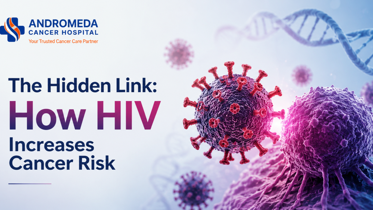
The Hidden Link: How HIV Increases Cancer RiskAs the world observes May 18 HIV Vaccine Awareness Day (HVAD), attention turns not only toward ongoing efforts to develop an effective HIV vaccine, but also toward understanding the long-term health challenges associated with HIV. The day honors scientists, healthcare professionals, researchers, and volunteers working toward a future free from HIV. While advances in treatment have transformed HIV into a manageable condition for many people, one critical issue still needs greater awareness: its strong connection with cancer. According to global health organizations, including the World Health Organization (WHO), people living with HIV are surviving longer due to improved treatment, but cancer remains one of the most important long-term health concerns in this population. Understanding this hidden link can help improve prevention, early detection, and overall outcomes.
How Does HIV Increase Cancer Risk?
HIV itself does not directly cause cancer. Instead, it weakens the bodys natural defense system, creating conditions where cancer can develop more easily. There are three main reasons this happens:
1. Weakening of the Immune System - HIV attacks important immune cells called CD4 cells, which help protect the body. A healthy immune system acts like security guards, finding and destroying abnormal cells before they become dangerous. When HIV weakens this system, those abnormal cells may grow unchecked.
2. Difficulty Fighting Other Viruses - Many cancers linked with HIV are actually caused by other viruses. Since immunity is lower, the body struggles to control these infections.
Some examples include:
HPV (Human Papillomavirus): linked with cervical and anal cancer
EBV (Epstein-Barr Virus): linked with certain lymphomas
HHV-8: linked with Kaposi Sarcoma
Hepatitis B and C: linked with liver cancer
3. Long-Term Inflammation -HIV creates ongoing low-level inflammation inside the body. Over time, this constant stress can damage cells and increase cancer risk.
<v:shapetype id="_x0000_t75"
coordsize="21600,21600" o:spt="75" o:preferrelative="t" path="m@4@5l@4@11@9@11@9@5xe"
filled="f" stroked="f">
<v:shape id="Picture_x0020_2" o:spid="_x0000_i1025" type="#_x0000_t75"
alt="Article content" style='width:450.6pt;height:300.6pt;visibility:visible;
mso-wrap-style:square'>
<v:imagedata src="file:///C:/Users/SHUBHA~1/AppData/Local/Temp/msohtmlclip1/01/clip_image001.png"
o:title="Article content"/>
Which Cancers Are More Common in HIV Patients?
Studies from major organizations like the National Cancer Institute show that some cancers occur much more frequently in people living with HIV.
Kaposi Sarcoma - People with HIV are over 200 times more likely to develop Kaposi Sarcoma. It often appears as purple or reddish patches on the skin and can also affect internal organs.
Non-Hodgkin Lymphoma - Risk is around 615 times higher. This cancer affects the lymphatic system and immune cells.
Cervical Cancer - Women with HIV have approximately 34 times greater risk because the immune system may not effectively clear HPV infections.
Other cancers with increased risk include:
Anal cancer
Hodgkin lymphoma
Liver cancer
Lung cancer
Why This Matters in Cancer Care
One important shift in modern medicine is that people living with HIV are now surviving longer because of better treatment. As life expectancy improves, doctors are seeing a rise in non-AIDS-defining cancers, including lung, liver, and anal cancers.
Another challenge is that cancer symptoms can sometimes be overlooked or diagnosed later in HIV-positive patients because fatigue, weight loss, or recurrent infections may initially be attributed to HIV itself. This can delay diagnosis.
Studies have also highlighted another concern: people living with HIV have historically been less likely to receive standard cancer treatments due to concerns about drug interactions and treatment complications. Today, integrated care involving HIV specialists and oncologists is helping overcome these barriers.
Can Cancer Risk Be Reduced?
Although HIV increases the risk of certain cancers, cancer is not inevitable. Early medical intervention and a proactive approach can significantly lower risk and improve outcomes.
Experts recommend:
✔ Consistent HIV treatment to maintain a stronger immune system✔ Regular cervical screening and HPV testing ✔ Hepatitis screening and vaccination ✔ Smoking cessation support ✔ Routine medical follow-ups ✔ Prompt evaluation of persistent symptoms such as unexplained weight loss, prolonged fever, unusual skin changes, or enlarged lymph nodes
For people living with HIV, regular monitoring becomes especially important because some cancers may develop silently in early stages.
Early detection remains one of the strongest tools in improving treatment success.
Advances in Cancer Care for People Living with HIV
Over the last decade, cancer care for HIV-positive patients has changed significantly. Earlier concerns about treatment limitations and complications have reduced with advances in medicine.
Today, oncologists increasingly use personalized treatment strategies that consider both cancer treatment and HIV management simultaneously. Improved supportive care, better understanding of drug interactions, targeted therapies, and multidisciplinary treatment planning have made cancer treatment safer and more effective.
The focus extends beyond treating cancer aloneensuring early diagnosis, comprehensive evaluation, and coordinated care that addresses the patient's overall health needs.
As awareness improves and medical science advances, the outlook for people living with HIV continues to become more encouraging.
Knowledge, timely screening, and expert care remain key pillars in protecting long-term health.
#HIVAwareness#HIVandCancer#CancerPrevention#PublicHealth#WorldHealth#MedicalAwareness

National Women’s Health Week 2026: Prevention, Innovation & the Power of Early ScreeningNational Womens Health Week 2026 (May 1016) arrives with a timely and urgent message: Prevention, Innovation, and Impact: A New Era in Womens Health. The theme is more than a sloganit reflects a global shift in healthcare. The key changes are:
From treating disease late to detecting risk early
Preventing illness proactively
Improving long-term quality of life for women.
For decades, womens health conversations were often limited to pregnancy or reproductive care. Today, the focus is broader and smarter: heart disease, cancers, bone health, hormonal changes, mental wellness, and lifestyle-related diseases are all part of the preventive healthcare landscape.
Yet despite advances in medicine, millions of women still miss essential screenings. In India alone, more than 15.23 crore cervical cancer screenings and 8.37 crore breast cancer screenings have been conducted through national health initiativesbut awareness, accessibility, and follow-up care remain major challenges.
The reality is simple: early screening saves lives.
Why Preventive Screening Matters More Than Ever
Breast cancer is now the most commonly diagnosed cancer among women globally. According to the World Health Organization, survival rates exceed 90% in many high-income countriesbut remain significantly lower in India, where delayed diagnosis is common.
Recent Indian screening campaigns revealed a concerning trend: nearly 4 out of 5 women screened required medical monitoring, follow-up, or intervention, despite many believing they were healthy.
The challenge is not only diseaseit is late detection.
Many women ignore warning signs because symptoms appear mild, responsibilities come first, or preventive care feels unnecessary when they feel fine. Unfortunately, cancers such as breast and cervical cancer often develop silently in early stages.
That is why modern womens healthcare is increasingly focused on risk prediction, early diagnosis, and personalized prevention.
Your Screening Checklist by Decade
In Your 20s and 30s: Build the Prevention Foundation
This is the decade where preventive habits have the greatest long-term impact.
Important Screenings Preventive Tests
HPV Vaccination (Vaccination offered to girls from the age of 9, recommended before HPV exposure, but can be taken at a later age)
Pap Smear / HPV Testing for cervical cancer screening
Blood pressure monitoring
Thyroid profile
Iron deficiency and anemia screening
Blood sugar testing for diabetes risk
Mental health and reproductive wellness checkups
Family history assessment for hereditary cancers
Women-Specific Cancer Focus
Cervical cancer remains one of the most preventable cancers because precancerous changes can often be detected years before cancer develops. WHO continues to emphasize HPV vaccination and routine cervical screening as major preventive tools.
Modern Advances
HPV DNA-based testing with higher sensitivity
AI-supported cervical screening interpretation
Digital reproductive health tracking apps
Teleconsultation-based preventive gynecology
In Your 40s and 50s: The Early Detection Decade
This is the stage where screening becomes criticalnot optional.
Essential Health Tests
Annual Mammograms
Clinical breast examination
Colorectal Cancer Screening
Lipid profile and cardiac risk assessment
Diabetes screening
Menopause and hormonal evaluation
Liver and kidney function tests
Vitamin D and bone health assessment
Women-Specific Cancer Focus
Breast cancer risk rises significantly after 40. The encouraging reality is that cancers detected at Stage 1 are associated with dramatically better treatment outcomes, less aggressive therapy, and improved survival.
Breakthroughs in Womens Health
Healthcare innovation is transforming screening:
AI-assisted mammography improves early detection of subtle lesions
3D mammography (Tomosynthesis) provides clearer imaging in dense breast tissue
Genetic risk profiling helps identify women at high hereditary risk
Liquid biopsy research is exploring cancer detection through blood samples
Personalized screening intervals based on individual risk
These advances are moving healthcare from one-size-fits-all screening toward precision prevention.
In Your 60s and Beyond: Focus on Longevity and Quality of Life
Healthy aging is not just about living longerit is about living independently, actively, and confidently.
Essential Screenings
Bone Density Scan (DEXA)
Breast and colorectal cancer surveillance
Cardiac health evaluation
Vision and hearing assessments
Cognitive and neurological screening
Osteoporosis monitoring
Fall-risk and mobility evaluation
Key Womens Health Concerns
Post-menopausal women face increased risks of:
Osteoporosis
Heart disease
Frailty fractures
Hormonal health complications
Late-stage cancer diagnosis
Preventive screening during this stage can significantly improve the quality of life and reduce the risk.
The Bigger Picture: Prevention Is Power
One of the strongest findings emerging globally is that lifestyle and preventive care can dramatically change outcomes. Recent international research suggested that more than a quarter of healthy years lost to breast cancer are linked to modifiable lifestyle factors such as smoking, physical inactivity, obesity, and poor diet.
At the same time, global reports estimate that nearly 1.5 billion women worldwide still lack access to essential preventive screenings.
That is why the 2026 themePrevention, Innovation, and Impactis so important. Prevention means encouraging women to prioritize routine checkups before symptoms appear. Innovation means using AI, precision diagnostics, and advanced screening technologies to detect disease earlier than ever before. Impact means ensuring that early detection translates into healthier families, reduced healthcare burden, and longer, healthier lives for women everywhere.
Women spend much of their lives caring for others. National Womens Health Week is a reminder that prioritizing their own health is not selfishit is essential. A mammogram was booked on time. A cervical screening is not postponed. A routine health checkup was completed early.
#Prevention, Innovation, and Impact: A New Era in Womens Health - Because in this new era of womens healthcare, prevention creates impact, innovation improves survival, and awareness becomes empowerment.
#NationalWomensHealthWeek2026#WomensHealth#EarlyDetectionSavesLives#PreventiveHealthcare#BreastCancerAwareness#MothersDay

The Pulse of Oncology: Why Nurses are the Future of Cancer CareThe image of a nurse is often one of a silent caregiver, a steady hand in a crisis, or the friendly face at the bedside. But as we move further into 2026, the narrative is shifting. Nurses are no longer just the "heart" of healthcare; they are its brain, its backbone, and its most critical future investment. The theme "Our Nurses. Our Future. Empowered Nurses Save Lives" is the multi-year global campaign established by the International Council of Nurses (ICN) for the year 2026.
The Global Pillar: Why Nurses Define Our Future
Nursing is the single largest healthcare profession in the world. According to the World Health Organization (WHO), nurses account for nearly 50% of the global health workforce. However, we are standing at a crossroads. Current data suggests a global shortage of nearly 4.5 million nurses by 2030.
The theme "Our Nurses. Our Future. Empowered Nurses Save Lives." is a call to action. It reminds us that the quality of our future healthcarefrom rural clinics to advanced urban hospitalsdepends entirely on how we support, educate, and empower the nursing community today. It serves as a strategic roadmap to address global health challenges and improve healthcare for everyone, everywhere.
The Multidisciplinary Impact
Nursing is not a monolith; it is a vast spectrum of expertise. From the high-pressure environment of the ICU and Emergency Care to the delicate precision of Pediatric nursing and the long-term emotional resilience required in Geriatric care, nurses are the primary navigators of patient outcomes. They are the scientists of care, managing complex medication regimes, interpreting real-time data, and acting as the vital link between technology and the human spirit.
The Oncology Angle: A Specialized Calling
While all nursing requires dedication, Oncology Nursing stands as one of the most intellectually and emotionally demanding specialties. In the fight against cancer, the "Pulse of Oncology" is found at the bedside.
Treatment Complexity: Oncology nurses manage advanced therapiesfrom traditional chemotherapy to cutting-edge immunotherapy and targeted biological agents. They must be experts in managing "cytokine storms" and complex side effects that require split-second clinical judgment.
Patient Advocacy Awareness: A crucial part of oncology nursing is patient education. Nurses empower patients to understand their diagnosis, debunking myths about cancer and encouraging early screening. They turn "medical jargon" into actionable hope.
The Emotional Anchor: Cancer is a journey of peaks and valleys. Oncology nurses provide the psychological scaffolding for families, navigating the transition from aggressive treatment to survivorship or palliative care with unmatched grace - helping patients navigate life after cancer, manage long-term side effects, and rebuild confidence beyond treatment.
Modern Challenges: The Burnout Crisis
To secure "Our Future," we must address the "Silent Epidemic" of nurse burnout. The physical and emotional toll of oncology care is immense. Modern healthcare must pivot toward providing nurses with better digital tools (AI-assisted monitoring) and robust mental health support. A supported nurse is a safe patient.
The Andromeda Standard: Leading the Way
At Andromeda Cancer Hospital, we don't just employ nurses; we celebrate them as the architects of our healing environment.
In our wards, the "Pulse of Oncology" is reflected through every dedicated professional who balances high-tech precision with high-touch compassion. We recognize that our nurses are the primary reason our patients feel seen, heard, and cared for. As we look to the future of cancer care, Andromeda is committed to being a place where nurses are empowered to lead, innovate, and continue saving lives every day.
Conclusion
Behind every successful treatment, every moment of reassurance, and every story of survival, there is a nurse whose dedication quietly shapes the future of healthcare. As medicine advances, technology evolves, and cancer care becomes increasingly sophisticated, one truth remains constant:
The future of healthcare will always depend on the strength, compassion, and leadership of its nurses.
#OurNursesOurFutureEmpoweredNursesSaveLives#OncologyNursing #InternationalNursesDay2026 #CancerCare #AndromedaCancerHospital

World Ovarian Cancer Day 2026: Early Signs, Myths & Modern Treatment AdvancesIn India, ovarian cancer is the third most common gynecological cancer, yet it remains one of the most misunderstood. Every year on May 8, the global community observes World Ovarian Cancer Day. In 2026, the theme is #NoWomanLeftBehind.
Because the symptoms of ovarian cancer are often vaguemimicking common digestive issues or age-related changesmany women are diagnosed only when the disease has reached an advanced stage.
Today, we are breaking the silence.
The Current Reality: India Global Burden (2026)
Over 320,000 women are diagnosed with ovarian cancer each year globally.
Over 47,000 new cases of ovarian cancer are diagnosed every year in India.
Among the highest incidence countries globally
The challenge isn't just the number of casesits the diagnostic gap.
The #NoWomanLeftBehind initiative aims to ensure that:
Women in metro cities
Women in semi-urban areas
Women in rural villages
👉 All have access to early detection, awareness, and precision medicine
Early Signs of Ovarian Cancer (Often Missed or Ignored)
Ovarian cancer is often called a silent disease, but in reality, it does show early warning signs. The issue is that these symptoms are subtle, repetitive, and easily dismissed.
Common Early Symptoms Include:
Persistent bloating or an increase in abdominal size
Feeling full very quickly while eating
Loss of appetite
Mild but continuous abdominal or pelvic discomfort
Frequent indigestion or gas-like symptoms
Unexplained fatigue or low energy
Back pain without a clear reason
Changes in menstrual cycle (in some cases)
⚠️ Important Pattern to Watch:
Symptoms occurring more than 1215 times per month
Symptoms lasting more than 23 weeks
👉 This is not normalthis is a signal to investigate
Busting the Myths: What Every Indian Woman Should Know
In our clinics, we encounter several myths that can lead to dangerous delays.
Lets set the record straight:
Myth 1: My Pap smear was normal, so I don't have ovarian cancer.
Fact: A Pap smear only screens for cervical cancer.
👉 There is currently no standard routine screening test (like a mammogram or Pap test) for ovarian cancer.
Myth 2: Its just gas or menopause bloating.
Fact: While bloating is common in the Indian diet and during hormonal changes:
👉 Persistent bloating is not normal, especially if it is frequent, progressive, or unusual for you
Myth 3: Only older women get it.
Fact:
More common after age 50
But certain types (like germ cell tumors) affect women in their 20s and 30s
When to See a Specialist: The BEAT Symptoms
We don't want to create panicbut we do want to create awareness.
If you experience these symptoms most days for 3 weeks, it is time for a check-up:
B Bloating that doesn't go away
E Eating difficulty (feeling full very quickly)
A Abdominal or pelvic pain
T Toilet changes (frequent or urgent urination)
👉 Key Insight: Persistence matters more than severity.
The Future is Here: Modern Trends in Treatment (2026)
The landscape of ovarian cancer care has shifted dramatically in the last few years.
We are moving away from one-size-fits-all chemotherapy toward Precision Oncology.
1. PARP Inhibitors (Targeted Therapy)
Smart medications like Olaparib:
Target cancer cell DNA repair mechanisms
Especially effective in BRCA1/2 mutation cases
Significantly increase remission duration
2. Liquid Biopsies
Advanced multi-omic blood tests (Multi-omic blood tests are advanced diagnostic tests that analyze multiple layers of biological information at the same timeinstead of looking at just one marker)
Detect cancer signals using lipids and proteins.
Shows promise for earlier detection than traditional methods
3. Advanced Ultrasound Assessment
Helps differentiate between benign cysts and malignant tumors
Improves diagnostic accuracy
Supports better clinical decision-making
Precautions and Prevention
While ovarian cancer cannot always be prevented, risk can be reduced and managed:
Know your family history (Breast/ovarian cancer in mother or sister increases risk)
Consider testing for the BRCA mutation if high-risk.
Oral contraceptive use (5+ years) Shown to reduce ovarian cancer risk
Maintain regular gynecological evaluations
Most importantly: 👉 Listen to your body 👉 If something feels different for more than 3 weeksinvestigate.
Final Thought
Ovarian cancer is no longer the death sentence it once was.
With the rise of:
Targeted therapies
Advanced diagnostics
Precision medicine
👉 The focus has shifted to long-term survival and quality of life
This World Ovarian Cancer Day, lets make a collective promise:
Stop adjusting to discomfort.
Start prioritizing early detection.
If you or a loved one is experiencing persistent symptoms, seek medical consultation.
Because in this fight #No woman should be left behind.
Disclaimer
This blog is for educational purposes and does not replace professional medical advice. Always consult a qualified oncologist for concerns regarding your health.
#NoWomanLeftBehind#WorldOvarianCancerDay#WomensHealthIndia#CancerAwareness#EarlyDetectionSavesLives

Melanoma Monday: A Gentle Reminder to Check Your SkinEvery year, the first Monday of May is recognized globally as Melanoma Monday. While it might sound like just another date on the calendar, for a country like Indiawhere the sun shines bright nearly year-roundit is a vital reminder to pay attention to our bodys largest organ: the skin.
In the Indian context, skin issues are incredibly common. From heat rashes and fungal infections to dust allergies, our skin goes through a lot. However, because we are so used to "minor" skin irritations, we often ignore things that could be more serious. This blog is a simple guide to understanding skin cancer without the panic, specifically tailored for our Indian climate and lifestyle.
What is Melanoma?
Melanoma is a type of skin cancer that starts in melanocytesthe cells responsible for giving our skin its color.
It isless common, but
More serious, because it can spread faster if not detected early
The positive side? 👉When caught early, it is highly treatable.
The Biggest Myth in India - We have more melanin, so we are safe.
While it is true that higher melanin provides some natural protection against UV rays, it doesnotmake us immune.
In fact, melanoma in Indians often appears in unexpected places:
Palms
Soles of feet
Under fingernails
Because we dont usually check these areas, diagnosis often happens late.
Risk Factors: Who Should Be Careful?
While anyone can develop skin cancer, certain factors increase the risk:
Prolonged Sun Exposure:People who work outdoors under the harsh afternoon sun without protection.
History of Sunburns:Even a few severe, blistering sunburns in childhood can increase the risk of skin cancer later in life.
Family History:If a close relative has had melanoma.
Weakened Immune System:Due to other medical conditions or long-term medications.
<v:shapetype
id="_x0000_t75" coordsize="21600,21600" o:spt="75" o:preferrelative="t"
path="m@4@5l@4@11@9@11@9@5xe" filled="f" stroked="f">
<v:shape id="Picture_x0020_4" o:spid="_x0000_i1026" type="#_x0000_t75"
alt="Article content" style='width:450pt;height:337.8pt;visibility:visible;
mso-wrap-style:square'>
<v:imagedata src="file:///C:/Users/SHUBHA~1/AppData/Local/Temp/msohtmlclip1/01/clip_image001.jpg"
o:title="Article content"/>
Allergy or Something More?
In India, "skin lesions" can be confusing. Is it a mosquito bite? A heat rash? An allergy to a new soap?
This is where most confusion happens.
Common Skin Issues:
Itchy
Sudden appearance
Triggered by food, weather, or products
Heal quickly
Suspicious Signs:
Painless
Dont go away
Slowly change over time
👉 The keyword is:Change
The ABCDE Rule for Screening:If you have a mole or a dark spot, check for these five signs:
A for Asymmetry:One half doesn't match the other.
B for Border:The edges are ragged, blurred, or irregular.
C for Color:The color is not the same all over (shades of black, brown, or even pink).
D for Diameter:The spot is larger than a pencil eraser (about 6mm).
E for Evolving:The spot is changing in size, shape, or color.
<v:shape
id="Picture_x0020_3" o:spid="_x0000_i1025" type="#_x0000_t75" alt="Article content"
style='width:451.8pt;height:451.8pt;visibility:visible;mso-wrap-style:square'>
<v:imagedata src="file:///C:/Users/SHUBHA~1/AppData/Local/Temp/msohtmlclip1/01/clip_image002.jpg"
o:title="Article content"/>
Precautions for the Indian Climate
Our temperatures often soar above 40C. Heres how to stay safe without living in fear:
The "Shadow Rule":If your shadow is shorter than you (usually between 10 AM and 4 PM), the suns UV rays are at their strongest. Seek shade.
Physical Barriers:In India, we have the best traditional defensetheDupatta,Gamcha, or an umbrella. Light-colored, long-sleeved cotton clothing is your best friend.
Sunscreen is for Everyone:Use a broad-spectrum sunscreen with at least SPF 30. Apply it 20 minutes before going out, and don't forget your ears and the back of your neck.
Check Your Extremities:Since Indian cases often appear on feet and hands, give yourself a "skin check" once a month after a bath. Look at your soles and between your toes.
When to See a Doctor?
Don't panic over every pimple or rash. However, you should visit a dermatologist if:
You have a sore thatdoesn't healwithin 3 or 4 weeks.
A new dark spot appears under a nail or on your palm/sole.
An existing mole starts toitch, bleed, or crust over.
Final Thought
Melanoma Monday isnt about creating fearits aboutawareness and action.
In a country where we endure so muchfrom heat to pollutionour skin quietly protects us every day. The least we can do is notice when it asks for help.
✨ Just 10 minutes a month. ✨ One small habit. ✨ One life-saving awareness.
BecauseEarly detectiondoesnt start in hospitals,it starts with you simply paying attention - it saves lives.
#MelanomaAwareness#SkinHealth#PreventiveHealthcare#HealthcareAwareness#EarlyDetection#ABCDEruleMelanoma#SkinCheckRoutine
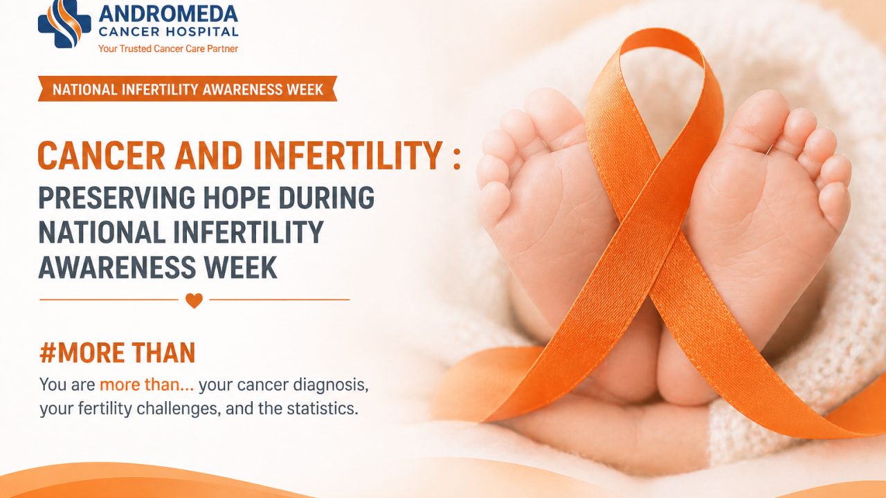
Cancer and Infertility: Preserving Hope During National Infertility Awareness WeekFrom April 1925, 2026, National Infertility Awareness Week (NIAW) is being observed globally to raise awareness about reproductive challenges and to break long-standing stigma surrounding infertility. Established by RESOLVE: The National Infertility Association, this years theme, #MoreThan, emphasizes a powerful message: individuals facing infertility are more than a diagnosis, more than a statistic, and more than a medical condition.
Infertility affects approximately 1 in 6 people worldwide, making it a significant public health concern. At our hospital, we believe a cancer diagnosis should be a fight for your lifenot a forfeit of your future.
As we observe National Infertility Awareness Week, we highlight a crucial yet often overlooked concern: fertility in cancer patients.
For many young individuals, one question quietly sits beside the diagnosis: Will I be able to have children?
Can Cancer Itself Cause Infertility?
Yesinfertility is not always just a side effect of treatment. Certain cancers can directly impair reproductive function.
1. Cancers Affecting Reproductive Organs
Testicular cancer damages sperm-producing cells
Ovarian cancer affects egg reserve and ovulation
2. Cancers of the Reproductive Tract
Cervical, uterine, and vaginal cancers may:
3. Hormone-Related Tumors
Pituitary gland tumors can disrupt the production of reproductive hormones like FSH and LH
How Cancer Treatments Affect Fertility
Most infertility in cancer survivors is due to treatment rather than the disease itself.
1. Chemotherapy
Drugs (especially alkylating agents like cyclophosphamide) target rapidly dividing cells
Unfortunately, reproductive cells are also fast-dividing
Effect:
Damage to ovarian follicles premature ovarian failure
Destruction of spermatogonia reduced or absent sperm production
2. Radiotherapy
Ionizing radiation causes direct DNA damage
Effects depend on location:
Pelvic radiation:
Testicular radiation:
3. Surgery
Removal of reproductive organs
Effect:
Immediate and permanent loss of natural fertility
Who is Most Vulnerable?
High-risk groups include:
Adolescents and Young Adults (AYAs)
Patients yet to complete their families
Prepubertal children
Even in children, treatments may damage stem cells, affecting future fertility after puberty.
The Power of Early Fertility Counseling
Recent data (20252026, ASCO) shows: Only ~50.7% of eligible cancer patients receive fertility counseling before treatment
Why Early Counseling Matters
Reduced regret later in life
Better mental health during treatment
Informed decision-making
Helps patients feel they still have control over their future
FERTILITY PRESERVATION OPTIONS
The best fertility preservation method depends on:
Age
Type of cancer
Urgency of treatment
Marital status
Hormonal sensitivity of tumor
FOR WOMEN
1. Egg Freezing (Oocyte Cryopreservation)
Preserving healthy eggs for future use
Cost (India): ₹1.2 ₹2.5 lakh per cycle + ₹10,000₹30,000/year storage
Best Suited For:
Breast cancer (before chemotherapy)
Lymphoma patients
Women who need urgent treatment but can wait ~2 weeks
Unmarried patients
Success Chances:
Per cycle: ~1030% pregnancy rate
Under 35 years: up to 7080% cumulative success
2. Embryo Freezing
Fertilized embryos frozen for future use
Cost (India):
IVF + freezing: ₹2.5 ₹4.5 lakh per cycle (approximately depends on IVF center)
Best Suited For:
Married or partnered women
Breast cancer patients delaying treatment briefly
Patients with good ovarian reserve
Success Chances:
Higher than egg freezing
~4060% per IVF cycle (age-dependent)
3. Ovarian Tissue Freezing
Ovarian tissue preserved for future fertility
Cost (India):
₹2 ₹5 lakh (limited availability, specialized centers)
Best Suited For:
Children (pre-pubertal girls)
Patients needing immediate chemotherapy (no delay possible)
Aggressive cancers
Success Chances:
Still evolving
Live birth rates improving (~3040% in advanced centers globally)
4. Oocyte Maturation (IVM)
Maturing eggs in the lab before freezing
Cost (India):
₹1.5 ₹3 lakh (varies widely)
Best Suited For:
Patients who cannot take hormone injections
Hormone-sensitive cancers (e.g., estrogen-positive breast cancer)
Success Chances:
Lower than IVF
~2040% (depends on lab expertise)
FOR MEN FERTILITY PRESERVATION OPTIONS
1. Sperm Banking
Freezing and storing sperm for future use
Cost (India):
₹5,000 ₹20,000 initial
₹5,000 ₹15,000/year storage
Best Suited For:
Testicular cancer
Before chemotherapy or radiotherapy
Any male cancer patient of reproductive age
Success Chances:
Very high
Comparable to normal IVF outcomes
2. Testicular Sperm Extraction (TESE)
Retrieving sperm directly from testicular tissue
Cost (India):
₹50,000 ₹1.5 lakh
Best Suited For:
When no sperm is present in semen
Testicular cancer patients
Post-chemotherapy azoospermia
Success Chances:
Depends on sperm retrieval success
Often used with IVF/ICSI
<v:shapetype
id="_x0000_t75" coordsize="21600,21600" o:spt="75" o:preferrelative="t"
path="m@4@5l@4@11@9@11@9@5xe" filled="f" stroked="f">
<v:shape id="Picture_x0020_2" o:spid="_x0000_i1026" type="#_x0000_t75"
alt="Article content" style='width:450.75pt;height:300.75pt;visibility:visible;
mso-wrap-style:square'>
<v:imagedata src="file:///C:/Users/ACH0080/AppData/Local/Temp/msohtmlclip1/01/clip_image001.png"
o:title="Article content"/>
Life After Cancer Treatment: Fertility Recovery Reality
Fertility after cancer is not always predictablebut it is often possible with the right support.
For Men
Sperm production may recover in 15 years after chemotherapy
Sperm banking before treatment is strongly recommended
Even a single sample can preserve future options
For Women
Menstrual cycles may returnbut this does not guarantee fertility
Important Considerations:
Reduced ovarian reserve (fewer eggs left)
Early menopause risk
Shorter reproductive window
Medical Tools to Assess Fertility Post-Treatment:
AMH (Anti-Mllerian Hormone) levels
Antral follicle count (AFC) via ultrasound
Pregnancy After Cancer: Is It Safe?
In many casesyes, with proper guidance.
General Recommendations:
Wait 6 months to 2 years after treatment (depends on cancer type)
Ensure disease remission
Close coordination between:
Modern Options for Parenthood After Cancer
Even if natural conception is difficult, options exist:
IVF using preserved eggs or embryos
Donor eggs or sperm
Surrogacy (when uterus is affected)
Adoption
Emotional Psychological Recovery
Fertility is not just biologicalit is deeply emotional.
Patients may experience:
Anxiety about future parenthood
Grief over fertility loss
Fear of recurrence
Support Matters:
Counseling
Support groups
Partner and family involvement
Conclusion: Hope Beyond Survival
Cancer treatment today is not just about survivalit is about quality of life after survival.
With early counseling, informed choices, and advancing medical science, parenthood remains a possibility for many cancer survivors.
#CancerAndFertility#Naionalinfertilityawarenessweek#FertilityPreservation#InfertilityAwareness
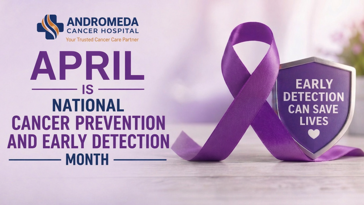
Cancer: How to Prevent It and Find It EarlyApril is National Cancer Prevention and Early Detection Month, serving as a critical reminder to adopt healthy habits and prioritize regular screeningsbecause cancer is most treatable when detected early. Cancer is a serious health problem in the world and in India. As per GLOBOCAN 2022, there were about 2 crore (20 million) new cancer cases and about 97 lakh (9.7 million) deaths worldwide. In India, the ICMR estimates around 15.6 lakh (1.56 million) new cases and more than 8.7 lakh (874,000) deaths in 2024.
Research clearly shows two things:
Large number of cancers can be prevented
Finding cancer early greatly improves chances of cure
This article shares proven ways to reduce cancer risk, clears some common myths, and explains why early check-ups and screening matter.
Cancer Prevention: Proven Ways to Reduce Risk
The World Health Organization (WHO) says about 3050% of cancers can be prevented by changing known risk factors. This is based on many years of research, not guesswork.
1. Tobacco (Smoking and Chewing): The Biggest Avoidable Risk
Tobacco is the number one cause of cancer that we can prevent.
It is linked to about 22% of cancer deaths worldwide.
It strongly increases the risk of:
In India, chewing tobacco (gutkha, khaini, zarda, paan with tobacco, etc.) is a major reason why mouth cancer is so common.
What happens in the body (simple): Tobacco has many harmful chemicals that damage our body cells and DNA. Over time, this damage can turn normal cells into cancer.
Stopping tobacco lowers riskeven if someone has used it for many years.
2. Being Overweight, Food Habits, and Diabetes-Related Factors
Being overweight and having an unhealthy diet can increase the risk of some cancers.
Extra body weight is linked with higher risk of:
Why this happens:
Being overweight leads to a state of ongoing inflammation.
It can lead to insulin resistance and poor blood sugar control.
It increased insulin like growth factor (IGF) levels that can support cancer growth.
Some food habits that can increase risk:
Eating a lot of processed meat (packed/processed meats) is linked to colon cancer.
Eating too little fibre (fruits, vegetables, whole grains) may reduce protection for the gut.
3. Alcohol: Increases Cancer Risk
Alcohol is a proven cause of cancer. (It is in the highest risk category, like tobacco.)
It is linked with higher risk of cancer of the:
Why alcohol increases risk (simple): When the body breaks down alcohol, it forms harmful substances that can damage DNA and make it easier for cancer to develop.
4. Infections That Can Cause Cancer (And Many Are Preventable)
In India, a good number of cancers are linked to certain infections.
HPV (Human Papillomavirus) can lead to cervical cancer (cancer of the mouth of the uterus).
Hepatitis B (HBV) can lead to liver cancer.
India has a large number of cervical cancer cases compared to many other countries.
Important fact: In India, about one woman dies from cervical cancer roughly every 8 minutes.
How to prevent these:
The HPV vaccine can prevent most high-risk HPV infections (more than 90% protection against the types it targets).
The Hepatitis B vaccine lowers the risk of long-term HBV infection and helps prevent liver cancer.
5. Not Being Physically Active
Sitting for long hours and not exercising can increase cancer risk because it can lead to:
Weight gain
Hormone changes
Weaker body defence (immune system) over time
Regular activity (like brisk walking, cycling, or any daily exercise) is linked with lower cancer risk and better health overall.
6. Pollution and Workplace Exposure
Research also shows risk from:
Air pollution (especially for lung health)
Some workplace chemicals (for example, asbestos and industrial chemicals)
In many Indian cities, pollution levels are high. Doctors now see more cases of lung cancer even in people who never smoked.
<v:shapetype
id="_x0000_t75" coordsize="21600,21600" o:spt="75" o:preferrelative="t"
path="m@4@5l@4@11@9@11@9@5xe" filled="f" stroked="f">
<v:shape id="Picture_x0020_2" o:spid="_x0000_i1025" type="#_x0000_t75"
alt="Article content" style='width:450.75pt;height:300.75pt;visibility:visible;
mso-wrap-style:square'>
<v:imagedata src="file:///C:/Users/ACH0080/AppData/Local/Temp/msohtmlclip1/01/clip_image001.png"
o:title="Article content"/>
Myths and Facts: Clearing Common Misconceptions
Wrong information can delay diagnosis and treatment. Lets correct a few common myths:
Myth 1: Cancer happens only because of family history (genes).
Fact: Only about 510% of cancers are truly hereditary. Most cancers are linked to lifestyle, environment, infections, or age.
Myth 2: If I dont smoke, I cant get cancer.
Fact: Non-smokers can also get cancer due to:
Air pollution
Unhealthy food habits and obesity
Some infections (like HPV, Hepatitis B)
Family history or genes (in some cases)
Myth 3: If I live healthy, I will never get cancer.
Fact: Healthy habits reduce the risk, but they cannot make the risk zero. Many factors play a role.
Myth 4: If I have no symptoms, I dont need screening.
Fact: Screening is done to find cancer before symptoms start, when treatment is often easier and the chance of cure is higher.
Myth 5: Biopsy or surgery spreads cancer.
Fact: There is no scientific proof that a biopsy spreads cancer. A biopsy is often necessary to confirm cancer and plan the right treatment.
Myth 6: Cervical cancer cannot be prevented.
Fact: Cervical cancer is one of the most preventable cancers with HPV vaccination and regular screening.
Early Detection: Why Finding Cancer Early Saves Lives
Early detection is not just adviceit is one of the strongest reasons people survive cancer.
Cancer usually starts small and then grows. If it is found early, before it spreads, treatment can be simpler and outcomes are better. Finding it early can improve:
How big the cancer is (tumor burden)
How strong the treatment needs to be (treatment intensity)
Chances of survival
Quality of life after treatment
How Early Detection Improves Results
In simple terms:
Early-stage cancer (Stage 1) is often limited to one area and can be treated successfully in most cases.
Late-stage cancer (Stage 34) may have spread, so treatment is usually more complex and outcomes may be poorer.
Example (breast cancer):
If found early, breast cancer patients have more than 9095% chance of survival at 5 years.
If found late, survival is much lower.
So, how early the cancer is found is one of the biggest factors that decides the result of treatment.
Two Ways to Find Cancer Early
There are two main ways:
1. Screening (Tests Done Before Symptoms)
Screening means doing certain tests in people who feel fine, so cancer can be found early.
Good screening programs need:
The right test (scan or lab test) that can catch early changes
Knowing who needs the test more (higher-risk people)
Quick follow-up if the test shows something suspicious
Examples include:
Mammography for breast cancer
Pap smear / HPV test for cervical cancer
Mouth check-up for people at high risk (especially tobacco users)
Screening can reduce deaths from some cancers, especially breast and cervical cancer.
2. Early Diagnosis (Not Ignoring Warning Signs)
If symptoms start, it is important to get checked quickly.
In India, cancer is often found late because:
People ignore symptoms or wait too long
Lack of awareness about warning signs
Getting treated first by someone who is not trained to check cancer
If any symptom continues for more than 23 weeks, it is safer to get a proper check-up. Warning signs can include:
Weight loss without trying
Cough that does not go away, or change in voice
Wound/ulcer that is not healing
Bleeding that is not normal
Any lump, especially new/growing
Getting checked early can lead to:
Finding the cancer at an earlier stage
Simpler treatment in many cases
Better chances of recovery
How Doctors Confirm Cancer (Simple Steps)
To confirm cancer, doctors usually follow a step-by-step process:
Doctor check-up (history and physical examination)
Scans/tests (to see where the problem is and how big it is)
Biopsy (taking a small tissue sample to confirm cancer)
This helps ensure:
Correct diagnosis
Less delay in starting treatment
Fewer unnecessary referrals and repeat tests
Early Detection at Andromeda Cancer Hospital: What We Offer
At Andromeda Cancer Hospital, we focus on finding cancer early through a team-based approach and modern testing facilities, in line with global standards.
1. Modern Tests and Scans
We have modern diagnostic facilities to help find cancer early and accurately, such as:
3D Mammography (very sensitive for small breast cancers)
High-quality ultrasound and elastography
Image-guided biopsy (biopsy done with scan guidance for accuracy)
PET-CT and other advanced scans (to check cancer spread)
These tests help doctors find:
Very small early changes
Early-stage cancers
Early signs of spread (when present)
This reduces the chance of missing a problem or finding it too late.
2. Screening and Preventive Care
We support screening and prevention through:
Breast cancer screening
Genetic counselling and testing (for families with strong cancer history)
Personalized screening plans based on your risk
This matters because in India, many people reach a hospital when the cancer is already at a later stage.
Many cancers are found in Stage 23 instead of Stage 1.
3. Team of Specialists (All in One Place)
One of the key differentiators is the multidisciplinary team (MDT) approach, involving:
Surgical oncologists
Medical oncologists
Radiation oncologists
Radiologists and pathologists
This ensures:
Comprehensive evaluation in a single setting
Faster decision-making
Evidence-based, individualized treatment planning
4. Integrated Diagnostic-to-Treatment Pathway
A critical issue in cancer care is delay between diagnosis and treatment.
At Andromeda:
Diagnostics, staging, and treatment planning are integrated under one roof
Minimizes patient movement between centers
Enables early initiation of treatment, which is crucial for outcomes
5. Full Spectrum Oncology Services Supporting Early Detection
The hospital provides a complete continuum of care, including:
Surgical Oncology (including minimally invasive and robotic surgery)
Medical Oncology (chemotherapy, immunotherapy, targeted therapy)
Radiation Oncology with advanced precision techniques
Oncopathology and radiology services for accurate diagnosis
Pain and palliative care for holistic patient support
Rehabilitation and physiotherapy services
This integrated model ensures that early detection seamlessly translates into effective treatment.
Closing Perspective: From Awareness to Action
Cancer care is no longer limited to treatmentit begins much earlier, with risk reduction and early identification.
The scientific evidence is clear:
A significant proportion of cancers are preventable
Early detection dramatically improves survival
Delays in diagnosis remain one of the biggest challenges in India
Improving cancer outcomes depends on timely detection, informed decisions, and access to the right care without delay.
At Andromeda Cancer Hospital, we are committed to providing comprehensive cancer carefrom early screening and accurate diagnosis to advanced treatmentunder one roof, supported by a multidisciplinary team and modern technology.
If you or your loved ones have any concerns or are due for screening, do not delay seeking medical advice. 📞 For appointments and consultations: 9138111625
Andromeda Cancer Hospital Your Trusted Cancer Care Partner.
#CancerPrevention#EarlyDetectionSavesLives#CancerAwareness#OncologyCare#PreventiveHealthcare
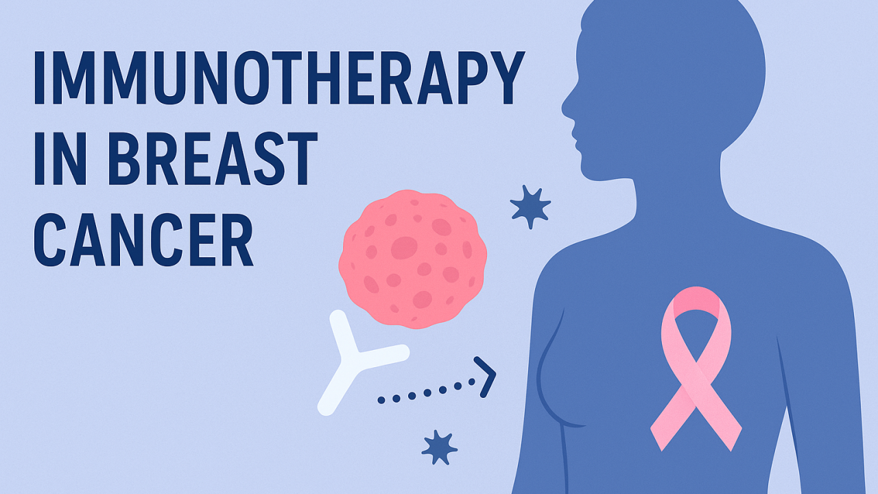
Immunotherapy in Breast Cancer: The New Era of HopeImmunotherapy in Cancer Treatment: A New Era of Hope
Understanding its role in Breast Cancer at Different Stages: Cancer treatment has evolved dramatically over the past decade. Among the most promising advancements is immunotherapy a treatment that uses the bodys own immune system to recognize and destroy cancer cells. Normally, our immune system is highly skilled at identifying anything that does not belong in the body. However, cancer cells can cleverly hide and suppress the immune response. Immunotherapy helps the immune system wake up, find cancer cells, and attack them more effectively. This approach is different from chemotherapy or targeted therapy, which directly act on cancer cells. Instead, immunotherapy gives power back to the bodys natural defense system. Immunotherapy drugs work by re-activating the immune systems ability to recognize and eliminate cancer cells. Tumors often evade immune destruction by exploiting regulatory pathways such as immune checkpoints for example, PD-1/PD-L1 and CTLA-4 which normally function to prevent excessive immune activation. Checkpoint inhibitor immunotherapies are monoclonal antibodies that block these inhibitory receptors or ligands, thereby releasing the brakes on cytotoxic T cells and allowing them to attack malignant cells more efficiently. Other forms of immunotherapy include CAR-T cell therapy, in which a patients T cells are genetically engineered to express specific receptors targeting tumor antigens, and cancer vaccines, which stimulate adaptive immune memory against tumor-associated markers. Additionally, cytokine-based treatments and immune stimulators enhance immune cell proliferation and activity within the tumor microenvironment. Together, these strategies aim to overcome tumor-induced immune suppression, achieve durable antitumor responses, and establish long-term immunologic surveillance to prevent relapse.
How Does Immunotherapy Work? Immunotherapy works in several ways, such as:
Checkpoint inhibitors: Cancer cells often use brakes on the immune system to avoid attack. These drugs release those brakes so immune cells can act freely.
Immunomodulators: They boost the overall activity of the immune system.
Cell-based therapies: Immune cells are enhanced or engineered outside the body and then infused back to target cancer more strongly.
Cancer vaccines: These help the body recognize cancer-specific markers and develop long-term defense.
These treatments have already improved outcomes in melanoma, lung cancer, kidney cancer, and many others.
Immunotherapy in Breast Cancer: Breast cancer is a very diverse disease, with different biological subtypes. Immunotherapy is becoming particularly valuable in Triple-Negative Breast Cancer (TNBC) a type that lacks hormone receptors and HER2 expression and tends to be more aggressive.
Heres how immunotherapy fits into different stages of breast cancer:
1. Early-Stage Breast Cancer
For patients with high-risk early-stage TNBC, immunotherapy is now used in combination with chemotherapy before surgery.
This approach increases the chance of completely eliminating cancer in the breast and lymph nodes.
It may also reduce risk of recurrence in the future.
2. Locally Advanced Breast Cancer
When the tumor is large or has spread to nearby nodes, but not distant organs, immunotherapy plus chemotherapy can shrink the cancer.
This helps make surgery more successful and improves long-term outcomes.
3. Metastatic / Stage IV Breast Cancer
Immunotherapy has shown meaningful benefit in patients whose TNBC expresses PD-L1, an immune-checkpoint marker.
It can help control cancer for longer, maintain better quality of life, and may extend survival.
In some selected patients, tumors shrink significantly, making ongoing treatment more manageable.
Why Immunotherapy Matters
More personalized treatment based on cancer biology
Better tolerance for many patients compared to traditional chemotherapy
Long-lasting immune memory, which can help keep cancer away even after treatment stops
It represents a major shiftfrom only attacking cancer to empowering the immune system to stay vigilant.
Side Effects of Immunotherapy in Breast Cancer Patients
Immunotherapy generally has a different side-effect profile compared to chemotherapy. Because it activates the immune system, the most common reactions come from the immune system attacking healthy tissues these are called immune-related adverse events (irAEs).
Some commonly seen side effects include:
Skin reactions Rash, itching, dryness, or changes in skin color. These are usually mild and manageable with creams or medicines.
Fatigue A common effect that can vary from mild tiredness to more significant weakness.
Gastrointestinal symptoms Diarrhea or inflammation of the bowel (colitis) can occur in some patients.
Hormone gland inflammation the thyroid, adrenal glands, or pituitary gland can get affected, leading to hormonal imbalance. Symptoms may include weight changes, hair loss, mood changes, or low energy, often requiring hormone replacement.
Lung inflammation (pneumonitis) Causes cough, breathlessness, or chest discomfort. This needs prompt medical attention.
Liver inflammation (hepatitis) Usually detected by blood tests before symptoms appear.
While many side effects are mild, some can be serious if not addressed early. The good news is that most irAEs respond very well to timely treatment, especially with corticosteroids or temporary pause of immunotherapy.
Why monitoring matters: During immunotherapy, patients are closely followed with regular check-ups and blood tests. Any new symptom even if small should be reported early so that doctors can act quickly and prevent complications. With proper monitoring, most patients continue treatment safely and benefit from its long-term effects.
Common Immunotherapy Drugs Used in Breast Cancer
At present, immunotherapy in breast cancer is mainly focused on Triple-Negative Breast Cancer (TNBC). The most commonly used drug is Pembrolizumab, a checkpoint inhibitor that targets the PD-1 receptor on immune cells. By blocking this receptor, the medicine allows T-cells to recognize and attack cancer cells more effectively. Pembrolizumab is now approved for use in early-stage high-risk TNBC along with chemotherapy before surgery, as well as in metastatic TNBC where the tumor expresses the PD-L1 marker. Another drug used in selected metastatic TNBC cases is Atezolizumab, which targets the PD-L1 protein on cancer cells and immune cells. These medicines have shown meaningful benefits in controlling disease and improving long-term outcomes in eligible patients.
The Road Ahead: Research is rapidly progressing to:
Expand immunotherapy beyond TNBC to other breast cancer subtypes
Identify which patients benefit the most
Improve combinations with targeted therapy, hormone therapy, and radiation
Immunotherapy is not yet for all breast cancer patients, but it is a strong and growing pillar of modern cancer care.
Landmark Trials in Immunotherapy for Breast Cancer
Landmark trials related to the immunotherapy in breast cancer: The introduction of immunotherapy in breast cancer has been driven by major clinical trials, particularly in triple-negative breast cancer (TNBC). The KEYNOTE-522 trial was a breakthrough in early-stage disease, showing that adding pembrolizumab to neoadjuvant chemotherapy significantly increased the rate of pathological complete response and improved event-free survival, leading to global approval for high-risk early TNBC. In metastatic settings, KEYNOTE-355 demonstrated that pembrolizumab combined with chemotherapy offered a meaningful survival benefit in PD-L1 positive metastatic TNBC, establishing it as a standard first-line option. Another key trial, IMpassion130, evaluated atezolizumab with nab-paclitaxel in metastatic TNBC and showed improved progression-free survival in PD-L1 positive patients, marking the first immunotherapy approval in advanced breast cancer. Although later trials such as IMpassion131 did not replicate the same benefit, the collective evidence from these pivotal studies has opened a new and hopeful chapter, integrating immunotherapy as an important treatment pillar in selected breast cancer patients.
Final Takeaway:
These drugs are not for all breast cancers they benefit patients whose tumors show PD-L1 expression and are mostly used in TNBC.
They are often combined with chemotherapy for better effectiveness.
Selection of patients is done using specialized biomarker testing.
Immunotherapy has opened a new chapter in breast cancer treatment one filled with innovation, hope, and better outcomes. As research continues, more patients will have access to this powerful and personalized strategy to fight cancer.
#breastcancer #TNBC #immunotherapyforbreastcancer #breastcancersurvival

Breast Cancer Surgery Without Losing The BreastA century of change in a single operation
In the early 1900s, Halsteds radical mastectomyremoving the entire breast, pectoral muscles, and extensive nodeswas the unquestioned standard for operable breast cancer. As systemic therapies emerged and our understanding of tumour biology matured, it became clear that more surgery wasnt always better. Modified radical mastectomy (MRM) preserved the pectoral muscles, reduced morbidity, and laid the foundation for the next leap: breast conservation surgery (BCS)tumour-focused excision with clear margins, combined with radiotherapy. The central insight was profound: survival depends as much on biology and systemic control as on extent of local surgery. Randomized trials across decades have since confirmed that appropriately selected patients can keep their breast without compromising survival.
From Mastectomy to BCS: What the Randomized Trials Proved
Two landmark randomized trials anchor the BCS evidence base. The NSABP B-06 study demonstrated, at 20-year follow-up, no difference in overall survival among total mastectomy, lumpectomy alone, and lumpectomy plus radiotherapy; radiotherapy, however, significantly reduced local recurrence after lumpectomy. Similarly, the Milan (Veronesi) quadrantectomy trial reported equivalent long-term survival between BCS and radical mastectomy, establishing oncologic safety for conservation. These data changed global practice and underwrite todays guidelines
The EBCTCG Meta-analysis: Why Radiotherapy Matters After BCS
The Early Breast Cancer Trialists Collaborative Group (EBCTCG) pooled individual patient data from 17 trials (10,801 women) and provided the most influential quantification of radiotherapys value after BCS: radiation halved the 10-year risk of any first recurrence (35.0% 19.3%) and reduced 15-year breast cancer mortality by about one-sixth. These proportional benefits were broadly similar across subgroups, though absolute benefit varied with baseline risk. This single overview codified the principle that BCS must be paired with high-quality radiotherapy for durable local control and survival benefit.
De-escalation Done Right
The shift from radical mastectomy to MRM to BCS is part of a larger oncology movement: de-escalation with precision. We aim to minimize treatment burden without sacrificing curesmaller operations, focused radiotherapy, omission of axillary dissection in node-negative or carefully selected post-neoadjuvant settings, and tailored systemic therapy. EBCTCG overviews across eras consistently show that better local control translates to fewer deaths, but beyond a certain point, more tissue removal does not improve survival. The art is matching treatment intensity to disease biology and patient values.
BCS vs Mastectomy: The Modern Data (And Why BCS Often Wins)
While RCTs established equivalence in survival between BCS+RT and mastectomy, large contemporary population studies (reflecting advances in systemic therapy, imaging, pathology, and radiotherapy) frequently show a survival advantage for BCS+RT over mastectomy in early breast cancer. For example, a nationwide Dutch analysis showed improved 10-year overall and relative survival with BCS+RT compared with mastectomy (with caveats about residual confounding). More recently, a 2024 meta-analysis again suggested a survival advantage for BCS+RT in early disease. These findings should be presented carefully to patientsas observational data subject to selection effectsbut they reinforce that BCS is not a compromise; it is often the best option for eligible patients.
BCS After Neoadjuvant Chemotherapy: Expanding Eligibility
Neoadjuvant systemic therapy (NST) downstages tumours, increasing the proportion of women eligible for conservationespecially in HER2-positive and triple-negative subtypes that can achieve pathologic complete response. Meta-analyses focusing on BCS after NST show comparable survival to mastectomy and acceptable local control when margins are clear and radiotherapy is optimized, though some reports note higher positive-margin rates and emphasize meticulous imaging, clip placement, and pathologic handling. The message: in experienced multidisciplinary programs, BCS after NST is both feasible and safe for many, provided we adhere to rigorous selection and technique.
Indias Journey: Acceptance, Access, and Oncoplastic Momentum
In India, uptake of breast conservation surgery historically lagged behind Western rates due to later stage at presentation, limited access to radiotherapy, variable training, and socio-cultural preferences. Earlier reports documented conservation rates between 11% and 34%. Yet over the last decade, comprehensive cancer centres (e.g., Tata Memorial Hospital, Mumbai) have documented steady increases in BCS utilization as awareness, imaging, pathology, radiotherapy capacity, and surgical expertise improved. Surveys of Indian surgeons show that specialized onco-surgical training and reliable access to radiotherapy independently drive greater BCS offering. The trajectory is positiveand oncoplastic integration is accelerating acceptance by delivering better shape and symmetry without compromising margins.
The Rise of Oncoplastic Breast Surgery
Oncoplastic techniques merge oncologic resection with plastic surgical principles to maintain or improve cosmesis while ensuring negative margins. Level I approaches (rearrangements within the breast) and Level II techniques (therapeutic mammoplasty, volume replacement) allow larger tumours relative to breast size to be safely treated with conservation. The clinical impact is twofold: more women become candidates for BCS, and fewer require re-excision for close margins when planning anticipates tissue movement and clips mark the cavity for precise radiotherapy boosts. Indian experts outline pragmatic pathways for building oncoplastic programseven in resource-constrained settingsthrough training, patient education, and team-based care.
What Patients Feel and Report: PROMs After BCS
In an era of shared decision-making, patient-reported outcome measures (PROMs) are as essential as survival curves. Tools like BREAST-Q and EORTC QLQ-BR23 capture satisfaction with breasts, psychosocial and sexual well-being, and treatment side-effects over time. Multiple comparative studies show that women treated with BCS plus radiotherapy often report equal or higher long-term satisfaction and better psychosocial/sexual well-being than those undergoing mastectomy (with or without reconstruction). Importantly, contemporary data suggest that at 10 years, satisfaction with breasts can be similar between BCS+RT and mastectomy + reconstruction, but psychosocial/sexual domains tend to favour BCS. For hospital teams, routinely embedding PROMs in follow-up is a practical way to individualize counseling and continuously improve technique.
<div class="ivm-view-attr__img-wrapper
">
Practical Take-Home for Clinicians and Patients
BCS is oncologically safe for the majority of women with early breast cancer when combined with radiotherapy, with randomized trials confirming equivalent survival to mastectomy and superior local control versus lumpectomy alone.
Modern datasets frequently show better survival with BCS+RT than mastectomylikely reflecting advances in imaging, radiotherapy, systemic therapy, and careful selectionreinforcing BCS as a first-choice for eligible patients.
After neoadjuvant therapy, BCS is feasible and safe in many; meticulous clip placement, margin assessment, and tailored radiotherapy are non-negotiable.
Oncoplastic surgery expands conservation to more women while enhancing cosmetic outcomescritical for long-term quality of life and confidence.
In India, acceptance is rising as radiotherapy access improves and specialist training spreads. Education, pathway standardization, and PROMs should be routine.
Our Commitment
At Andromeda Cancer Hospital, breast conservation is not just a surgical techniqueits a philosophy of care. Every woman is evaluated in a multidisciplinary tumour board; we place markers at biopsy, use pre- and post-neoadjuvant imaging to define the target precisely, perform oncoplastic resections tailored to body habitus and tumour location, and coordinate with radiation oncology for accurate boosts to the tumour bed. We also try to integrate PROMs into follow-up, because how our patients feelabout their body, relationships, and daily lifematters as much as what their scans show. When conservation is safe, we advocate for it. When mastectomy is necessary or preferred, we ensure access to immediate or delayed reconstruction and survivorship support.
For many women, keeping the breast and curing the cancer go hand-in-hand. If youor someone you lovehas been diagnosed with breast cancer, ask your care team whether breast conservation is right for you. Evidence-based, oncoplastic, patient-centered breast surgery is availableand it changes lives.

Bone Health in Breast Cancer Survivors: Often Neglected Part of Survivorship CareModern Breast Cancer Management
Care Beyond the CancerHolistic Care for Life
Breast cancer is the commonest cancer affecting women globally as well as in India. Last few decades have seen significant advancements in the understanding and the diagnostic and treatment options for breast cancer. As a result, the outcome of the disease is improving day by day. As a result of improvement in the survival outcome, the quality-of-life concerns of the breast cancer survivors have come in the sharp focus. Long term bone health is one crucial aspect of long-term wellbeing of the breast cancer survivors, sadly this often goes unnoticed. For many women, especially those undergoing hormonal therapies or chemotherapy-induced menopause, bone loss is a silent companion to their cancer journey. Protecting bone strength is, therefore, not merely about preventing fracturesits about preserving quality of life, independence, and confidence in survivorship.
Why Bone Health Matters in Breast Cancer
Bone is a living tissue, continuously renewed through a balance between bone formation and bone resorption. Several factors in breast cancer disturb this balance:
Natural aging and menopause reduce estrogen levels, leading to accelerated bone loss.
Cancer therapiesespecially those that reduce estrogen or ovarian functionfurther intensify this process.
Metastatic disease may directly involve the bone, causing pain and structural weakness.
Thus, the risk of osteopenia, osteoporosis, and fractures is significantly higher in breast cancer survivors compared to women of similar age without cancer.
Causes and Risk Factors for Bone Loss
Bone health in breast cancer patients is influenced by a mix of disease-related, treatment-related, and lifestyle-related factors.
1. Treatment-Induced Factors
Aromatase Inhibitors (AIs): Drugs like letrozole, anastrozole, and exemestane suppress estrogen production, leading to a 23% loss in bone density each year in postmenopausal women.
Chemotherapy-Induced Ovarian Failure: Premenopausal women receiving chemotherapy may experience premature menopause, drastically reducing estrogen levels.
Ovarian Suppression / Ablation: Medical (GnRH analogues) or surgical ovarian suppression also accelerates bone loss.
Corticosteroids: Used intermittently in chemotherapy or antiemetic regimens, they can impair bone formation.
Radiation Therapy: Radiation to the chest wall or spine can affect local bone integrity, especially in the ribs or vertebrae.
2. Disease-Related Factors
Bone Metastases: Common in advanced breast cancer, causing pain, pathological fractures, hypercalcemia, and reduced mobility.
Cytokine-mediated Bone Resorption: Tumour-secreted factors (e.g., RANKL, IL-6) promote osteoclast activation.
3. Lifestyle Nutritional Factors
Sedentary lifestyle, reduced physical activity during and after completion of the treatment
Low calcium and vitamin D intake
Smoking and alcohol consumption
Low body weight and malnutrition
Impact of Breast Cancer Treatment on Bone Health
Breast cancer treatments save livesbut they often come at the cost of accelerated skeletal aging.
AIs vs. Tamoxifen: While aromatase inhibitors cause bone loss, tamoxifen actually preserves or increases bone density in postmenopausal women (though it can reduce it in premenopausal women).
Chemotherapy: In younger women, chemotherapy-induced menopause can cause bone density loss equivalent to 10 years of natural aging within just 612 months.
Endocrine Therapy Duration: With adjuvant endocrine therapy now extending up to 10 years in some cases, the long-term skeletal effects are substantial.
Unmanaged bone loss can lead to osteoporotic fractureswhich not only impair mobility but also reduce survival and independence. Having such fractures is not only physically debilitating, it also leads to loss of mobility, dependence on others, nervousness, loss of confidence in performing day to day self-care activities and eventually depression.
Diagnosis and Assessment
Early identification of bone loss is crucial. Guidelines recommend a baseline bone mineral density (BMD) assessment before starting aromatase inhibitors or ovarian suppression therapy.
1. Bone Mineral Density (DEXA Scan):
Gold standard for diagnosing osteopenia and osteoporosis.
T-score 1.0 = Normal
T-score between 1.0 and 2.5 = Osteopenia
T-score 2.5 = Osteoporosis
2. Laboratory Tests:
Serum calcium, phosphate, alkaline phosphatase
25-hydroxy vitamin D
Renal and thyroid function (to rule out secondary causes)
3. Bone Turnover Markers (Optional):
Used in research or specialized settings to monitor therapy response.
4. Imaging for Metastases:
If bone pain or high ALP levels are present, bone scan, PET-CT, or MRI should be done to detect metastases early.
Prevention and Lifestyle Precautions
Bone health preservation begins from the start of cancer therapy. The following preventive steps can make a substantial difference:
1. Nutrition
Calcium: 10001200 mg/day (preferably from dietmilk, curd, paneer, ragi, almonds).
Vitamin D: 8001000 IU/day; exposure to sunlight for 1520 minutes daily.
Protein: Adequate intake supports bone matrix maintenance.
2. Exercise
Weight-bearing exercises (brisk walking, stair climbing, dancing) and resistance training improve bone strength and balance.
Avoid high-impact activities if bone metastases or fractures are present.
3. Lifestyle Modifications
Stop smoking and reduce alcohol.
Maintain a healthy BMI.
Prevent fallsgood lighting, supportive footwear, and home safety measures, bed railings, anti-skid floor in the bathrooms and other living areas.
Medical Management
For women at significant risk of bone loss (T-score 2.0, or 1.5 with additional risk factors), pharmacologic intervention is recommended alongside lifestyle measures.
1. Bisphosphonates
Examples: Zoledronic acid (IV yearly or 6-monthly), Alendronate, Risedronate.
Benefits: Prevent osteoporosis, reduce skeletal events, and may lower bone metastasis risk in postmenopausal women.
2. Denosumab
A monoclonal antibody against RANKL (60 mg subcutaneous every 6 months).
Effective alternative to bisphosphonates; improves bone density and reduces fractures.
3. Hormone Replacement Therapy (HRT)
Routinely not used in breast cancer patients due to potential recurrence risk.
4. Analgesics and Supportive Care
For metastatic bone disease: pain management, radiation therapy, and Orthopaedic interventions as needed.
Follow-up and Monitoring
Long-term follow-up is key to maintaining skeletal integrity and preventing late complications.
DEXA scan: Every 12 years to monitor bone density trends.
Vitamin D and calcium levels: Check annually.
Treatment adherence: Ensure bisphosphonate or denosumab schedules are followed.
Dental evaluation: Before starting bisphosphonates or denosumab, to prevent osteonecrosis of the jaw.
Symptom review: Ask about back pain, height loss, or new pain sites during follow-up visits.
Special Considerations
Premenopausal women: Monitor for chemotherapy-induced menopause and initiate early bone protection if required.
Metastatic disease: Bisphosphonates or denosumab are mandatory to prevent skeletal-related events.
Elderly patients: Focus on fall prevention and functional independence.
<div class="ivm-view-attr__img-wrapper
">
Conclusion: Building Strength from Within
Bone health is a vital yet often overlooked aspect of breast cancer care. By integrating routine screening, preventive nutrition, exercise, and timely medical interventions, we can protect the skeletal foundation that supports every woman through and beyond her cancer journey.
The message is simple yet powerful
We treat the cancer, but we also care for the woman who undertakes that treatment journey.
As we celebrate survivorship, lets ensure that every breast cancer warrior stands strongnot just cancer-free, but bone-healthy and life-ready.

Young Women with Breast Cancer: Preserving Fertility, Preserving HopeYoung Women with Breast Cancer: Preserving Fertility, Preserving Hope
Breast cancer is increasingly being diagnosed in younger women a group for whom life goals often extend beyond cancer survival to dreams of motherhood, career, and personal fulfilment. The diagnosis, while daunting, no longer needs to mean the end of these dreams. Advances in oncology and reproductive medicine now make it possible for many young women to preserve their fertility and even achieve pregnancy after successful treatment.
This article explores key aspects of fertility preservation, the role of GnRH analogues, data supporting their use, and the concept of planned interruption or tamoxifen holiday for women on long-term hormonal therapy who wish to conceive.
Breast Cancer in Young Women: A Unique Challenge
Approximately 510% of breast cancers occur in women under 40, and this number is rising in several countries, including India. These women face unique biological, emotional, and social challenges. Cancers in younger women tend to be biologically more aggressive, often hormone receptor-positive, and require systemic treatments like chemotherapy and hormonal therapy that can impact ovarian function.
For these women, discussions around fertility are not a luxury they are an essential part of comprehensive cancer care. Preserving fertility is not merely about having children; it is about preserving choices, hope, and a sense of normalcy in life after cancer.
Impact of Cancer Treatment on Fertility
Chemotherapy-induced ovarian toxicity is the major concern. Alkylating agents like cyclophosphamide can cause irreversible damage to ovarian follicles, leading to premature ovarian insufficiency. The degree of risk depends on:
Age at treatment (younger women have a higher ovarian reserve)
Type and dose of chemotherapy
Duration of therapy
Use of concurrent gonadotoxic agents
Hormonal therapy, especially tamoxifen (commonly prescribed for 510 years in hormone receptor-positive cancers), delays childbearing further, increasing the chance of age-related infertility.
Thus, early fertility counselling ideally before starting systemic therapy is critical. Oncologists, reproductive specialists, and patients should work together to discuss and plan available fertility preservation options.
Fertility Preservation Options
Fertility preservation can broadly be classified into established and experimental methods. The choice depends on time available before starting chemotherapy, patients age, and personal circumstances.
1. Embryo Cryopreservation
This is the most established and successful method. It involves ovarian stimulation, egg retrieval, and fertilization with partners or donor sperm before freezing embryos. Modern vitrification techniques ensure excellent survival rates and outcomes comparable to natural conception.
2. Oocyte Cryopreservation (Egg Freezing)
For unmarried women or those without a partner, oocyte cryopreservation is the preferred option. Recent improvements in freezing techniques have made this an equally effective approach, with live birth rates approaching those from fresh eggs.
3. Ovarian Tissue Cryopreservation
A promising technique, especially when chemotherapy must begin immediately or in prepubertal girls. A part of ovarian cortex containing primordial follicles is surgically removed, frozen, and later reimplanted. While still considered somewhat experimental, over 200 live births worldwide have been reported with this method.
4. Ovarian Suppression with GnRH Analogues
The simplest and least invasive option and one that can be implemented immediately is the use of GnRH analogues (gonadotropin-releasing hormone agonists) during chemotherapy.
Role of GnRH Analogues in Fertility Preservation
GnRH analogues (like goserelin or leuprolide) act by temporarily suppressing ovarian function, placing ovaries in a resting state during chemotherapy. The hypothesis is that dormant follicles are less susceptible to cytotoxic damage.
Clinical Evidence
For years, the role of GnRH analogues was debated, but high-quality randomized trials and meta-analyses have now clarified their benefit.
The landmark POEMS/S0230 trial, published in the New England Journal of Medicine (2015), provided strong evidence. In this study involving premenopausal women with hormone receptor-negative breast cancer, those who received goserelin along with chemotherapy had:
70% lower rate of premature ovarian failure
Double the rate of post-treatment pregnancies compared to those who did not receive goserelin
No compromise in disease-free or overall survival
Subsequent meta-analyses have confirmed these findings, showing that GnRH analogues significantly reduce the risk of chemotherapy-induced ovarian failure, irrespective of hormone receptor status.
Thus, current ASCO and ESMO guidelines recommend the use of GnRH analogues during chemotherapy for ovarian function preservation in young premenopausal women who are receiving gonadotoxic chemotherapy especially when other fertility preservation methods are not feasible or as an adjunct to them.
Conceiving Naturally After Treatment Completion
One of the most frequent questions asked by young survivors is:
Can I conceive naturally after breast cancer treatment?
The reassuring answer is yes; many can.
Studies suggest that up to 5060% of women who resume menses after chemotherapy are capable of natural conception, depending on their age, baseline ovarian reserve, and type of treatment received.
Importantly, pregnancy after breast cancer does not increase the risk of recurrence even in hormone receptor-positive disease provided an appropriate disease-free interval is observed before attempting conception.
Tamoxifen and the Pregnancy Pause The POSITIVE Trial
For women with hormone receptor-positive breast cancer, tamoxifen or aromatase inhibitors are prescribed for 510 years as adjuvant endocrine therapy. This prolonged course can delay pregnancy until a womans mid or late 30s a critical issue for fertility.
The recently published POSITIVE (IBCSG 48-14) trial, presented at San Antonio Breast Cancer Symposium 2022 and later published in NEJM 2023, addressed this question directly.
The study enrolled over 500 young women who wished to become pregnant but were on adjuvant endocrine therapy. Participants paused their therapy for up to two years to allow conception and childbirth, after completing at least 18 months of treatment.
Key results were encouraging:
74% achieved pregnancy, and 64% had at least one live birth.
After a median follow-up of 3 years, no increase in cancer recurrence was observed compared to matched controls continuing endocrine therapy.
The findings affirm that planned interruption of endocrine therapy (tamoxifen holiday) can be safely considered under close medical supervision for selected women desiring pregnancy.
After childbirth and breastfeeding, endocrine therapy can be resumed to complete the planned duration.
A Multidisciplinary Path Forward
Fertility preservation is a rapidly evolving field that demands seamless coordination between oncology, reproductive medicine, and psychosocial support teams.
Key steps for best outcomes include:
Early referral to a fertility specialist, ideally before starting systemic therapy
Discussion of all available options, including GnRH analogues during chemotherapy
Individualized counselling about timing and safety of pregnancy
Emotional and psychological support through survivorship
Oncologists must normalize these discussions. For a young woman, knowing that motherhood may still be possible can transform the entire outlook of cancer treatment turning fear into hope.
A Message of Hope
Today, breast cancer in young women is no longer synonymous with lost fertility or lost dreams. With timely counselling, use of GnRH analogues, cryopreservation techniques, and structured pregnancy planning, many women go on to experience motherhood after cancer.
The message to every young woman diagnosed with breast cancer is clear: Pause, dont give up. Cancer may challenge you, but it doesnt have to take away your future.

“Breaking the Delay: The Impact of Awareness in Breast Cancer Care"Why breast cancer in India is still diagnosed lateand how awareness can close the gap
Breast cancer is now the most commonly diagnosed cancer among Indian women. It has surpassed cervical cancer to take the number one position in the list of cancers affecting women in India. In 2022, India recorded an estimated 192,020 new breast cancer casesover one in four cancers in womenand 98,337 deaths. For example, an assessment of breast cancer diagnosis in women in AIIMS, one of the largest tertiary-care cancer centres in India between 2014 and 2019 showed that out of 977 patients, only 40 patients were detected with stage I (4%), 326 patients with stage II (33%), 419 with stage III (42%) and 212 with stage IV (21%) breast cancer. Despite improvements in treatment, too many women still arrive late in the care pathway, when cure is harder and cost, complexity, and distress are higher. This newsletter outlines where delays occur, why they persist, what the latest data show, and how well-designed awareness efforts can meaningfully reduce time to diagnosis.
What do we mean by delay?
Clinically, delays can be separated into three linked intervals:
Patient delay: time from first noticing a symptom to first contact with a health worker.
Diagnostic (system) delay: time from first presentation to tissue diagnosis.
Treatment delay: time from diagnosis to initiation of therapy.
In Indian studies from rural and semi-urban settings, median patient delays around 45 days and system delays around 19 days are typical, with wide variation (interquartile ranges often spanning weeks to months). These lags allow tumours to grow and spread, contributing to Indias persistently high burden of locoregionally advanced stage at diagnosis reported across registry-linked analyses.
Why are Indian women still presenting late?
1) Low baseline awareness and screening uptake
Population-representative data remain stark: only ~0.9% of Indian women aged 3049 report ever having been screened for breast cancer (under the governments NCD program), reflecting negligible coverage of clinical breast examination (CBE) and opportunistic mammography. Beyond screening, awareness of symptoms and risk factors is low in community surveysmany women cannot name a warning sign or risk factor, and breast self-examination (BSE) practice is uncommon. A large majority of women report to have noticed the changes in the breast but ignore them because most of the times the changes are painless to begin with. Many cancerous breast lumps are ignored as hormonal changes, clogged milk duct. They simply wish that the lump will go away in few days. Many women feel embarrassed to consult a doctor when they notice breast lumps.
2) Cultural and social barriers
Fear of a cancer label, stigma around breast symptoms, modesty concerns, and competing family/work responsibilities commonly defer help-seeking. Qualitative work from Indian cohorts repeatedly surfaces fatalism, embarrassment, and reliance on home remedies as early steps in a wait-and-watch trajectory that amplifies patient delay. In the largely patriarchal society, womens health is not a priority. Women themselves also put their health on the backburner when it comes to family responsibilities.
3) Access and affordability
In many districts, women must travel long distances for clinical evaluation, imaging, and biopsy. Public systems face bottlenecks for ultrasound, mammography, core needle biopsy, and pathology reporting, stretching diagnostic timelines. For the uninsured or under-insured, out-of-pocket costs and lost wages further deter timely care.
4) Program design and capacity gaps
Indias population-based screening uses CBE delivered by ASHAs/ANMs from age 30 at 5-year intervals, with referral to primary and district facilities. Implementation assessments highlight low screening priority at Health Wellness Centres, workforce overload, supply gaps, and weak tracking of screen-positive womenall of which limit impact.
5) Provider-side delays
Even after first contact, navigation through imaging, biopsy, and surgical/oncology consults can add weeks. Mixed-methods studies depict fragmented pathways and referral loops that extend system delay; where BSE/SBE is absent, the initial presentation can be to non-oncology providers, further prolonging time to diagnosis.
What do the numbers say?
Burden: Breast cancer is Indias top cancer in women by incidence and mortality.
Stage at diagnosis: Multi-registry analyses report a majority diagnosed beyond very-early stages, with consequences for survival and cost.
Delays: Typical median patient delay 45 days; system delay 19 days, with substantial variation by setting and socioeconomic status.
Screening coverage: 0.9% ever screened for breast cancer among women 3049 in NFHS-5; participation is better in a few southern states but remains low nationally.
Can awareness activities really reduce delay?
Yeswhen awareness is targeted, practical, and paired with access. Three streams of evidence matter:
Community CBE trials show downstaging and mortality benefit. In Mumbais 20-year cluster RCT (151,538 women, 3564 years), biennial CBE by trained health workers plus awareness led to significant downstaging and a ~30% reduction in breast cancer mortality among women 50 (overall mortality reduction ~15%, borderline significance). This demonstrates that awareness + simple examination can shift disease stage and outcomes in real-world Indian communities. Complementary results from Trivandrum show earlier stage at detection with triennial CBE.
Awareness campaigns improve knowledge and early-help behaviours. Well-structured campaignsin workplaces, colleges, and community groupssustainably raise knowledge and self-examination practice at 612 months, a prerequisite for shrinking patient delay. Recent focus-group evaluations from North India echo this, showing movement from hesitation to I should get checked when messages are culturally attuned and delivered by trusted messengers.
Breast-self-awareness links to shorter delays. Mixed-methods studies find women who regularly check their breasts or who recognize a change earlier reach providers sooner, with measurable reductions in both patient- and system-level delay.
Breast cancer awareness that actually shortens time to diagnosis
Breast cancer awareness can be seen as a pathway. Awareness programs that hope to make a difference in India might be more impactful if they are shaped around four guiding principles:
A. Meet women where they are
Consider running community sessions through ASHAs, SHGs, Anganwadi workers, or even factory floors.
Messages in local languages, addressing common myths (e.g., a painless lump is harmless), and giving clear next steps such as clinic days or phone numbers could help.
Involving men and family decision-makers may also ease issues like transport, time, and funds.
Evidence suggests that when messages are credible and immediately relevant, women are more likely to seek help earlier.
B. Pair messages with services
Awareness days could be turned into screen-and-refer camps that include on-site CBE, ultrasound triage, and direct biopsy scheduling.
In resource-limited areas, exploring newer technologies being piloted by public programs might help expand reach and reduce late-stage diagnoses.
C. Build fast lanes
Hospitals might think about setting up one-stop breast clinics, where CBE, imaging, and biopsy scheduling happen in a single visit.
Nurse navigators and simple tools like WhatsApp/SMS reminders could shorten diagnostic intervals.
Pre-booked radiology/biopsy slots during awareness drives may prevent backlogs.
Studies from India point out that navigation and streamlined referrals are often critical for reducing delay.
D. Close the loop with data
Hospitals could also choose to track and reflect on:
Median patient delay (symptom first visit)
Median diagnostic delay (first visit diagnosis)
Stage distribution (stage III vs IIIIV)
Time-to-treatment initiation
Loss-to-follow-up after a positive CBE
Looking at these numbers regularly can help turn awareness into a quality-improvement cycle, in line with national screening goals.
The bottom line: India faces a large and growing breast cancer burdenand delay is the enemy. But the evidence is clear: awareness that is paired with simple, accessible examination (CBE), rapid referral, and navigated diagnostics can downstage disease and save livesas demonstrated by Indias own cluster randomized trials.
For Andromeda Cancer Hospital, this is not just outreach; it is mission to improve the breast health in our society. We organize programs to educate women about the critical knowledge related to breast cancer. If we are able to shorten the journey from first symptom to first consult and from first consult to tissue diagnosis, we will change the survival curve for thousands of Indian womenone timely diagnosis at a time.

Childhood Cancers: A Comprehensive Overview"Small Warriors, Big Battles: Understanding Childhood Cancer
Childhood cancers, though relatively rare, represent a profound challenge in paediatric healthcare, affecting thousands of families across the globe each year. Unlike adult cancers, they often arise from unknown causes and progress rapidly, demanding swift diagnosis and specialized treatment. From leukaemia and brain tumours to neuroblastoma and Wilms tumour, these diseases strike at vulnerable ages, often before children can articulate their symptoms. Understanding the risk factors, common types, and evolving treatment landscape is crucial not only for clinicians but also for caregivers and policymakers. This newsletter explores the global and Indian context of childhood cancers, highlighting advances that offer renewed hope.
1. Risk Factors
Unlike adult cancers, most childhood cancers do not have well-established environmental or lifestyle risk factors. However, some known contributors include:
Genetic mutations: Around 510% of childhood cancers are linked to inherited genetic changes.
Infections: Chronic infections like HIV, Epstein-Barr virus, and malaria are associated with increased risk, especially in low- and middle-income countries (LMICs).
Radiation exposure: Prenatal or early-life exposure to ionizing radiation may elevate risk.
Congenital syndromes: Conditions like Li-Fraumeni syndrome, Down syndrome, and neurofibromatosis are associated with higher cancer susceptibility.
Despite extensive research, most childhood cancers arise spontaneously without identifiable external triggers.
2. Common Types of Childhood Cancers
Childhood cancers differ significantly from adult cancers in origin and behaviour. The most prevalent types include:
Leukaemia: Especially Acute Lymphoblastic Leukaemia (ALL), accounting for nearly 30% of cases.
Brain and central nervous system tumours: Including medulloblastomas and gliomas.
Lymphomas: Both Hodgkin and non-Hodgkin types.
Neuroblastoma: A cancer of nerve tissue, often affecting infants.
Wilms tumour: A kidney cancer typically seen in children under 5.
Bone cancers: Such as osteosarcoma and Ewing sarcoma.
Retinoblastoma: A rare eye cancer, often diagnosed before age 5.
3. Incidence: Global and Indian
Globally, an estimated 400,000 children and adolescents (019 years) are diagnosed with cancer annually. The burden varies widely:
Region
Estimated Cure Rate
Key Challenges
High-income countries
80%
Early diagnosis, access to care
LMICs (including India)
30%
Delayed diagnosis, treatment abandonment
In India, approximately 50,000 new cases are reported each year. Childhood cancers represent about 4% of all cancers in the national registry. Delhi shows the highest age-adjusted incidence rates: 203.1/million (boys) and 125.4/million (girls).
4. Common Age at Risk
The paediatric age group is typically defined as 014 years, though some centres extend it to 18 years. Specific cancers tend to peak at different ages:
Infants (1 year): Neuroblastoma, retinoblastoma
Toddlers (14 years): Wilms tumour, ALL
School-age (59 years): Brain tumours, lymphomas
Adolescents (1019 years): Bone cancers, Hodgkin lymphoma
5. Common Symptoms
Detecting cancer in children is challenging due to symptom overlap with common illnesses. Key warning signs include:
Persistent, unexplained weight loss
Frequent headaches with morning vomiting
Swelling or pain in bones/joints
Lumps in abdomen, neck, or chest
Whitish appearance in pupil or vision changes
Recurrent fevers without infection
Excessive bruising or bleeding
Fatigue and paleness
Awareness among caregivers and primary physicians is crucial for early detection.
6. Genetic Testing
Genetic testing plays a growing role in:
Identifying predisposition: For families with history of cancer or syndromic features.
Guiding treatment: Certain mutations (e.g., TP53, ALK) influence therapy choices.
Risk stratification: Helps determine prognosis and recurrence risk.
However, access to genetic counselling and testing remains limited in many Indian settings.
7. Diagnosis
Diagnosis involves a multidisciplinary approach:
Clinical evaluation: History and physical examination.
Imaging: MRI, CT scans, PET scans for tumour localization.
Biopsy: Essential for histopathological confirmation.
Blood tests: Including complete blood count, tumour markers.
Bone marrow aspiration: Especially for leukaemia.
Timely and accurate diagnosis is often hindered in LMICs due to lack of infrastructure.
8. Treatment
Treatment depends on cancer type, stage, and patient age. Modalities include:
Chemotherapy: Backbone of paediatric cancer treatment.
Surgery: For localized solid tumours.
Radiotherapy: Used selectively due to long-term side effects.
Stem cell transplant: For high-risk leukaemia and relapsed cancers.
Targeted therapy: Emerging options for specific mutations.
In India, specialized paediatric oncology units are essential but not uniformly available.
9. Prognosis
Prognosis varies by cancer type and healthcare access:
High-income countries: Survival rates exceed 80% for many cancers.
India: Estimated survival around 70%, but varies widely.
Poor prognosis factors: Late-stage presentation Treatment abandonment Toxicity-related deaths Relapse
Long-term survivors may face challenges like growth delays, fertility issues, and secondary cancers.
10. Recent Advances
Exciting developments are reshaping childhood cancer care:
Immunotherapy: CAR-T cell therapy shows promise in relapsed leukaemia.
Precision medicine: Genomic profiling enables personalized treatment.
Minimally invasive surgery: Reduces recovery time and complications.
Digital health tools: Improve follow-up and symptom tracking.
Global initiatives: WHOs Global Initiative for Childhood Cancer aims to raise survival to 60% worldwide by 2030.
11. Psychosocial Impact and Support Systems
Emotional toll: Childhood cancer affects not just the child but the entire family. Anxiety, depression, and post-traumatic stress are common.
Support services: Play therapy, counselling, and parental support groups are vital but underprovided in many Indian hospitals.
12. Healthcare Infrastructure Gaps in India
Less than 50% of tertiary hospitals in India offer essential services like brachytherapy, safe chemotherapy preparation, or daycare beds.
Only 41.6% of public tertiary hospitals have dedicated paediatric oncology departments.
13. Data Systems and Policy Needs
India lacks robust childhood cancer registries and data-driven policies, which hinders planning and resource allocation.
WHO emphasizes that data systems are crucial for improving care quality and informing national strategies.
14. Long-Term Survivorship and Late Effects
Survivors often face growth delays, cognitive impairments, fertility issues, and increased risk of secondary cancers.
There's a growing need for survivorship clinics and long-term follow-up protocols.
15. Prevention and Public Awareness
While most childhood cancers can't be prevented, public awareness can reduce delays in diagnosis and treatment abandonment.
Campaigns tailored to rural and underserved communities are especially critical.
In India, efforts are underway to strengthen paediatric oncology infrastructure, data systems, and training.
Childhood cancers, though daunting, are increasingly treatable with timely diagnosis and evolving therapies. Global collaboration, improved access, and scientific innovation offer renewed hope for young patients. By raising awareness and strengthening care systems, we can transform outcomesensuring that every child, regardless of geography, has a fighting chance at life.

Prostate Cancer Today: Statistics and Practical PearlsProstate Cancer: Risk, Diagnosis, and Management
Introduction
Prostate cancer is the most common cancer in men worldwide, and its incidence increases with age. While many cases grow slowly and may not cause harm, others can be aggressive and life-threatening. Understanding risk factors, early signs, diagnostic methods, and treatment options is essential for effective management and improved outcomes.
Epidemiology and Age-Related Risk
Prostate cancer incidence rises steeply with age, with men over 65 at the highest risk. Around six in ten cases are diagnosed in men aged 65 or older, with the median age at diagnosis being 6768 years.
Rare before age 40.
Sharp rise after age 50.
Nearly 43% of new cases occur between 6574 years, and another 18% between 7584 years.
This strong association with increasing age underscores prostate cancer as primarily a disease of older men.
Risk Factors
Established Risk Factors
Age: The single most significant factor.
Family History: Having a first-degree relative with prostate cancer doubles the risk. Risk is higher if multiple relatives are affected or diagnosed at a young age.
Ethnicity: Black men have the highest incidence and mortality.
Genetics: BRCA1 and BRCA2 mutations, Lynch syndrome, and other inherited genetic changes increase risk.
<div class="ivm-view-attr__img-wrapper
">
Lifestyle and Environmental Factors
Obesity: Moderately increases risk and is associated with more aggressive disease.
Diet: High intake of red meat and dairy, and low intake of fruits and vegetables, may play a role.
Lifestyle changes: Sedentary habits and urbanization contribute to rising incidence in developing countries.
Prostate Cancer in India
In India, incidence is very low below age 55 but rises sharply after this age, peaking in men over 65. Age-specific incidence rates from cancer registries show:
Below 1 per 100,000 in men under 55.
Rising to 1015 per 100,000 in those over 65.
Recent trends indicate a gradual rise among men aged 5564, and to a lesser extent in younger groups (3544) in large cities, reflecting lifestyle changes. Young-onset prostate cancer, while still rare, is increasing in urban India and often presents at a more advanced stage, with higher mortality due to delayed diagnosis.
Symptoms and Early Detection
Early Stage
Most early prostate cancers cause no symptoms and are detected through screening. When present, symptoms often involve urinary changes:
Difficulty starting urination.
Weak or interrupted urine flow.
Frequent urination, especially at night.
Feeling of incomplete bladder emptying.
Blood in urine or semen.
Painful urination or erectile dysfunction.
<div class="ivm-view-attr__img-wrapper
">
These are not specific to cancer and may also occur in benign prostate conditions.
When to Consult a Doctor
Men should seek medical evaluation if urinary symptoms persist, especially if accompanied by:
Bone pain.
Unexplained weight loss.
Erectile dysfunction.
Men over 50or younger men with family history or high riskshould discuss screening with their doctors.
Diagnosis
Initial Evaluation
Digital Rectal Exam (DRE): Detects abnormalities in prostate size and texture.
Prostate-Specific Antigen (PSA) Test: Elevated levels may indicate cancer but can also occur in benign conditions.
Imaging and Biopsy
Multiparametric MRI: Identifies suspicious areas for targeted biopsy.
Biopsy: Transrectal or transperineal ultrasound-guided sampling is required for confirmation and grading.
Staging
Cancer staging determines the extent of disease using:
PSMA PET-CT (highly sensitive).
Bone scans, CT, and MRI. Staging is crucial for treatment planning.
<div class="ivm-view-attr__img-wrapper
">
Biomarkers
Beyond PSA, several biomarkers aid diagnosis and prognosis:
PCA3: A urine test highly specific for prostate cancer.
AMACR: A tissue marker often positive in biopsy pathology.
PTEN loss and TMPRSS2-ERG fusion: Associated with prognosis and therapy response.
Emerging biomarkers: Exosomal proteins, microRNAs, and DNA methylation profiles offer potential for precision medicine.
Treatment Options
Treatment depends on cancer stage, aggressiveness, and patient health.
Active Surveillance
For low-risk localized cancers, close monitoring avoids or delays treatment. Regular PSA tests, MRIs, and biopsies are done, with curative treatment initiated if disease progresses. This strategy balances effective control with minimizing side effects.
Surgery
Radical Prostatectomy removes the prostate gland and sometimes nearby tissues.
Best suited for healthy men with localized cancer.
Risks include urinary incontinence and erectile dysfunction.
Requires hospitalization but offers excellent long-term control.
Radiation Therapy
Radiation destroys cancer cells using external beams (EBRT) or internal sources (brachytherapy).
Less risk of urinary incontinence compared to surgery.
More likely to cause bowel-related issues.
Highly effective for localized disease.
Hormone Therapy
Androgen deprivation therapy reduces testosterone, slowing cancer growth. Used for advanced or recurrent disease, often in combination with other treatments.
Chemotherapy
Reserved for advanced or castration-resistant prostate cancer.
Novel Therapies
High-Intensity Focused Ultrasound (HIFU).
Immunotherapy in selected cases.
PARP inhibitors for men with BRCA mutations.
PSMA-targeted radioligand therapy for advanced disease.
Surgery vs Radiation: Key Comparisons
Both surgery and radiation offer similar long-term survival for localized cancer.
Surgery: Higher risk of urinary incontinence and erectile dysfunction but relieves obstruction.
Radiation: Fewer urinary side effects but more bowel issues; ED develops gradually over time.
Choice depends on cancer stage, patient health, and personal preference.
Side Effects of Treatment
Urinary Incontinence
After Surgery: Occurs in 622% of men, especially older or obese patients. Improves with time; pelvic floor therapy or surgical correction may be required.
After Radiation: Less common (10% at 3 years) but can occur late due to bladder damage.
Erectile Dysfunction
After Surgery: Very common due to nerve injury; recovery may take up to 18 months. Nerve-sparing techniques and younger age improve outcomes.
After Radiation: Gradual onset, with 1726% affected at 2 years.
Bladder Function After Radiotherapy
Reduced bladder compliance and capacity.
Increased urgency and frequency of urination.
Persistent outlet obstruction since the prostate remains in place.
Risk of late complications such as urethral stricture and radiation cystitis.
Role of PSMA PET in Management
PSMA PET is a breakthrough imaging tool that detects prostate cancer with high sensitivity.
Applications:
Accurate staging at diagnosis.
Detection of recurrence when PSA rises after treatment.
Improved treatment planning with tailored strategies.
Lower radiation exposure and fewer inconclusive results compared to CT or bone scans.
FDA-approved tracers and global guidelines now endorse PSMA PET as a standard in prostate cancer care.
Advances in Prostate Cancer Treatment
Recent innovations have improved outcomes and reduced side effects:
Robotic-assisted surgery offers precision with faster recovery.
Advanced radiotherapy techniques minimize collateral damage.
Novel systemic therapies (androgen receptor inhibitors, PARP inhibitors).
PSMA-targeted radioligand therapy for advanced disease.
Immunotherapy trials show promise in early-stage and aggressive cases.
These advances reflect a move toward personalized and targeted treatment strategies.
<div class="ivm-view-attr__img-wrapper
">
Prognosis
Prostate cancer prognosis depends on stage, grade, and health at diagnosis.
Localized disease: Excellent, with nearly 100% 5-year survival.
Advanced disease: Poorer, with around 38% 5-year survival in metastatic cases.
Other important prognostic factors include:
Gleason score
PSA levels
Tumour volume
Surgical margins
Molecular markers (p53, Ki-67)
Younger men and those in good overall health have better outcomes. Despite many cases being slow-growing, risk stratification is essential to tailor therapy.
Conclusion
Prostate cancer is predominantly a disease of older men but is rising in younger age groups in India and worldwide due to lifestyle changes and better detection. Early diagnosis through PSA, DRE, and advanced imaging like PSMA PET allows timely and tailored treatment. With evolving therapiesranging from active surveillance to precision medicineoutcomes are improving. Individualized care remains the cornerstone of balancing cancer control with quality of life.
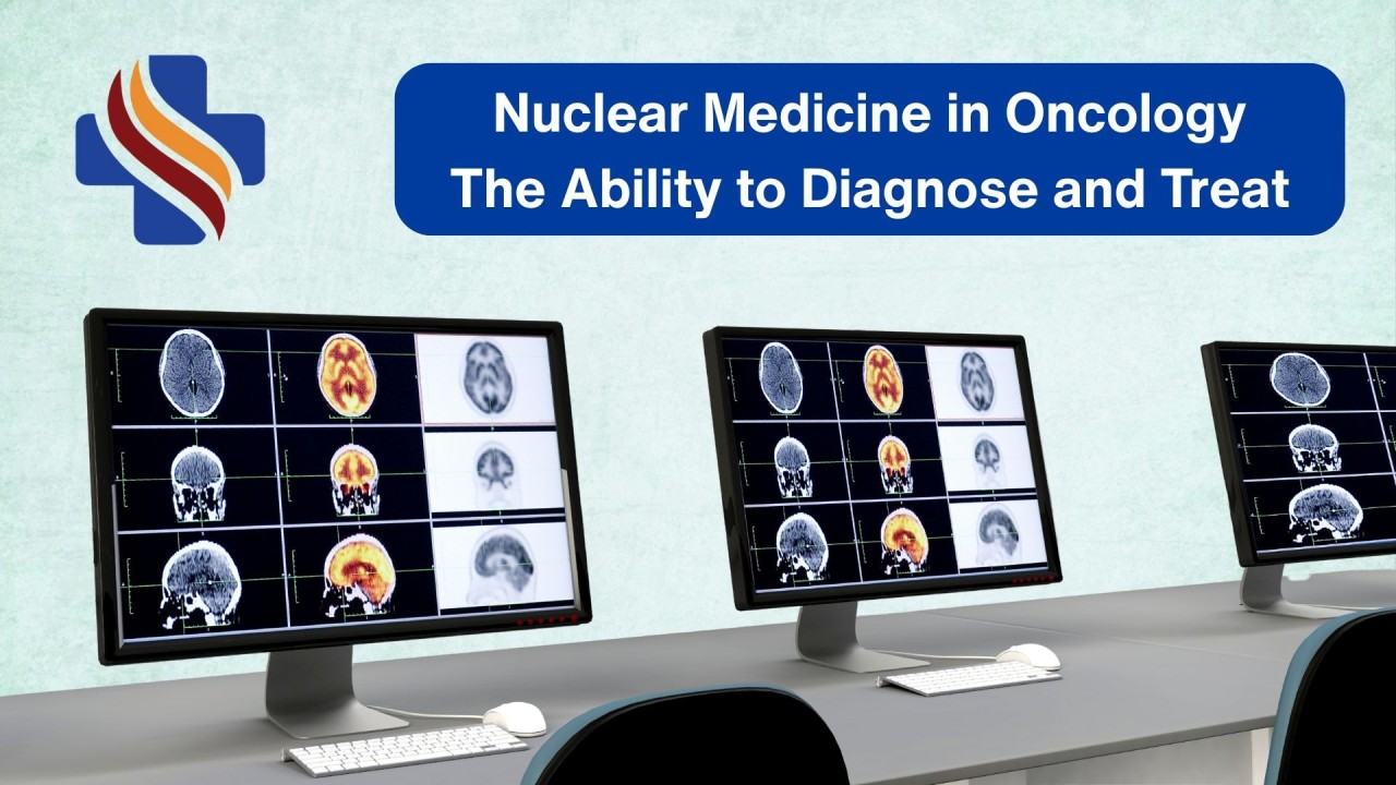
Nuclear Medicine in Oncology: Lighting the Way in Cancer Diagnosis & TherapyThe Story Begins with a Tracer
The foundation of nuclear medicine goes back to a groundbreaking idea from Hungarian chemist George de Hevesy in the early 20th century, a Nobel Prizewinning scientist and also widely recognized as the "father of nuclear medicine"
Hevesy discovered that minute quantities of radioactive compounds (radiopharmaceuticals) / radioactive tracers could be used to follow chemical processes inside living beingsjust like adding a drop of dye to water to see where it flows.
In his famous experiment, he used tiny amounts of radioactive isotopes to study how plants absorbed nutrients.
Thistracer principle is still the basis of nuclear medicine today: ➡️ Use a safe radioactive compound (called a radiotracer) that behaves like natural substances in the body. ➡️ Track its path with special cameras. ➡️ Learn how organs and diseases functionfrom the inside out.
<div class="ivm-view-attr__img-wrapper
">
The Fundamental Principle of Nuclear Medicine
What is Nuclear Medicine?
Nuclear medicine is a branch of medical imaging and therapy that uses small amounts of radioactive materials (called radiotracers) to diagnose and treat diseases, especially cancer. These tracers are injected, swallowed, or inhaled, and they target specific organs or tissues. Special cameras detect the radiation emitted, creating images that show how your body is functioning at a molecular levelnot just its structure.
Unlike X-rays or MRI that show body structures, nuclear medicine looks at how the body works at a cellular level - like how cells are metabolizing or if a tumor is active.
Small amounts ofradioactive tracers (safe, medical-grade) are used.
Special scanners detect these tracers and create detailed images.
The result? Doctors dont just seewhere something is, but also how active it is.
It has two main arms:1. Imaging using:
Conventional Nuclear Medicine (Gamma Camera / SPECT)
Molecular Imaging (PET-CT)
2. NM therapy or now popularly known as Theranostics (Diagnosis + Therapy with the same molecule)
Conventional Nuclear Medicine The Gamma Camera Era
Uses gamma cameras to detect radioactive tracers and Produce images of organ function.
Common tracers: 99mTc (Technetium-99m) based RPs most widely used isotope globallyThe workhorse tracer, used in over 80% of procedures. Used for bone scans, renal scans, cardiac scans, thyroid scans. Iodine I-123 / I-131: Benign and malignant thyroid diseases - Targets thyroid tissue. In diagnosis, it scans for thyroid cancer; in therapy, higher doses destroy cancerous cells.
Helps in cancer by: Detecting bone metastases (spread of cancer to bones). Assessing thyroid cancer.
PET-CT The Molecular Imaging Revolution
PET-CT combines positron emission tomography (PET) with CT.
Shows both function (PET) and structure (CT) in one scan.
Uses radiotracers that mimic natural body chemicals or target receptors in various physiological and patholigcal conditions.
Common PET tracers in cancer:
18F-FDG (Fluorodeoxyglucosewith F-18) Mimics sugar, detects sugar-hungry cancer cells and used in most of the cancers including lung, lymphoma, breast and colorectal cancers.
68Ga-PSMA prostate cancer.
68Ga-DOTATATE neuroendocrine tumors.
FES (Fluoroestradiol) breast cancer (estrogen receptor imaging).
Role in Cancer Diagnosis 👉PET-CT is often called the GPS for cancer, guiding doctors at every step:
Early detection: Using various metabolic and functional processes such as FDG PET scan targetting glucose metabolism: Cancer cells consume more energynuclear scans pick this up early.
Accurate staging: Shows if cancer has spread.
Treatment planning: Helps doctors decide surgery, chemotherapy, or radiation approaches.
Treatment Response Assessment: Shows if cancer is responding or not to treatment
Follow-up: Detects recurrence earlier than most tests.
Role in Cancer Therapy: 🎯 Theranostics Treat What You See and Diagnose and Treat in One Go
Nuclear medicine is not just about diagnosisit can also treat cancer.
Theranostics = Therapy + Diagnostics.
Uses the same molecule for both diagnosis and treatment, just with different isotopes, For e.g. 68GaPSMA for Prostate cancer staging using PET CT and 177LuPSMA PRLT therapy for progressive and metastatic CRPC prostate cancer. It's personalized medicine at its best: "See it, then zap it."
<div class="ivm-view-attr__img-wrapper
">
Two Aspects of Nuclear Medicine
Targeted therapy: Radioactive medicines deliver treatment directly to cancer cells while sparing most healthy tissue.
Used inthyroid cancer, prostate cancer, lymphomas, and neuroendocrine tumors.
Think of it as asmart bombprecise and powerful.
<div class="ivm-view-attr__img-wrapper
">
Past, Present and Future of Theranostics
Clinical impact:
Lu-177 PSMA therapy has shown 40% reduction in risk of death in advanced prostate cancer (VISION trial).
NETTER-1 trial showed 79% progression-free survival improvement with Lu-177 DOTATATE in neuroendocrine tumors.
In India, more than 10,000 Lu-177 therapies have been performed in the last 5 years, positioning the country as a regional hub for theranostics.
The Numbers Nuclear Medicine Worldwide : Nuclear medicine is booming, driven by aging populations and cancer rises.
Diagnostic Nuclear Medicine: ~40 million (4 crore) procedures annually worldwide.
USA: ~20 million scans/year.
India: ~0.51 million scans/year (rapidly growing).
Therapeutic Nuclear Medicine:
In 2023,over 100,000 theranostic procedures were performed globally, with rapid growth in prostate and neuroendocrine treatments.
Growing at1015% annually worldwide.
Theranostics centers expanding rapidly, especially in prostate and neuroendocrine cancer treatment.
Market value:
$10.19B in 2024, projected to $42B by 2032 (CAGR ~19%). Theranostics segment growing fastest at 13-15% CAGR.
USA: $5.12B in 2023, to $16.85B by 2033 (CAGR 12.6%). High adoption of PET-CT (over 2,000 scanners).
India: Rapid growth with 300+ centers. ~1-2 million procedures/year. Market to $1.07B by 2030 (CAGR 10.9%), fueled by affordable tech and rising cancer cases (1.4M new/year).
Therapeutic procedures (like radioiodine therapy) grew 20% globally post-COVID, with India seeing 25% annual increase due to better access.
Future: Bright and Innovative
By 2030, theranostics could dominate cancer care. Prospects include:
Expansion to Lung, breast, brain, pancreatic, and ovarian cancers.
Alpha emitters (like Ac-225) for resistant cancers.
AI-integrated imaging for smarter imaging interpretation, predictive response modeling and faster, more accurate targeting.
Combination with immunotherapy.
ImmunoPET tracers: tracking immune checkpoint inhibitors in real time
Personalized dosimetry: tailoring therapy dose to maximize tumor kill minimize toxicity
Challenges: Supply chain for rare isotopes, but advancements in cyclotron production are helping.
Myths vs Facts
Myth: Nuclear medicine is unsafe because of radiation. ✅ Fact: Radiation doses are carefully regulated, often equal to or less than a CT scan. It is safe, regulated, and used worldwide for decades. Doses are low a PET scan is like 2-3 years of natural background radiation. Tracers decay fast (hours/days), and benefits outweigh risks.
Myth: The radioactivity stays in your body forever.✅Fact: Most tracers are eliminated naturally within days. No long-term buildup.Myth: It's painful or invasive.✅Fact: Just an IV injection or pillless invasive than surgery, no pain beyond a needle prick.
Myth: It causes cancer✅Fact: Risk is minimal (1 in 10,000 for diagnostics). It's used to fight cancer, not cause it.Myth: Only for end-stage cancer.✅Fact: Great for early detection and monitoring, improving survival rates by 20-30% in many cases.Myth: PET-CT replaces all other scans. ✅ Fact: Different scans answer different questions. PET-CT complements, not replaces.
Myth: Once treated with radiotheranostics, you glow or are radioactive. ✅ Fact: The radiation is medical-grade, controlled, and leaves the body safely.
Why It Matters for PatientsFor patients, nuclear medicine means:
Less invasive testsMore confidence in diagnosis
Tailored Personalised treatmentBetter chances of cure and survival
Take-Home Messages
Nuclear medicine is not science fictionitsscience saving lives daily.
It allows doctors tosee cancer earlier, track it better, and treat it smarter.
FromHevesys tracer principle to modern PET-CT and theranostics, the field has grown into a cornerstone of cancer care.
India is catching up fast with global trends, and access is expanding.
The future promisespersonalized, precise cancer care with nuclear medicine at its heart.
Bust the myths: It's safe, precise, and life-saving.
Nuclear medicine is like a torchlight in the dark tunnel of cancerhelping doctors see clearly, act precisely, and give patients hope.
Nuclear medicine is not just scienceits a blend of technology and compassion, guiding modern cancer care.
Next time you hear about PET scans or targeted radiotherapy, remembertheyre part of a powerful field called nuclear medicine that combines vision with cure.
Have you or your loved one ever had a PET-CT or heard about nuclear medicine therapy? Share your thoughts in the comments.By Dr. Aashish Gambhir, Director Head, Nuclear Medicine
Andromeda Cancer Hospital
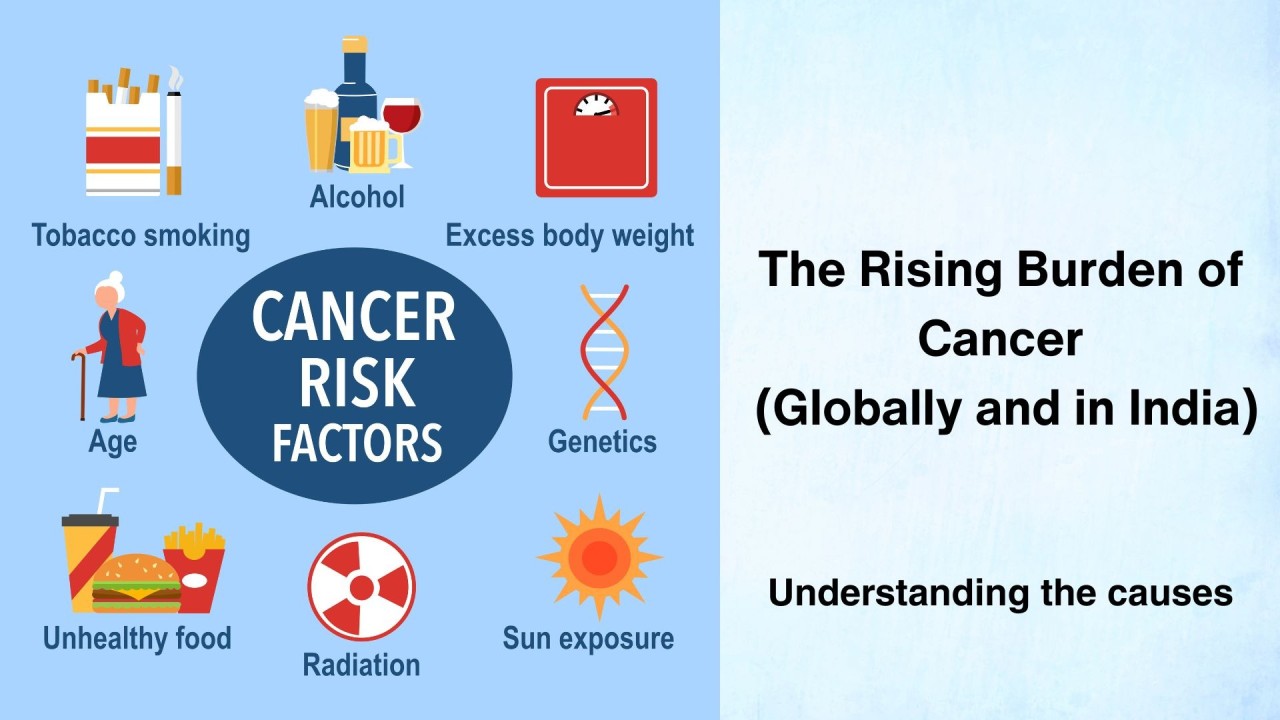
The (Rising) Burden of Cancer What is causing this silent epidemic?
Cancer is one of the leading non-communicable diseases globally. Further, the burden of cancer is rising continuously. While significant progress has happened in early diagnosis and treatment of cancer, the burden of death due to cancer is still high.
Cancer is the 2nd leading cause of death worldwide and causes nearly 15 to 16% of all deaths.
In India too, cancer is a major contributor to the causes of death, 2nd overall in urban areas and 4th overall in rural areas.
The cliched saying is A stitch in time saves nine but it may be highly relevant when it comes to cancer. Preventing is cancer is theoretically much easier than treating a cancer. However, each and every member of the society needs to be aware of the causes of cancer.
Individual action and commitment plays an important role in cancer prevention. Government policies and programs are important but can not work in isolation without public participation.
With this background, we are sharing the list of major causes of cancer globally and also highlight their importance based on gender.
Recognized Causes and Risk Factors for Human Cancer
A. Lifestyle-related
Tobacco use (smoked and smokeless)
Alcohol consumption
Dietary factors: processed/red meat, low fruits/vegetables, obesity-promoting diets
Obesity / overweight
Physical inactivity
B. Environmental and occupational
Air pollution (PM2.5, diesel exhaust, indoor coal smoke)
Occupational exposures: asbestos, silica, benzene, formaldehyde, wood dust, vinyl chloride, certain metals (arsenic, cadmium, chromium, nickel)
Radiation: ionizing radiation (X-rays, gamma rays, radon), ultraviolet radiation (sunlight, tanning beds)
C. Biological / infectious agents
Viruses: HPV, HBV, HCV, EBV, HTLV-1, KSHV, MCPyV
Bacteria: Helicobacter pylori
Parasites: Opisthorchis viverrini, Clonorchis sinensis, Schistosoma haematobium
D. Hormonal and reproductive
Endogenous hormones: prolonged estrogen exposure (early menarche, late menopause, nulliparity, hormone replacement)
Exogenous hormones/medications: oral contraceptives, menopausal hormone therapy, DES
Immunosuppressive drugs (tacrolimus, azathioprine)
E. Genetic and host-related
Inherited cancer syndromes (e.g., BRCA1/2, Lynch, Li-Fraumeni)
Family history (polygenic risk)
Ageing (strongest single risk factor, reflects cumulative mutations and immune decline)
Cancer Risk Factors Grouped by Gender
Predominantly male cancers / risks
Tobacco use (still higher in men globally lung, head neck, bladder, esophagus, pancreas)
Occupational exposures (asbestos, silica, diesel exhaust, metals historically more in men)
Alcohol use (higher consumption rates in men)
HPV oropharyngeal cancer risk increasingly seen in men
<div class="ivm-view-attr__img-wrapper
">
Predominantly female cancers / risks
Reproductive/hormonal factors (estrogen/progesterone exposure breast, endometrial, ovarian)
HPV cervical cancer (exclusively female)
Hormone therapy (HRT, OCPs, DES)
<div class="ivm-view-attr__img-wrapper
">
Both men and women
Tobacco, alcohol, obesity, diet, physical inactivity
Air pollution, radiation, environmental exposures
Infectious causes: HBV/HCV (liver), EBV (lymphoma/nasopharynx), H. pylori (stomach), parasites
Genetic predispositions
<div class="ivm-view-attr__img-wrapper
">
Relative Contribution to Cancer Burden (Attributable Fraction)
Global estimates (WHO / IARC / GLOBOCAN; varies by region):
Tobacco ~22% of all cancer deaths worldwide (8 million deaths annually). Causes lung, head neck, bladder, esophagus, pancreas, stomach, kidney.
Infections ~1315% of cancers globally, higher in developing countries. HPV, HBV, HCV, H. pylori dominate.
Alcohol ~56% of cancers worldwide. Strong links: oral cavity, pharynx, larynx, esophagus, liver, breast, colorectum.
Obesity/overweight physical inactivity ~58% globally. Strong for breast (postmenopausal), colorectal, endometrial, pancreas, kidney, liver.
Diet (low fruits/vegetables, high processed meat, low fiber, high salt) ~5%.
Occupational exposures ~35% of cancers (but higher in men in industrialized areas).
Air pollution (ambient + household) ~23% globally; larger share in Asia.
Radiation (ionizing + UV) ~2%. UV is major for skin cancers (melanoma, squamous, basal).
Genetic predisposition ~510% of cancers due to inherited mutations.
Ranking by Estimated Impact (Global cancer burden)
Tobacco (22%)
Infections (1315%)
Alcohol (56%)
Obesity / overweight / inactivity (58%)
Dietary factors (5%)
Occupational exposures (35%)
Air pollution (23%)
Radiation (2%)
Genetic predisposition (510% but varies by cancer type)
<div class="ivm-view-attr__img-wrapper
">
Key Takeaways:
The largest preventable causes globally are tobacco, infections, alcohol, and obesity-related factors.
Men: Major risk related to tobacco, alcohol, and occupational risk factors.
Women: Major risk related to reproductive/hormonal factors, HPV, obesity-linked cancers.
Both sexes: Diet, infections, air pollution, and radiation.

The Challenges of Breast Cancer in IndiaIntroduction
Breast cancer is the commonest cancer of women globally and in India. According to GLOBOCAN 2022, breast cancer accounts for nearly 23 lakh new cases each year. In India, nearly 2 lakh cases of breast cancer are diagnosed every year. In India, 1 in 28 women will develop breast cancer during their lifetime.
Due to intense research efforts, breast cancer has become highly curable. Unfortunately, 5060% of breast cancer cases in India are still diagnosed at Stage 3 or beyond, compared to less than 10- 20% in high-income countries. Five-year survival rate in western countries is nearly 85 to 90%. However, India lags behind significantly in this regard: the five-year survival figure is around 60% only in India. The poor outcomes of breast cancer treatment in India, as compared to developed countries, stem from a combination of medical, social, infrastructural, and economic factors which can be broadly grouped into two as patient factors and system factors.Reasons for Worse Survival of Breast Cancer Patients in India
Late diagnosis of breast cancer
Lack of access to standardized high quality breast cancer care for a large proportion of breast cancer patients
The problem of delay in diagnosis can be managed at the patient (society) level by spreading awareness, guiding them to early symptoms and signs of breast cancer, educating them about breast self-examination, encouraging them to seek medical help early.The problem of delay in diagnosis at the level of healthcare system needs multi-pronged efforts. It is important for the clinicians who see patients with breast symptoms to understand that early diagnosis and prompt initiation of treatment are critical for successful outcome of breast cancer treatment. Every clinician must know that for early and accurate diagnosis, the most reliable step is triple assessment clinical examination, imaging, and pathological confirmation of diagnosis by biopsy.
Clinical ExaminationA detailed history (age, family history, duration, changes in size) and systematic clinical examination of the breasts and axillae to detect lumps, skin changes, nipple retraction, discharge, or palpable nodes.
Imaging
Mammography: gold standard for women over 40
Ultrasound: especially useful for younger women with dense breasts
MRI: reserved for complex cases, multi-focal disease, or inconclusive findings.
Pathological Assessment
Image guided core needle biopsy, which gives not just diagnosis but tumour grade, receptor status (ER, PR, HER2), all this information is essential for treatment planning.
When all three correlate, the diagnostic accuracy exceeds 99% minimizing false negatives and false positives.
<div class="ivm-view-attr__img-wrapper
">
Triple Assessment for Breast Cancer (Examination, Imaging, Biopsy)
Challenges in Breast Cancer Care in India
In this article, we would like to describe some of the challenges we perceive (as breast specialists) which define the breast cancer care in our country.
Lack of Trained Breast Specialists
Breast cancer care is now a recognized specialty in India, and many young doctors are taking it up. However, most women with breast symptoms first visit healthcare providers who are not trained in breast examination or cancer screening.
Many healthcare providers lack training in proper breast examination.
Triple assessment (clinical exam + imaging + biopsy) is not routinely followed.
Important signs like skin changes or nipple retraction are missed.
Breast lumps are often dismissed as
Breast pain is treated casually with Vitamin E or evening primrose oil without proper tests.
Advanced cancers are mistaken for mastitis/breast infection.
Breast cancers during pregnancy are mostly misdiagnosed as pregnancy associated breast changes.
Many doctors manage breast cancers themselves despite not having the right skills.
This casual approach, poor clinical examination skills, lack of awareness about triple assessment (clinical exam, imaging, and biopsy), and wrong interpretation of symptoms lead to diagnostic delays and worse outcomes. It is necessary to train and sensitize healthcare providers to detect and refer suspected cases early.
Problems in Breast Imaging and ReportingStandards of breast imaging in India vary widely, especially between urban and rural centres.
Many reports dont follow BIRADS guidelines or assign wrong categories.
Indian women have denser breasts, making mammograms harder to interpret.
Poor communication between doctors and radiologists, outdated machines, and shortage of trained breast radiologists add to the problem.
In rural areas, lack of modern imaging and expert reporting means more women are diagnosed late.
Younger women (more likely to have triple-negative cancers) often get benign-sounding reports, leading to missed diagnoses.
Continued Use of FNAC (Fine Needle Aspiration Cytology) for diagnosis and planning treatmentAlthough once common, FNAC has major drawbacks:
Less accurate than core needle biopsy (more false negatives).
Does not give enough tissue for ER/PR/Her2/Ki67 testing.
Cannot confirm invasion or grade the tumour.
No information on margins, lymphovascular invasion, or surrounding tissue.
Sampling errors are common, leading to missed cancers.
Overuse of Direct Excision Biopsies without Proper AssessmentMany women are advised to have their lump removed directly without imaging or core biopsy because breast surgery is wrongly thought to be easy.
Myths about biopsy spreading cancer make patients agree to excision.
Sometimes the removed lump is not sent for pathology, denying diagnosis.
Specimens are often removed into pieces or not oriented for margins, making accurate size and margin clearance assessment impossible.
Poorly placed surgical incisions may prevent breast conservation later.
Axillary node assessment is not done, requiring another surgery.
These issues can delay treatment and reduce chances of cure.
Starting Treatment without knowing IHC Results (Biological profile of the individuals breast cancer)Today, breast cancer treatment is personalized. Information on tumour biology (ER/PR/Her2/Ki67) and stage is essential for planning.
Patients with certain subtypes (triple-negative, Her2-positive) or large tumours benefit from neoadjuvant systemic therapy before surgery.
Without these details, outdated mastectomy first approaches deny patients the advantages of modern treatment.
Overuse of MastectomyMany women undergo mastectomy solely based on FNAC results, sometimes even when its a false-positive.
Breast conservation surgery (BCS) is equally safe for early cancers, but lack of surgeon training, old beliefs, and ignorance mean that 2/3 of breast cancer surgeries in India are still mastectomies.
Mastectomy can cause lifelong psychological distress. Although, breast reconstruction can be safely performed after mastectomy, it requires advanced surgical skills, extra days of hospitalization and post-surgical recovery and significantly additional cost compared to the breast conservation surgery.
Incomplete MastectomiesWhen performed by untrained surgeons, mastectomy may leave significant breast tissue or lymph nodes behind.
This leads to high recurrence risk and rapid disease progression.
Completion surgery is technically difficult and may prevent breast reconstruction.
Scarring and poor incision placement complicate future treatments.
Delays in adjuvant therapy worsen prognosis.
Very limited availability of resources and skills for sentinel node biopsyWithout SLNB, many women with early breast cancer undergo full axillary lymph node dissection, even when nodes are not involved.
This causes high risk of complications such as arm swelling (lymphedema), shoulder stiffness, numbness, chronic pain, and higher risk of wound problems.
It prolongs recovery, lowers quality of life, and leaves lasting disability.
SLNB is a safer, less invasive, guideline-recommended procedure.
Many Indian patients face avoidable harm and overtreatment due to this.
Issues related to systemic Therapy for Breast Cancer
Drug availability cost barriers: Newer targeted drugs and supportive medicines may be unaffordable or unavailable for many patients.
Incomplete biological profiling: ER, PR, Her2, and Ki67 tests are sometimes not done before starting chemotherapy, leading to non-personalized treatment.
Use of outdated regimens: In smaller centres, older protocols may still be used instead of evidence-based modern regimens.
Risk of errors high toxicity: Incorrect dosing, poor monitoring, and lack of proper supportive care can cause preventable side effects.
Infrastructure limitations: Many places lack dedicated day-care chemotherapy units, proper infection control, and trained oncology nurses.
Inadequate patient counselling: Patients may not receive enough guidance on side effects, fertility preservation, or the importance of completing all cycles.
Poor adherence to treatment: Due to side effects, cost, or lack of awareness, some patients discontinue therapy early, reducing chances of cure.
Issues related to Radiotherapy for Breast CancerLimited availability of advanced technology: Many centres still use outdated cobalt machines instead of modern linear accelerators with 3DCRT, IMRT, or IGRT capabilities.
Geographic and access barriers: Radiotherapy facilities are concentrated in larger cities, forcing rural patients to travel long distances daily for several weeks.
Long waiting times: High patient load and limited machines lead to delays in starting treatment, which can worsen outcomes.
Lack of breast-specific techniques: Inadequate use of advanced methods like deep inspiration breath-hold (DIBH) to protect the heart and lungs in left-sided cancers.
Inconsistent treatment quality: Variation in contouring, planning, and dose delivery between centres due to lack of standard protocols or quality audits.
Shortage of trained staff: Limited numbers of radiation oncologists, physicists, and technologists with specialized breast cancer training.
Side effect management gaps: Insufficient counselling and follow-up for managing skin reactions, fatigue, lymphedema, and late toxicities.
Financial burden: High cost of advanced radiotherapy techniques and travel/accommodation expenses during prolonged treatment courses.
Challenges in Genetic Counselling and Testing for Breast Cancer patients.Low awareness: Many patients and even healthcare providers are unaware of the role of BRCA and other genetic mutations in breast cancer risk.
Limited availability of trained counsellors: Very few centres have qualified genetic counsellors to guide patients and families.
High cost of testing: Genetic tests are expensive, often not covered by insurance, and unaffordable for many patients.
Access barriers: Testing facilities are concentrated in urban centres, limiting availability for rural populations.
Cultural and social stigma: Fear of discrimination, marriage-related concerns, and family pressure discourage many from testing.
Poor integration into routine care: Genetic risk assessment is not consistently incorporated into breast cancer evaluation and follow-up.
Lack of pre- and post-test counselling: Inadequate explanation of test implications can lead to anxiety, misinterpretation, or misuse of results.
Limited cascade testing: Family members at risk are rarely offered or encouraged to undergo testing.
Data privacy concerns: Fear of genetic information misuse due to weak legal safeguards.
Missed prevention opportunities: Without testing, high-risk women lose the chance for preventive measures such as enhanced screening, chemoprevention, or prophylactic surgery.
Concerns Related to Recurrence Score Testing in Breast CancerHigh cost: These tests are expensive and often not covered by insurance, making them unaffordable for many patients.
Limited availability in India:Most samples are sent abroad, increasing cost and turnaround time.
Delay in treatment decisions: Waiting for results can postpone the start of adjuvant therapy.
Lack of awareness: Many oncologists and patients are unfamiliar with the availability, benefits, and limitations of these tests.
Unclear applicability in all populations: Most validation studies are from Western populations; Indian-specific outcome data is limited.
Infrastructure and logistics: Sample collection, preservation, and international shipping may be challenging, especially in smaller centres.
Patient anxiety: Misunderstanding the meaning of recurrence scores can cause unnecessary worry or false reassurance.
Limited use in public sector: Government hospitals rarely offer or recommend such tests due to cost constraints.
Ethical and equity concerns: Only wealthier patients can access these tools, creating disparities in personalized cancer care.
In summary: Improving breast cancer outcomes in India requires:
Training doctors in proper clinical and diagnostic pathways.
Enforcing quality standards in imaging and reporting.
Phasing out FNAC for diagnosis in favour of core biopsy.
Working towards establishing early diagnosis and prompt initiation of treatment.
Making breast cancer treatment available and affordable to all the patients
Avoiding unnecessary or poorly performed surgeries.
Multi-disciplinary approach to ensure that the treatment decisions are made after full pathological and biological assessment.
Ensuring oncological safety and working towards preserving long term quality of life of the survivors.
<div class="ivm-view-attr__img-wrapper
">
Multi-disciplinary Board Meeting

Obesity and Cancer — What Everyone Should Know?(Eat and exercise your way to a healthy life.)
Welcome from Andromeda Cancer Hospital
At Andromeda Cancer Hospital, our mission is to bring useful health information straight to our community. Today, we tackle an important, often misunderstood topic: the connection between obesity and cancer. With obesity rates rising and cancer cases growing in Haryana and across India, its the right time to learn and take charge of our health.
Obesity is assessed by BMI but normal weight obesity also occurs and truncal fat is more harmful.
What Causes ObesityObesity develops from a mix of different factors working together. While eating more calories than the body burns is a major cause, theres much more to it. Here are some of the key contributors:
Unhealthy diet and overeating: Consistently eating more calories than needed, especially from high-sugar and processed foods.
Lack of physical activity: Sedentary lifestyles make it easier to gain weight.
Genetics: Family history can influence how your body stores fat and uses energy.
Hormonal imbalances: Conditions like hypothyroidism or changes in hormones can affect weight.
Medical conditions and medications: Some illnesses and medicines can contribute to weight gain.
Emotional stress: Stress or emotional eating may lead to consuming excess calories.
Environmental and lifestyle factors: Easy access to unhealthy foods, busy routines, and lack of resources for exercise also play a part.
Obesity has multiple underlying causes
In summary, obesity is usually the result of a complex interplay between lifestyle habits, biology, medical issues, and our environment.
Quick Facts:
The burden of obesity is rising all over the world.
In India, over 135 million people are affected by obesity. The burden has increase by nearly 200% in last two decades and is expected to double in the next 10 to 15 years.
How Are Obesity and Cancer Connected?Many people dont realize that carrying extra weight can actually increase ones risk of developing cancer. Research has shown that obesity is a risk factor for several types of cancers. WHO and IARC have identified 13 different cancers whose risk is increased by obesity. These include.
Uterine (endometrial) cancer
Breast cancer (especially after menopause)
Ovarian Cancer
Cancer of oesophagus
Colon and rectum cancer
Liver cancer
Gallbladder cancer
Pancreatic cancer
Stomach cancer
Kidney cancer
Multiple myeloma
Thyroid cancer
Meningioma
See the Connection Between Obesity and Cancer:Why Does Obesity Increase Cancer Risk?
You dont need to be a scientist to understand the basic idea: Think of your body like a car engine. If its overloaded and running too hot, problems will follow.
Extra Fat Increases Inflammation: Chronic, low-level inflammation caused by obesity can damage cells and lead to cancer.
Growth factors: Excess fat in the body leads to insulin resistance and increases the release of insulin and related growth factors in response to food. These growth factors increase the cancer risk.
Hormone Changes: Excess fat converts some hormone released from the adrenals to active oestrogens and the higher levels of postmenopausal oestrogens are associated with higher risk of cancers such as breast cancer, endometrial cancer, etc.
Impact on Immunity: Obesity can weaken your immune system, making it harder to detect and fight off cancer cells.
What Can You Do? Actionable TipsYou have more power than you think! Start with small, lasting changes:
Choose local, seasonal fruits and vegetables. Try to add millet (bajra, jowar) and pulses to your meals. Limit processed foods and sugary drinks.
Move more every day. Even a 30-minute walk in your neighbourhood or some light yoga can make a big difference.
Exercising is important for obesity control. Diet alone will not be sufficient in the long run.
Regular health checkups. Early detection saves lives! Dont hesitate to consult our team for advice.
Family support.Involve your loved ones in healthy habits together, its easier.
What the other harms in cancer patients who are obese?The problems dont stop at increasing the risk of cancer. Cancer treatment is more problematic in obese patients, complications of treatment are higher, and the treatment may be less effective. Obese patients may have higher risk of cancer recurrence.
Is it possible to get out of this vicious cycle?Yes, if you can control your weight, you can reduce your risk of cancer. Reduction in weight after cancer diagnosis and treatment also reduces the side effects of cancer treatment, improves long term survival and improves quality of life.Various approaches including exercise, dietary modifications, anti-obesity medications, bariatric surgery can all be used depending upon the individual patients needs and choices.
At Andromeda Cancer Hospital, we offer nutrition guidance, tailored cancer screenings, and weight management support. Our doors are always open for you and your family.
QA Busting Common MythsLets clear up some common misunderstandings:Myth 1: Only junk food makes you obese.Reality: While diet matters, other factors like low physical activity, genetics, and even stress play important roles.Myth 2: Thin people dont get cancer, and overweight people always do.Reality: Anyone can get cancer but being obese increases risk for certain cancers. Maintaining a healthy weight lowers (but doesnt eliminate) your risk.Do you have questions or want to share your story? Wed love to hear from you. Write to us or visit Andromeda Cancer Hospital lets fight cancer together.
For free health talks, screenings, and support, contact us at 9138111625.
Catch our next newsletter in your LinkedIn feed! Subscribe to the newsletter.
Stay informed. Follow Andromeda Cancer Hospital here on LinkedIn for trusted tips, inspiring stories, and upcoming events.Have a question or a topic youd like us to cover? Comment below or message us directly!
Together, lets build a healthier India, healthier Haryana and healthier Sonipat. Let us fight cancer at all levels, prevent it, diagnose it early, treat it better and help patients live a happy and healthy cancer free life.
Please visit our YouTube channel for videos on Obesity and Cancer.
You can also visit and subscribe to the YouTube channel for regular informative videos about cancer awareness.

Immune Therapy in Cancer: mRNA Vaccines.....What is mRNA?
mRNA (messenger RNA or ribonucleic acid) is polymeric molecule produced in the cells by a process of transcription using DNA. mRNA is a template used for synthesis of proteins. The proteins then get modified in various ways and serve their diverse functions. Some of the proteins serve as antigens that are recognized by immune cells of the body and body can mount an immune defence against these antigens.
What are mRNA vaccines and how do they work?
An mRNA vaccine is a revolutionary type of immunization that uses mRNA prepared in the laboratory and injected. It instructs the body cells to produce a viral or tumor-related protein. This stimulates the immune system to build both antibody (humoral) and T-cell (cellular) responses.Steps in the process for mRNA vaccine preparation and usage
Antigens have to be identified from the viral pathogens or cancer cells
mRNA are produced in the laboratory using equipment that can generate mRNA molecules in the instructed sequence.
The mRNA molecules are packaged in lipid nanoparticles (LNPs).
The vaccine is administered via intramuscular injection.
Cells translate the mRNA to produce the target antigen (e.g., SARS‑CoV‑2 spike protein).
When the antigen appears on the cell surface, the immune system recognizes it as foreign and activates adaptive immunity.
The mRNA is degraded afterward (and does mRNA vaccines across different diseases.
Infectious diseases: Viral Targets: COVID‑19 mRNA vaccines (PfizerBioNTech Comirnaty Moderna Spikevax) are the only currently authorized mRNA vaccines.
There are many others in development/testing: influenza, RSV, CMV, EBV, Zika, HIV, norovirus, Hepatitis C, genital herpes, malariamany already in Phase 13 clinical trials
Bacterial targets: A novel candidate vaccine against Yersinia pestis (plague) recently showed 100% efficacy in mice; human trials still pending.How many mRNA vaccines are approved?
COVID‑19: PfizerBioNTech Comirnaty Moderna Spikevax These are the only licensed mRNA vaccines worldwide to date
No mRNA vaccines have yet received approval for other diseases; many are in varying stages of clinical trials.
mRNA vaccines in cancer: current landscape
Oncology research in mRNA vaccines is rapidly advancing:
Clinical trial status: Over 120 clinical trials are exploring mRNA vaccines for lung, breast, prostate, melanoma, pancreatic, brain cancers and more.
Safety tolerability: Multiple trials have found mRNA cancer vaccines to be well tolerated with manageable side-effects, sometimes less than traditional chemotherapy.
Promising efficacy signals: A Phase I personalized neoantigen mRNA vaccine for pancreatic cancer (16 patients) generated specific T-cell responses; responders remained recurrence-free for up to 18 months.
Personalized vaccine platforms (e.g., Autogene cevumeran) are entering Phase II trials
Moderna/Mercks mRNA‑4157/V940 combined with pembrolizumab showed a 44% reduction in melanoma recurrence in Phase II, now progressing into Phase III.
"Universal" vaccine approach: Preclinical studies in mice (University of Florida) indicate a general mRNA booster that primes the immune system when used with checkpoint inhibitors, though human trials are still in planning. This particular vaccine could work against multiple different type of cancers and not be dependent on cancer specific antigens.
Looking Ahea
Challenges:The field of mRNA vaccines is relatively new. The vaccines against Covid received emergency use approval. Long term safety data is necessary. However, the concept underlying the mRNA vaccines is very promising. However, scientific research has to complete all phases of testing and show evidence of benefit with acceptable toxicity profile. Regulatory issues, cost of therapy, scaling personalized vaccine production, delivery methods, etc are issues to be handled.
Hopeful future: If ongoing trials confirm efficacy, mRNA vaccines could become vital in adjuvant cancer therapy to prevent recurrence and possibly treat ongoing cancers.
Conclusions
mRNA vaccines work by instructing cells to produce antigens that drive immune responseoffering rapid, flexible platforms.
Today, only COVID‑19 mRNA vaccines are licensed.
mRNA tech is in active trials for infectious diseases and is showing very encouraging progress in oncology, with multiple phase II/III human trials underway.
Several cancer-specific mRNA vaccines have successfully entered human trials, with early data indicating both safety and potential efficacy.

De-escalation of Treatment in Breast Cancer Balancing Cure and Quality of Life
Breast cancer treatment has transformed dramatically over the last five decades.It used to be one-size-fits-all approach. It was dominated by radical mastectomy/modified radical mastectomy in nearly all patients and chemotherapy and radiotherapy in selected cases.
Mammographic imaging is a game changer for screening and early diagnosis
It is now a highly personalized approach with individualized decision making. At the heart of this evolution lies a powerful idea: de-escalation of treatment.De-escalation means deliberately reducing the intensity or extent of surgery, chemotherapy, or radiotherapy without compromising cure. The aim is not only to save lives but also to preserve quality of life, minimize side effects, and avoid long-term harm.This represents a shift from maximum tolerated treatment to minimum effective treatment.
There are multiple approaches or modalities used in breast cancer treatment today.
Why De-escalation MattersSurvival rates have improved with earlier detection and better systemic therapies. But aggressive treatments often leave lasting scars:
Chronic lymphedema after axillary dissection
Disfigurement and trauma after mastectomy
Infertility and menopause from chemotherapy
Cardiotoxicity, fatigue, and secondary cancers after radiotherapy
These burdens have led oncologists worldwide to ask: Can we treat less and still cure? Increasingly, the answer is yeswhen patients are carefully selected.
Areas of De-escalation
1. Surgery
Breast Surgery Breast Conservation after NAST: In large operable and selected locally advanced cancers, neoadjuvant systemic therapy (NAST) often shrinks tumours, making breast conservation surgery (BCS) possible. With proper imaging, margin control, and radiotherapy, outcomes are comparable to mastectomy, with superior cosmetic and psychological benefits. Omission of Surgery: Trials are exploring whether patients achieving complete response after NAST can safely avoid surgery. While promising, this approach requires rigorous imaging, biopsy confirmation, and close follow-up in clinical trial settings before wider adoption.
Axillary Surgery Sentinel Lymph Node Biopsy (SLNB): SLNB has replaced axillary lymph node dissection (ALND) in node-negative patients, reducing lymphedema while providing accurate staging. Recurrence rates remain equivalent to ALND. Avoiding ALND in Limited Nodal Disease: Trials such as ACOSOG Z0011, IBCSG 23-01, AMAROS, SENOMAC, and SENODAR show that patients with minimal nodal disease on SLNB can avoid ALND, sometimes substituting axillary radiation. Omission of Axillary Surgery Altogether: The SOUND trial demonstrated that early breast cancer patients with negative axillary ultrasound can avoid even SLNB without compromising safety. This major advance reduces morbidity, shoulder dysfunction, and preserves body image.
2. Radiotherapy
Hypo-fractionated Schedules: Delivering higher doses in fewer sessions is now standard, offering equal efficacy, lower toxicity, and more convenience.
Partial Breast Irradiation: In very low-risk patients undergoing BCS, targeting only the tumour bed instead of the entire breast achieves safe outcomes.
Omission of Radiotherapy: In carefully chosen elderly, low-risk patients, omission does not compromise survival and spares them from toxicity.
3. Systemic Therapy
Genomic Assays: Tests like Oncotype DX and MammaPrint help identify hormone-receptor positive patients who can avoid chemotherapy, receiving only endocrine therapy.
Shorter Chemotherapy Courses: Selected regimens with reduced cycles show comparable results, minimizing cumulative toxicity.
Targeted Therapy: For HER2-positive disease, studies suggest that shorter durations of trastuzumab may suffice, lowering cardiotoxicity risk.
Various approaches are combined based on finding the disease biology through molecular testing
4. Endocrine TherapyEndocrine therapy improves survival but prolonged use causes menopausal symptoms, bone loss, and adherence problems. Evidence indicates that in some low-risk patients, five years of therapy may be adequate compared to ten years.Risks of De-escalationDe-escalation carries challenges that must be weighed carefully:
Undertreatment: Lower-intensity therapy may increase recurrence in some patients.
Tumour Heterogeneity: Low-risk appearance does not always equal indolent biology
Compensatory Overtreatment: Less surgery is often offset by more systemic therapy or radiation, shifting rather than reducing toxicity.
Psychological Concerns: Some patients equate less treatment with less cure, creating anxiety.
Limited Long-Term Data: Many de-escalation trials have short follow-ups; survival data over decades are awaited.Thus, de-escalation must be evidence-driven, guided by tumour boards, and aligned with patient preferences.
Benefits of De-escalationDespite the risks, the advantages are substantial:
Reduced Toxicity: Lower rates of lymphedema, cardiotoxicity, infertility, and secondary malignancies.
Improved Quality of Life: Better cosmetic outcomes, body image, and emotional recovery.
Cost Savings: Particularly valuable in low-resource settings like India.
Faster Recovery: Enables earlier return to family, work, and normal life.
Patient-Centred Care: Aligns treatment with individual biology and personal values.
Long-Term Advantages
Personalized Medicine: Molecular profiling and AI tools will refine risk stratification, allowing precise tailoring of therapy intensity.
Healthcare Sustainability: Avoiding overtreatment conserves resources, improving access to effective care.
Survivorship Focus: With rising survival rates, quality of life and long-term well-being take centre stage. De-escalation prevents chronic complications, ensuring survivors thrive beyond cancer.
Global Relevance: In resource-limited countries, evidence-based de-escalation provides safe, affordable care without compromising outcomes.
The Way ForwardDe-escalation is not a universal formula. Success requires:
Careful patient selection using clinical, pathological, and molecular tools
Multidisciplinary tumour board decision-making
Shared decision-making, incorporating patient preferences
Robust clinical trial participation and long-term data generation
Ultimately, de-escalation reflects the art of modern oncology: treating smarter, not harder.
ConclusionBreast cancer care has entered an era of precision and compassion. De-escalationacross surgery, radiotherapy, and systemic therapybrings together the twin goals of cure and quality of life.For patients, it means fewer scars and a fuller life. For oncologists, it represents evidence-based, humane medicine. For society, it ensures sustainable, affordable cancer care.The challenge now is to refine our tools and judgment so that every woman receives not just the best chance of survival, but the best chance of living well.Andromeda Cancer Hospital now offers world class cancer treatment to patients of Haryana, Delhi and Northern India. For breast cancer management, it has a specialized centre of excellence "Andromeda Breast Cancer Centre".Andromeda Cancer Hospital Youtube Channel: http://www.youtube.com/@andromedacancerhospitalYou will find a treasure trove of informational videos at this channel. Visit this page and find out for yourself. Also request you to follow and share the link to the youtube channel.