
Introduction
A cough that sticks around long after the season ends. A small lump that feels wrong under your fingers. Fatigue so deep it makes ordinary mornings hard.
Most people shrug these off. In Haryana, where cancer cases are climbing year after year, ignoring them is a gamble you don’t have to take. Early screening catches disease when treatment is simpler and survival odds are highest.
Yet most Indians who qualify for free screening never get it. The country’s screening rates for cervical and breast cancer sit below 10%, even with government programs running for years. That gap is not about a lack of technology, it is about awareness, access, and knowing which facility actually delivers a diagnosis instead of just running a scan.
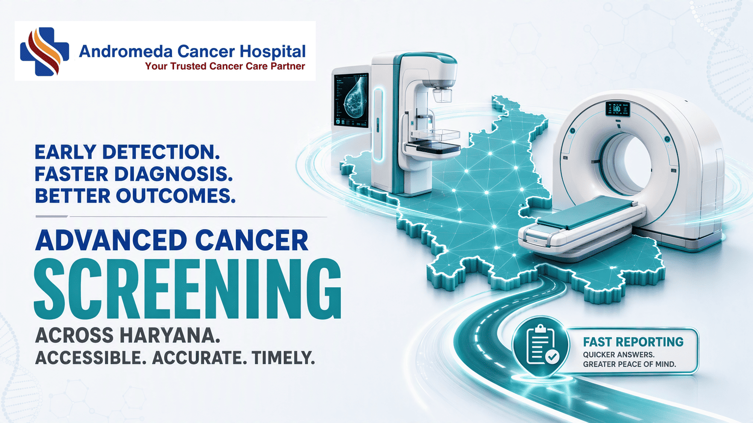
Advanced diagnostic centers are opening across Haryana now. Andromeda Cancer Hospital in Sonipat and Oncare Cancer Centre in Gurugram operate the same imaging platforms you see in Mumbai or Delhi: full-field digital mammography, high-speed CT, and integrated reporting workflows that compress the wait between a suspicious finding and a treatment plan. This article walks you through which technologies exist, what they do, and how to tell the difference between a hospital that owns a machine and one that has built a real diagnostic system around it.
Key Takeaways
- Advanced cancer screening in Haryana runs on three pieces of technology, but the real difference is whether they sit inside a hospital system that acts on the result.
- A center qualifies as 'advanced' when it combines digital PET-CT metabolic imaging, 3D mammography (tomosynthesis), and a linear accelerator like the Varian TrueBeam. Each technology picks up what the others miss.
- Digital PET-CT flags malignant cell activity by tracking a radioactive tracer. 3D mammography slices breast tissue into thin layers, cutting down the false positives that plague older 2D scans. The TrueBeam then delivers treatment with accuracy measured in millimeters.
- A whole-body PET-CT in Haryana costs between ₹12,000 and ₹25,000 depending on the tracer. That is indicative, your actual bill will shift with the facility and the specific protocol.
- Speed varies by center. Andromeda Cancer Hospital coordinates PET-CT and IHC testing so results land in front of a tumor board within one week.
- Here is the distinction that matters: an integrated hospital feeds your imaging straight into a multidisciplinary tumor board, pathology, surgery, and radiation review it together. A stand-alone scan gives you a report and a referral slip, and the orchestration is on you.
What Defines an ‘Advanced’ Cancer Screening Center
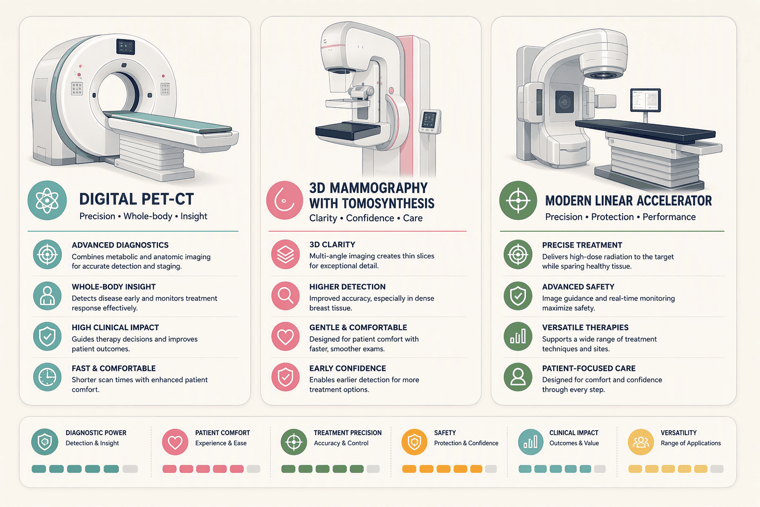
An 'advanced' cancer screening center comes down to what's bolted to the floor, not what's written on the website. You need a digital PET-CT scanner that picks up metabolic hot spots, a 3D mammography unit with tomosynthesis to see through overlapping breast tissue, and a modern linear accelerator like the Varian TrueBeam. That last one matters even before treatment starts because a center investing in millimeter-precision radiotherapy has already built the physics and quality-assurance culture that sharpens every earlier screening step.
| Technology | What It Does | Key Specification |
|---|---|---|
| Digital PET-CT | Injects a radioactive tracer to create images of metabolic activity, exposing malignant cells that consume glucose at higher rates. | Scan duration of 20 to 30 minutes after a 45 to 60-minute tracer uptake period. |
| 3D Mammography (FFDM / Tomosynthesis) | Captures multiple X-ray slices of breast tissue to construct a three-dimensional image, reducing tissue overlap that hides tumors or creates false positives. | Classified by the FDA as a Class II (special controls) device requiring extensive laboratory testing including spatial resolution, noise analysis, and phantom testing. |
| Varian TrueBeam Linear Accelerator | Delivers high-energy X-rays to destroy cancer cells, using on-board cone-beam CT imaging to verify tumor position before each treatment fraction. | Generates high-quality 3-D cone-beam CT images 60% faster than earlier generations of Varian imaging technology and can create images using 25% less X-ray dose compared to earlier image-guided technologies. |
The table hits the three machines that define the category. But a center that owns these machines and runs them in silos is still just a collection of equipment. The TrueBeam's ability to deliver a treatment plan is only as good as the imaging and pathology data that feeds it.
The Core Technologies: Digital PET-CT and Modern Mammography
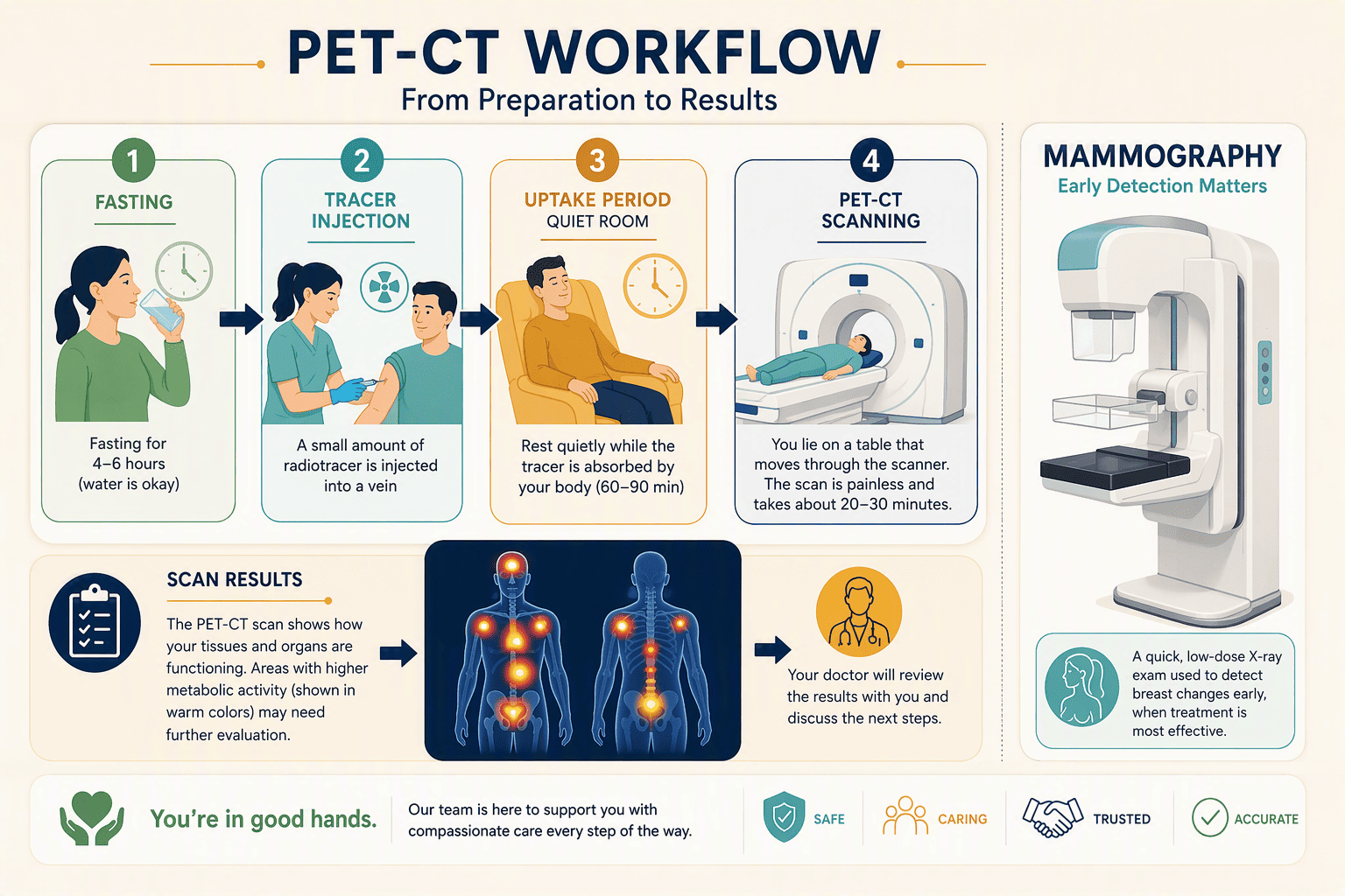
A digital PET-CT scan is not a simple X-ray. It is a functional imaging procedure that traces metabolic activity. After a patient fasts for 4 to 6 hours, a radioactive glucose tracer is injected into the bloodstream.
Cancer cells absorb the tracer in higher concentrations than normal tissue. The patient then rests in a quiet uptake room for 45 to 60 minutes before the scan itself, which lasts 20 to 30 minutes. The resulting image shows hot spots of potential malignancy anywhere in the body, from a primary site to distant metastases.
Modern mammography has moved decisively beyond the flat, two-dimensional images that could hide lesions behind overlapping tissue. Full-Field Digital Mammography (FFDM) with tomosynthesis acquires a series of thin, high-resolution slices through the breast and reconstructs them into a 3D volume a radiologist can scroll through layer by layer. That means fewer false positives and fewer women called back for unnecessary follow-up scans. Andromeda Cancer Hospital operates full field digital mammography with tomosynthesis as part of its breast oncology pathway. The technology requires strict calibration: under FDA special controls, manufacturers validate spatial resolution, signal-to-noise ratio transfer, and automatic exposure control performance.
Inside Andromeda Cancer Hospital, Sonipat: Equipment and Integrated Pathways
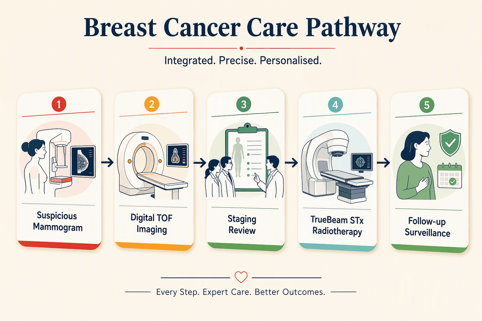
A woman walks into Andromeda Cancer Hospital in Sonipat with a suspicious mammogram from a local clinic. What happens next separates this facility from most cancer centers in India.
Andromeda Cancer Hospital runs a Varian TrueBeam STx linear accelerator for radiation oncology https://andromedahospital.in/treatments/radiation-therapy and digital TOF imaging for cancer staging, treatment response assessment, and recurrence surveillance. Its breast oncology unit pairs full-field digital mammography with tomosynthesis alongside an onco-pathology lab that processes tissue from biopsies to surgical resections. But the machine list isn't the story.
The hospital holds multidisciplinary tumor board meetings twice weekly. PET-CT and IHC testing turn around within one week for tumor board review, compressing the anxious waiting period between a suspicious finding and a treatment decision. Patients also get cost estimates and documentation support for crowdfunding campaigns, a practical acknowledgment of the financial reality of cancer care in India.
The diagnostic pathway folds imaging directly into treatment planning. After a positive screening, the radiologist's findings are routed into the twice-weekly tumor board where surgical oncologists, medical oncologists, and pathologists review cases concurrently.
For a newly diagnosed breast cancer patient, clinical examination, imaging, and pathology are discussed under one roof. The hospital's radiation oncology team can then apply the https://andromedahospital.in/treatments/radiation-therapy TrueBeam STx delivery modes (IMRT, IGRT, volumetric arc therapy, SRT, SRS, and SBRT) using the same digital PET-CT dataset that flagged the disease.
Other Key Centers Offering Advanced Diagnostics in Haryana
Oncare Cancer Centre in Gurugram has built its model around multidisciplinary expertise rather than a single flagship machine. Its physicians have 15+ years of experience and training from AIIMS and TMH, bringing the decision-making framework of those large institutions into a community clinic. The center reports having served 10,000+ patients and performed 5,000+ treatments, figures that suggest a substantial clinical volume. Its lead medical oncologist, Dr. Pooja Babbar, brings over 15 years of experience and is ESMO certified by the European Society of Medical Oncology. The hematology-oncology coverage is anchored by a clinician with 11+ years of experience in stem cell transplantation.
Fortis Cancer Institute FMRI in Gurugram represents a different scale of advanced diagnostics. Its campus houses South Asia's first Gamma Knife Esprit for non-surgical brain tumor treatment, an MR Linac that combines a linear accelerator with an MRI scanner for real-time adaptive radiation planning, and a dedicated team of 75 expert consultants providing multidisciplinary care. Max Healthcare’s molecular oncology speciality in the NCR adds another diagnostic layer with next-generation sequencing, liquid biopsies, and bioinformatics for tumor profiling. These centers represent the expanding upper boundary of Haryana’s cancer diagnostic capability.
Stand-Alone Screening vs. Hospital-Integrated Care: The Critical Difference
A patient can walk into a diagnostic lab, pay ₹15,000 for a PET-CT, and leave with a USB drive containing DICOM images and a typed report. That is a stand-alone scan. A hospital-integrated diagnostic process is a fundamentally different experience. In an integrated model, from Andromeda Cancer Hospital to the large Fortis and Max campuses, the steps are sequential but tightly linked.
- Tracer injection and uptake: The patient completes the standard 45 to 60-minute quiet uptake phase after tracer injection, as with any PET-CT.
- Scan acquisition: The 20 to 30-minute scan captures whole-body metabolic data across all organ systems.
- Rapid radiologist interpretation: The images are read, often within hours, and a preliminary report is generated. The speed here is critical; a center claiming 'advanced' capability should not take days to produce a report on a scan that supports same-day reading.
- Direct feed to a multidisciplinary tumor board: The radiologist’s findings are not emailed to the referring physician. They are presented at a scheduled tumor board meeting where a surgeon, a medical oncologist, a radiation oncologist, and a pathologist review the images alongside any biopsy results. At Andromeda, this happens twice weekly.
- Immediate treatment planning: If the tumor board confirms a malignancy and radiation is indicated, the planning can begin using the same imaging dataset. The TrueBeam system’s cone-beam CT and gated RapidArc capability illustrate the downstream payoff: in a lung SBRT case, gated RapidArc combined with High Intensity Mode delivery at 2400MU/min can deliver a full, 20Gy lung SBRT fraction in 7 minutes.
The Cost and Step-by-Step Process for a Whole-Body PET-CT Scan in Haryana
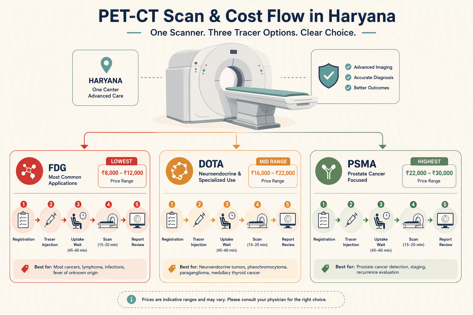
A whole-body PET-CT scan in Haryana costs between ₹12,000 and ₹25,000. The price depends almost entirely on the radioactive tracer used. Standard FDG scans, the workhorse for most cancer staging, fall at the lower end. When the clinical question points to a neuroendocrine tumor or prostate cancer, the facility switches to a specialized tracer like DOTA or PSMA, and the cost rises to the upper end of that band.
These figures are indicative. Not every center stocks every tracer. A lab that only runs FDG scans will miss cancers that need PSMA or DOTA imaging, so the referral matters as much as the price.
The scan follows a fixed protocol. It starts with the nuclear medicine physician. They review the clinical question, check recent blood work, and go through the patient's current medications. Then comes a 4-to-6-hour fast. Plain water is allowed; anything else interferes with tracer uptake.
At the center, a technician places an IV line and injects the tracer. The patient rests in a dim, quiet room for 45 to 60 minutes while the tracer circulates and pools in metabolically active tissue. Movement during the uptake phase can ruin the scan.
The technologist positions the patient on the PET-CT table and the acquisition runs for 20 to 30 minutes, skull base to mid-thigh. The machine collects a low-dose CT for anatomy and a PET emission scan for metabolic activity, then fuses the two into color-coded images. At a hospital-integrated center like Andromeda Cancer Hospital, an oncologist can review the scan before the patient leaves the building, collapsing the separate appointments a stand-alone lab would require into one visit.
How to Verify Coordinated, Multidisciplinary Diagnostics at Any Center

Marketing brochures that list five oncology departments do not prove that those departments ever sit in the same room to discuss a patient’s case. Verification is a practical, in-person exercise, and it begins with one direct question.
Ask the center’s patient coordinator how often the tumor board meets and whether a radiologist physically attends. The correct answer includes a specific frequency, weekly or twice-weekly, and confirmation that the radiologist participates in person or by live video, not by sending a PDF report ahead of time. Hospitals that cannot answer this question may list multidisciplinary departments without implementing multidisciplinary care pathways. Andromeda Cancer Hospital, for example, states that its multidisciplinary tumor board meetings are convened twice weekly.
The second verification step targets report speed. Same-day or next-day PET-CT reporting is a technical capability of any modern scanner; the bottleneck is institutional will. A center that takes five days to issue a routine PET-CT report is signaling disconnected workflow.
Finally, verify physical collocation. A collocated pathology lab means the biopsy taken after a suspicious PET-CT finding is processed in-house, not dispatched to a third-party reference lab in another city where slides wait in a queue.
Onco-pathology capabilities, including immunohistochemistry, must sit on the hospital campus. When a hospital’s imaging, pathology, and clinical oncology teams share the same building and the same electronic medical record system, the coordination is structural rather than aspirational.
Conclusion
An 'advanced' cancer diagnostic centre in Haryana is not defined by owning a PET-CT scanner. It is defined by whether the centre can layer digital time-of-flight imaging with tomosynthesis and a treatment platform like the Varian TrueBeam STx, then route every finding through a multidisciplinary tumor board that meets on a set calendar.
That integration is the hard line between a centre that issues reports and one that builds a treatment plan. For someone working through the weeks between a first symptom and a confirmed diagnosis, that line is more consequential than any equipment badge.
The centres in Sonipat and Gurugram are not chasing a label. They are assembling the pieces a patient actually needs: early detection and a single, unbroken handoff from imaging to intervention.
Frequently Asked Questions
Which cancer screening centers in Haryana have advanced diagnostic equipment like digital PET-CT and 3D mammography?
Several centers in Haryana provide leading cancer imaging and oncology services:- Andromeda Cancer Hospital https://andromedahospital.in/ (Sonipat): Offers digital TOF imaging and full-field digital mammography with tomosynthesis.- Oncare Cancer Centre (Gurugram): Provides expert oncology with physicians trained at AIIMS and TMH.
- Fortis Cancer Institute FMRI (Gurugram): Houses digital PET, Gamma Knife, and MR Linac technology.
What advanced imaging technologies are used for cancer screening and staging in Haryana?
The core technologies are digital PET-CT for whole-body metabolic imaging, 3D mammography with tomosynthesis for breast tissue, and linear accelerators like the Varian TrueBeam STx that deliver IMRT, IGRT, SRS, and SBRT. Cone-beam CT and gated RapidArc allow for precision treatment of moving tumors.
How does Andromeda Cancer Hospital in Sonipat perform advanced cancer diagnostics?
The hospital operates a Varian TrueBeam STx and full-field digital mammography with tomosynthesis. It coordinates PET-CT and IHC testing with a turnaround within one week for its twice-weekly multidisciplinary tumor board review. An on-site onco-pathology lab processes biopsies and surgical specimens.
What makes a cancer screening center 'advanced' in terms of diagnostic equipment and reporting speed?
An advanced center is defined by three integrated components:
- Digital PET-CT scanner: Provides high-resolution imaging for detecting cancerous lesions.
- 3D mammography with tomosynthesis: Offers detailed breast imaging to improve diagnostic accuracy.
- Linear accelerator: Delivers precise radiation therapy to target tumors.
How can I verify if a cancer hospital in Haryana really offers coordinated multidisciplinary diagnostics?
Ask for the tumor board’s specific meeting frequency and whether a radiologist physically attends. Confirm same-day or next-day PET-CT reporting capability. Verify that a full pathology lab, including immunohistochemistry, is physically collocated on the hospital campus rather than outsourced to a third party.
What is the cost and process for getting a whole-body PET-CT scan for cancer screening in Haryana?
Key particulars for a PET-CT scan in Haryana:
- Cost: Ranges from ₹12,000 to ₹25,000 depending on the tracer.
- Procedure: Includes a pre-scan consultation, a 4-to-6-hour fast, tracer injection, a 45-to-60-minute quiet uptake period, and a 20-to-30-minute scan.
- Integration advantage: Hospital-integrated centers may bundle the scan with an immediate oncologist review.
Sources
- Breast Oncology | Andromeda - www.andromedahospital.in
- Full Field Digital Mammography System - Class II Special Controls Guidance for Industry and FDA Staff | FDA - www.fda.gov
- Varian TrueBeam | Cancer Center | SUNY Upstate - www.upstate.edu
- Oncare Cancer Centre Gurugram – Advanced Cancer Care - oncarecancer.com
- Fortis Cancer Institute FMRI | Fortis Healthcare - www.fortishealthcare.com
- Oncare Cancer Centre Gurugram – Advanced Cancer Care - www.oncarecancer.com